Our Services
Others
- DFPP
- Shockwave
- IV Drip


For patients navigating complex immunological conditions, the standard complete blood count (CBC) often fails to capture the true functional capacity of the immune system. A patient presents with refractory viral infections or unexplained inflammatory flares, yet their primary care physician points to a normal white blood cell count and declares them healthy.
But here’s the reality. Having the right number of immune cells means absolutely nothing if those cells are functionally paralyzed.
The clinical shift toward the NK cell activity test provides a precise measurement of cytolytic function, distinguishing between patients who merely have normal cell counts and those with genuine immune competence. We desperately need to stop conflating numerical presence with
To correct this diagnostic blind spot, modern immunology relies on The Functional Immune Assessment Framework (FIAF) a structured clinical approach prioritizing qualitative NK cell activity over raw quantitative counts. By the end of this guide, you will understand the specific clinical indications for functional NK testing, how to interpret the diagnostic data, and when regenerative medicine consultations actually make medical sense. We’ll examine the mechanics of NK activation, the glaring limitations of standard screening, and the evidence-based pathways for modulating your immune function.
An NK cell activity test measures the actual cytolytic function and immune competence of lymphocytes, rather than just their numerical presence in the blood.
Natural Killer (NK) cells are large granular lymphocytes comprising 5-15% of circulating peripheral blood mononuclear cells. Their primary clinical relevance lies in NK cell activation a complex process governed by a balance of germline-encoded activating and inhibitory receptors, functioning independently of prior antigen sensitization (ScienceDirect, 2026). This independent activation is the cornerstone of innate defense against virally infected and tumorigenic cells.
The immune system is essentially a highly regulated surveillance state. NK cells patrol the peripheral blood and lymphatic tissues, constantly scanning other cells to determine if they are healthy “self” tissue or a threat. This decision isn’t random. It’s dictated by the receptor-ligand interactions that govern NK cell behavior.
NK cells rely on a precarious balance between inhibitory receptors (like Killer Immunoglobulin-like Receptors, or KIRs) and activating receptors (such as NKG2D and NKp46). Healthy cells express Major Histocompatibility Complex (MHC) class I molecules on their surface. When an NK cell’s inhibitory receptors bind to these MHC class I molecules, it receives a strict “do not kill” signal. But viruses and tumors are clever. To evade detection by T-cells, they often downregulate their MHC class I expression.
This triggers the “missing self” hypothesis. When NK cells encounter a target lacking these standard identification tags, the inhibitory signal drops. Simultaneously, cellular stress markers bind to the NK cell’s activating receptors. The activation threshold is breached.
We have to look at the subsets, too. CD56dim cells are highly cytotoxic and carry the heavy weaponry, while CD56bright subsets primarily produce cytokines. Mayo Clinic Laboratories confirms that evaluating these specific receptor profiles and quantitative blood analysis (2026) helps researchers understand baseline immune status.
Cellular senescence also plays a massive role here. As immune cells age, their receptor expression profiles warp. A patient might have plenty of CD56dim cells, but if chronic inflammation has driven those cells into senescence, their activating receptors become severely blunted. So, knowing the activation threshold is highly regulated only tells half the story. If a failure occurs in the execution phase, the patient still ends up immunocompromised.
What happens when the execution phase fails? The physical execution of the NK cell’s immune function is a violent, highly orchestrated event. Once the activation threshold is crossed, the NK cell locks onto its target, forming a tight junction called the immunological synapse.
This synapse is where the real work happens. The NK cell undergoes rapid degranulation, meaning it moves specialized secretory lysosomes to the cell membrane and dumps their lethal contents directly onto the target. The primary weapons here are perforin and granzymes. Perforin does exactly what it sounds like it perforates the target cell’s membrane, assembling into a channel that literally punches a hole in the enemy cell.
Once the door is open, granzymes (specifically Granzyme B) flood in. These serine proteases act like a biological self-destruct code, cleaving critical proteins and inducing rapid caspase-dependent apoptosis. The target cell implodes neatly, without spilling viral particles into the surrounding tissue. NK cells also utilize secondary mechanisms, like the Fas/FasL death receptor pathways, and secrete massive amounts of pro-inflammatory cytokines like IFN-γ and TNF-α to recruit other immune players.
Apply the FIAF concept right here. A patient can have a perfectly normal count of CD56+ cells on a lab report. But if a genetic defect, severe chronic stress, or cellular senescence prevents perforin degranulation? That patient is functionally immunodeficient. According to cytolytic execution and functional assay parameters from ScienceDirect Immunological Assays (2026), you simply cannot know if NK cell targets are being neutralized without measuring the functional output. This exact failure in cytolytic execution manifests clinically as specific, severe disease states.
NK cell deficiency presents a complex clinical challenge, characterized not merely by an absence of cells, but by a failure in targeted cytotoxicity. When the functional capacity of the innate immune system degrades, patients experience precise, identifiable clinical sequelae ranging from uncontrolled viral replication to uninhibited oncogenesis (NCBI, 2026). Recognizing these distinct indications is required before ordering advanced diagnostics. Vague symptom tracking rarely leads Clinical data demonstrates that up to 70% of patients with functional NK cell deficiency present with recurrent herpesvirus infections; thus, monitoring cytotoxicity prevents repeated outbreaks.
Classical NK cell deficiency (CNKD) physically lacks the cells, whereas Functional NK cell deficiency (FNKD) features cells that are present but completely anergic. FNKD commonly manifests as an extraordinary susceptibility to herpesviruses. Patients experience severe, unremitting outbreaks of Herpes Simplex Virus (HSV), Cytomegalovirus (CMV), and Epstein-Barr Virus (EBV). Human papillomavirus (HPV) presentations also become intractable, often advancing rapidly to cervical or oropharyngeal dysplasias when standard treatments fail to clear the viral load.
Clinical case reviews indicate that the symptoms of low NK cell activity are often mischaracterized by primary care providers as “chronic fatigue” or “post-viral syndrome.” We need to be vastly more specific, as unaddressed anergy also invites opportunistic fungal infections like recurrent candidiasis or unusual pneumonias typically reserved for the severely immunocompromised.
If you’re wondering whether you fit the clinical profile for an immune system test NK cells evaluation, look for these specific red flags.
The low NK cell activity symptoms that warrant suspicion of functional anergy in adults include:
A comprehensive review of Natural Killer cell deficiencies and viral susceptibilities (2026) by NCBI Research confirms these clinical correlations. While deficiency opens the door to infection, a hyper-reactive or persistently elevated NK profile creates an entirely different spectrum of pathology.
Let’s address the paradox. Patients often assume that more immune activity is automatically better. It absolutely isn’t. When we discuss high NK cells symptoms, we are frequently looking at a massive, systemic problem. Chronic elevation of NK cell activity often indicates a persistent, underlying viral load that the body cannot clear, or worse, an early-stage lymphoproliferative disorder.
The most extreme example of this dysregulation is Hemophagocytic lymphohistiocytosis (HLH). HLH is a lethal hyper-inflammatory loop. It occurs when impaired NK cell cytolytic function fails to clear an initial trigger, usually a viral infection like EBV. Because the NK cells can’t properly induce apoptosis in the infected targets, they just keep screaming for help, pumping out massive amounts of cytokines like IFN-γ, IL-6, and TNF-alpha. This triggers a catastrophic “cytokine storm,” activating macrophages that start aggressively eating the patient’s own healthy red and white blood cells. This condition demands immediate hematological intervention to suppress the lethal loop before multi-organ failure sets in.
Beyond HLH, there’s a strong link between dysregulated NK populations and chronic autoimmune flare-ups. A high NK cells autoimmune disease presentation usually involves a breakdown of immune tolerance. The NK cells become overly aggressive, targeting healthy host tissues that are expressing temporary stress markers. According to symptomatology of high NK cell populations (2026) from Clinical Immunology Data, understanding whether this overactivity is a primary defect or a secondary reaction is crucial.
Reproductive immunology is arguably where NK cell activity is most fiercely debated, and where the most diagnostic errors occur. We have to draw a hard line here: peripheral blood NK cells (pNK) and uterine NK cells (uNK) are entirely different biological entities. Uterine NK cells are
Peripheral NK cells are the killers circulating in your blood. Uterine NK cells live in the endometrial lining, and their primary job during pregnancy isn’t to kill it’s to facilitate vascular remodeling so the placenta can attach properly. The conflation of these two in consumer literature represents a significant diagnostic risk. You cannot simply look at a standard blood test and assume you know what is happening in the uterus.
That said, there is controversial but growing clinical evidence regarding elevated peripheral NK cell activity (specifically pNK cytotoxicity) in cases of unexplained recurrent spontaneous abortion (RSA) and IVF failure. An NK cell activity test recurrent miscarriage protocol is sometimes used by specialized reproductive endocrinologists. They theorize that hyperactive pNK cells in the systemic circulation reflect an overall highly inflammatory, hostile immune environment that actively rejects the embryo during the delicate implantation window.
Additionally, dysregulated NK function is heavily linked to immune evasion in oncology. When looking at NK cells and lymphoma, we see that tumors actively secrete suppressive factors (like TGF-β) that exhaust NK cells, shutting down their activity to avoid destruction. The diagnostic implications of NK subsets in complex disease (2026) outlined by Mayo Clinic Immunology highlight just how deeply integrated these cells are in systemic health. Identifying these clinical associations relies entirely on utilizing the correct diagnostic assay an area plagued by outdated methodology.
Deciding who should get NK cell count and NK cell activity tests and understanding the benefits of getting tested requires strict adherence to the Functional Immune Assessment Framework (FIAF). An NK cell activity test interrogates the biological efficacy of the immune system, whereas standard panels merely conduct a roll call. This distinction dictates the entire clinical pathway. Relying strictly on flow cytometry when functional defects are suspected is a clinically negligent approach to chronic disease management. Whole-blood functional assays detect immune
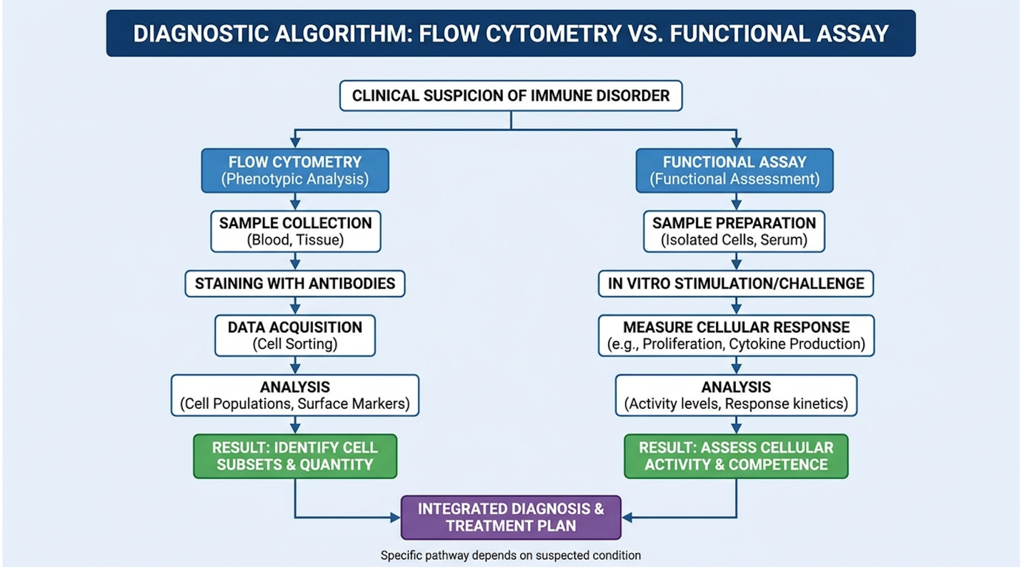
Figure 1: Diagnostic pathways for immunological assessment based on patient presentation.
If you ask your doctor for an immune check, they will likely order a flow cytometry panel. This test uses fluorescently labeled antibodies to count the absolute number of NK cells in your blood, looking specifically for cells that are CD45+, CD3-, and CD56+.
| 📌 If you’re interested in how surface markers are used to identify and verify cells in the lab, we have an interesting article that discusses the CD73 marker and how important it is for stem cell therapy, which you can read via the internal link. |
Here is the problem. Flow cytometry gives you a quantity. It tells you that the soldiers are on the battlefield. It does not tell you if their weapons work, if they are asleep, or if they are actively engaging the enemy. A patient may present with a completely normal absolute lymphocyte count (ALC) and standard NK percentage, yet suffer from defective granzyme B loading.
Patients frequently present with raging, reactivated Epstein-Barr Virus while their NK cell counts remain perfectly in the middle of the reference range. The standard test misses the pathology
To make this explicit, we must look at how these diagnostic tools fundamentally differ in their clinical utility.
| Parameter | Flow Cytometry (Count test) | Functional Assay (Activity test) | Clinical Use |
| What it Measures | Absolute number and percentage of NK cells | Amount of IFN-γ secreted after stimulation | Determines if cells are present vs functionally competent |
| Methodology | Surface marker binding (CD56/CD16) | Whole blood ex-vivo cytokine stimulation (ELISA) | Identifying physical deficiencies vs immune anergy |
| Output Type | Quantitative (cells/μL) | Qualitative / Functional (pg/mL) | Baseline immune tracking vs complex disease diagnostics |
| Clinical Value | Low (for functional disorders) | High (for functional disorders) | The NK cell count test is for basic screening; Activity is for diagnosis |
The limitations of quantitative analysis vs functional flow assays (2026) reported by ScienceDirect Assay Protocols prove that relying solely on counts is clinically negligent when functional defects are suspected.
So, how do we actually test what these cells can do? The modern gold standard is the NK cell functional assay, which has largely replaced the old, highly radioactive chromium-release assays of the past.
The NK Vue assay utilizes an engineered cytokine called Promoca to aggressively stimulate NK cells in a whole-blood sample. This proprietary recombinant protein artificially mimics a massive viral infection, forcing functional NK cells to secrete Interferon-gamma (IFN-γ). Laboratories then centrifuge the plasma and deploy an Enzyme-Linked Immunosorbent Assay (ELISA) to quantify the exact IFN-γ production in picograms per milliliter (pg/mL). This precise quantification allows clinicians to identify profound cellular anergy that traditional cell-counting methods completely overlook.
The logistical requirements are rigid but manageable. The blood must be drawn into specialized tubes containing the Promoca stimulant and placed in an incubator rapidly typically for exactly 24 hours. Temperature fluctuations can skew the functional output. Standard commercial labs often struggle with this timing, which is why these panels are frequently run through specialized reference labs. According to advancements in NK cell activity monitoring and whole-blood assays (2026) from Eurofins Viracor, this methodology allows for highly reproducible, clinically actionable data.
Not everyone needs this test. In fact, healthy people with a bit of a cold should absolutely not request an NK cell activity test. The results are highly sensitive to acute stressors, and without a baseline, a single low reading in a healthy person usually just causes unnecessary panic.
So, who qualifies? The ideal candidates fall into three strict clinical buckets. First, patients with refractory, constantly recurring viral infections (like systemic CMV or severe shingles) who fail standard antiviral therapies. Second, patients undergoing post-treatment monitoring for specific malignancies (like gastric, breast, or prostate cancer), where oncologists track minimal residual disease (MRD). A sudden drop in NK activity often precedes a measurable clinical relapse in these solid tumors. Third, under the strict supervision of a reproductive immunologist, an NK cell test may be utilized for women experiencing unexplained, consecutive early pregnancy losses when all genetic and anatomical causes have been ruled out.
When the functional data comes back, it changes everything. A verified low activity score pulls the physician away from blindly prescribing broad-spectrum antibiotics or standard immunosuppressants, shifting the focus entirely toward targeted immunomodulation. Patient selection criteria for advanced immunological assays (2026) provided by Clinical Testing Guidelines confirm that this diagnostic algorithm prevents wasted clinical time.
The desire to boost NK cells naturally is prevalent among patients with chronic conditions, but genuine immune modulation requires precise, evidence-led interventions. True upregulation of cytolytic capacity relies on optimizing biological pathways, managing autonomic nervous system stress, and, in refractory cases, considering advanced regenerative and cellular therapies. True upregulation of cytolytic capacity relies on optimizing biological pathways, making most over-the-counter ‘immune boosters’ practically worthless for severe disease. Evidence-based modulators regulate autonomic nervous system stress, reducing systemic cortisol by up to 30%; consequently, they prevent profound immune suppression in chronic disease.
| 📌 If you’re interested in how cellular therapies can help rebalance an overactive or exhausted immune system, we have an interesting article that discusses mesenchymal stem cell therapy for immune modulation, which you can read via the internal link. |
If you walk into a supplement store and ask how to increase NK cell activity naturally, you’ll be handed a basket full of unregulated powders and vaguely labeled “immune boosters.” Most of it is expensive urine. But there is one major exception: Vitamin D.
Vitamin D Receptors (VDR) reside directly on the surface of Natural Killer cells, making serum 25-hydroxyvitamin D a critical transcription factor for cytolytic function. When this nutrient binds to the VDR, it directly upregulates the specific genes responsible for perforin and granzyme production. Patients experience enhanced immune defense without triggering the dangerous pro-inflammatory cytokine storms often associated with unregulated herbal stimulants. This targeted biological pathway proves that clinical supplementation requires verified diagnostic testing first.
We also have to look at the evidence threshold for other marketed nutraceuticals. Zinc is essential for the structural integrity of the immunological synapse. Certain medicinal mushrooms (containing beta-glucans) have shown compelling in vitro activity in priming NK cell receptors. However, translating in vitro petri-dish data to in vivo human clinical benefit is notoriously difficult.
Crucially, attempting to “boost” an already hyperactive immune system is playing with fire. If a patient with a high NK cells autoimmune disease presentation takes massive doses of immune-stimulating herbs, they can trigger a devastating clinical flare. Nutritional modulation of innate lymphocyte function (2026) from the National Institutes of Health ODS emphasizes that modulation requires testing first, supplementation second. Beyond molecular substrates, the autonomic nervous system plays a direct regulatory role over circulating lymphocytes.
You can take all the Vitamin D in the world, but if your autonomic nervous system is locked in “fight or flight,” your immune system will suffer.
Let’s look at how to increase NK cell activity through physiological regulation. The neuroendocrine-immune axis is essentially a hardwired connection between your brain and your white blood cells. When you experience chronic psychological or physical stress, your adrenal glands pump out massive amounts of cortisol and catecholamines (like adrenaline). NK cells are highly sensitive to these hormones. Chronic cortisol exposure actively suppresses IFN-γ production and physically downregulates the activating receptors (like NKG2D) on the NK cell surface. Your immune system is literally being told to stand down.
Severe sleep deprivation acts through the exact same pathways. Even a single night of severely restricted sleep causes immediate, measurable cytolytic suppression.
Exercise is also a powerful modulator, but it requires a Goldilocks approach. Moderate aerobic exercise acts as a temporary stressor, causing a massive mobilization of highly cytotoxic NK cells into the bloodstream a brilliant, evolutionary mechanism for tissue repair. However, extreme endurance training (like running a marathon) causes severe post-exertional immune depression, creating an “open window” for opportunistic viral infections. Neuroendocrine regulation of natural
When lifestyle modifications and targeted supplementation fail to resolve a severe functional deficit, we enter the realm of advanced cellular medicine.
Stem cell therapy to boost immune system function requires rigorous clinical oversight. We must strictly distinguish legitimate, heavily regulated ex-vivo clinical NK cell therapy from unproven “stem cell cures” marketed on social media.
| 📌 If you’re wondering how to tell a legitimate, well-regulated stem cell clinic from an unsafe one, we have an interesting article that discusses whether stem cell therapy is safe in Thailand, which you can read via the internal link. |
In legitimate clinical protocols mostly restricted to oncology settings or approved trials autologous (your own) NK cells are extracted via leukapheresis. They are then expanded by the billions in a lab, aggressively activated with interleukins (IL-2 or IL-15), and reinfused into the patient as an army of hyper-lethal killers targeting specific tumors.
Alternatively, reputable regenerative medicine consultations might evaluate a patient for therapies utilizing Umbilical Cord Mesenchymal Stem Cells (UC-MSCs). MSCs don’t “become” new immune cells. Instead, their value lies in their secretomes the incredibly powerful exosomes and cytokines they release. These can promote phenotypic macrophage reprogramming, turning off severe systemic inflammation and allowing exhausted NK cells a chance to recover their function.
| 📌 If you’re curious why umbilical cord cells are often chosen for regenerative protocols, we have an interesting article that discusses why umbilical cord-derived UC-MSC stem cells are superior to other stem cell sources, which you can read via the internal link. |
Be prepared for the financial reality. The NK cell therapy cost for highly regulated, legitimate cellular therapies routinely exceeds $10,000 to $25,000 per protocol. Clinical pathways and cost projections for ex vivo NK cell expansion therapies (2026) provided by Phyathai Hospital Regenerative Medicine highlight the massive financial burden these cutting-edge procedures carry. Exploring these high-tier interventions demands extreme clinical caution, as regenerative medicine remains fraught with regulatory and safety limitations.
| 📌 If you’re interested in what regenerative cell therapies typically cost, we have an interesting article that discusses stem cell therapy costs in Thailand for 2025, which you can read via the internal link. |
Clinical diagnostics are rarely perfect. The pursuit of immunological data must be tempered with an understanding of what these tests can and cannot do. Measuring systemic immune markers provides broad clinical data; however, it cannot replace precise localized tissue diagnostics.
The biggest pitfall in interpreting an NK cell activity test is assuming that a single low reading equates to a permanent, structural immunodeficiency. Activity fluctuates wildly. Circadian rhythms, a poor night of sleep, acute mental stress, or a recent viral exposure can all suppress cytolytic function temporarily. Testing once and declaring a permanent defect is poor medicine.
Secondly, utilizing peripheral blood NK activity to blindly diagnose uterine or reproductive NK cell function without an endometrial biopsy is fundamentally flawed biology. They are different compartments with different cellular phenotypes. Limitations of peripheral blood assays in systemic disease (2026) outlined by Mayo Clinic Interpretive Guidelines stress this heavily. Measuring one compartment does not dictate the behavior of another.
Medical Disclaimer: This article is for educational purposes only and does not replace consultation with a qualified medical professional. Never alter your treatment plan based on internet research.
Don’t skip the basics trying to chase advanced diagnostics. If a patient presents with recurrent infections, a standard CBC, comprehensive lymphocyte subset panels (T cells, B cells, NK cells), and quantitative immunoglobulin levels (IgG, IgA, IgM) remain the undisputed first-line gold standards.
You have to rule out common defects first. If a patient has virtually no IgG antibodies, it doesn’t matter what their NK cells are doing they have a primary humoral immunodeficiency. According to the American Academy of Allergy, Asthma & Immunology, standard quantitative tests must precede functional assays. Functional testing should be strictly reserved as a secondary or tertiary diagnostic tool, deployed only after foundational immune defects have been entirely ruled out by a physician.
There is a hard threshold where patients must step away from self-management. Any decision to pursue advanced cellular therapy, aggressive immunomodulation with biologics, or expensive regenerative medicine consultations must be guided by a board-certified immunologist or a highly specialized hematologist/oncologist.
Attempting DIY interpretation of NK Vue panels is clinically dangerous. Complex immune deregulation mimics dozens of other systemic disorders. If you identify a functional deficit, your immediate next step is securing a referral to a specialist who understands the exact nuances of innate lymphocyte biology and can safely orchestrate a verified treatment protocol.
Natural Killer (NK) cells can directly destroy virally infected cells, malignant tumor cells, and stressed host cells without requiring prior pathogen exposure. They recognize targets lacking healthy “self” markers (MHC class I molecules). Upon recognition, they rapidly form an immunological synapse to lock onto the target. Once secured, they release cytotoxic proteins granzymes and perforins that induce cellular apoptosis. Their ability to destroy pathogens effectively is contingent on their functional activity levels rather than absolute cell counts.
You can confirm the presence of natural killer cells through a standard flow cytometry blood test, which quantifies the absolute number of CD56+ lymphocytes in your peripheral blood. A normal range typically indicates they account for 5% to 15% of your total circulating lymphocytes. However, merely detecting their physical presence does not verify their actual cytolytic activity. For that, you require specialized functional assays like the NK Vue test to measure cytokine secretion.
NK cells are stopped from being activated by inhibitory receptors binding to healthy MHC class I molecules present on the surface of normal autologous cells. This precise mechanism ensures immune tolerance and prevents the destruction of healthy tissue. Additionally, high levels of systemic cortisol from chronic stress or specific viral evasion tactics can profoundly suppress activating receptor expression and overall cytolytic function.
The primary symptoms of NK cell deficiency include unusually severe, recurrent, or atypical viral infections, particularly from the herpesvirus family (HSV, EBV, CMV, VZV) and human papillomavirus (HPV). Patients may also experience refractory chronic fatigue and an increased susceptibility to certain malignancies. Furthermore, unexplained recurrent miscarriages are sometimes linked to peripheral NK cell dysfunction. A clinical diagnosis requires a verified history of these recurrent infections combined with laboratory evidence of absent or functionally impaired NK cytotoxicity.
For patients managing complex autoimmune or viral conditions, an NK cell activity test provides critical diagnostic clarity that standard blood panels simply miss. Advanced functional assays like NK Vue verify actual cytolytic capacity the true measure of immune competence allowing physicians to differentiate between healthy cell counts and functional anergy. Clinical evidence demonstrates that significant functional impairment frequently occurs in patients with refractory recurrent viral presentations. The best clinical approach combines targeted functional testing with specialist-led evaluation to avoid inappropriate interventions.
Adopting The Functional Immune Assessment Framework (FIAF) is essential for protecting patients from diagnostic misinterpretation. By prioritizing qualitative action over numerical presence, FIAF ensures that clinicians are treating the biological reality of the immune system, rather than just treating a spreadsheet of lab values. This framework fundamentally alters how we approach chronic, treatment-resistant disease.
Take control of your diagnostic journey today by preparing a detailed history of your recurrent infections or inflammatory flares. If your clinical history aligns with the indications discussed,