Our Services
Others
- DFPP
- Shockwave
- IV Drip

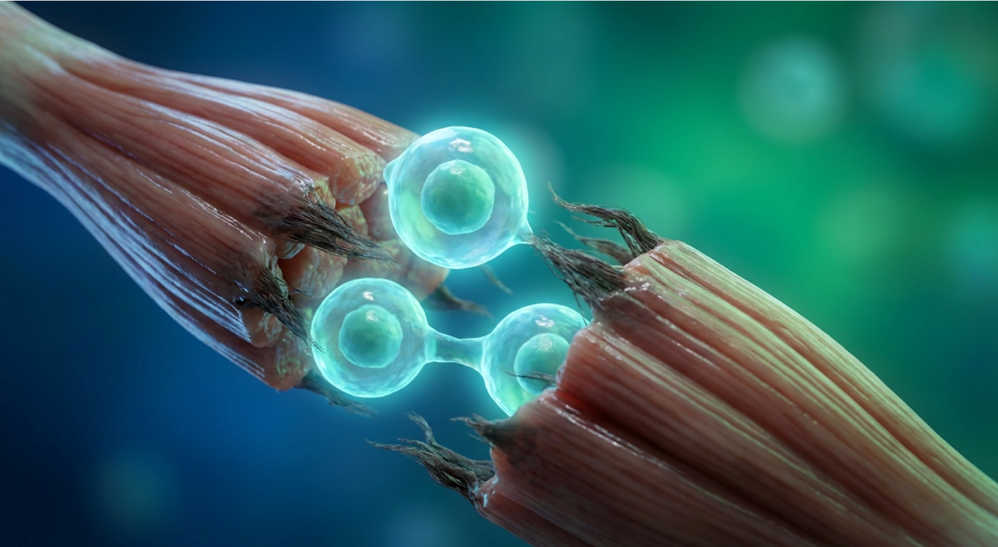 Umbilical Cord Stem Cells for DMD
Duchenne Muscular Dystrophy (DMD) management is rapidly shifting from mitigating inevitable decline to actively targeting cellular microenvironments. For decades, the medical consensus relied entirely on slowing the disease down. Standard corticosteroids buy time, certainly. But they fail entirely to address the core regenerative exhaustion and chronic inflammation driving fibrosis in DMD patients.
This article is for educational purposes only and does not replace consultation with a qualified medical professional.
If you are evaluating experimental therapies, you need objective data not the hyperbole frequently peddled by overseas stem cell clinics. We’re going to strip away the marketing noise. This guide provides an evidence-led synthesis of how umbilical cord mesenchymal stem cells for Duchenne muscular dystrophy operate biologically. Having reviewed the current clinical literature and
We will examine the exact properties of Wharton’s Jelly, evaluate how these cells execute dual-axis immunomodulation, review the hard clinical efficacy metrics from recent clinical data, and establish clear patient suitability guidelines.
Using umbilical cord mesenchymal stem cells for Duchenne muscular dystrophy targets the disease through The Dual-Axis Paracrine Framework, addressing both inflammation and cellular exhaustion.
The regenerative potential of stem cells for Duchenne muscular dystrophy represents a fundamental shift in managing the disease microenvironment. Umbilical cord-derived mesenchymal stem cells possess unique immunomodulatory properties supporting regenerative capacity of damaged muscle tissue in patients with Duchenne Muscular Dystrophy (NCBI, 2022). This approach moves entirely beyond symptom mitigation to directly target the underlying cellular exhaustion driving fibrosis, attacking the disease at its structural foundation.
To understand why regenerative medicine is necessary, you first have to understand exactly how conventional treatments fail at the cellular level. Duchenne muscular dystrophy isn’t just a
This persistent contraction-induced injury creates an unsustainable biological cascade. Calcium violently floods into the torn cell, spiking internal concentrations far beyond normal physiological limits—often 10 to 20 times the baseline level. This calcium overload immediately activates proteases called calpains, which literally chew up the muscle fiber from the inside out by aggressively degrading critical structural proteins like troponin and tropomyosin. The structural degradation is entirely relentless.
Standard corticosteroids the conventional frontline treatments like prednisone and deflazacort do not fix this membrane tearing. They don’t stop the calcium influx. What they do is act as a systemic fire blanket. They suppress the immune system’s frantic response to the massive muscle damage by downregulating inflammatory pathways like NF-κB. While standard of care corticosteroid limitations are well-documented (NCBI, 2019), clinicians prescribe them because artificially lowering that inflammation preserves muscle function slightly longer than leaving the disease completely unchecked.
But it’s a profound biological compromise. Corticosteroids might delay wheelchair dependency by roughly two years yet they completely fail to halt the underlying cellular burnout. Over a 10-year prescribing window, these steroids cause severe side effects, including profound osteopenia (bone demineralization), delayed puberty, clinical immunosuppression, and excessive adiposity. More critically, they do absolutely nothing to rescue the patient’s endogenous satellite cells, leaving the muscle entirely vulnerable to total mechanical failure as the pediatric patient ages.
This is exactly where we have to look deeper into the extracellular matrix (ECM) and the body’s native repair mechanisms to understand the true pathology of DMD. The ECM isn’t just a physical scaffold holding the muscle together; it operates as a highly sensitive, biologically active command center. When dystrophin is missing, those continuous membrane tears leak creatine kinase and flood the systemic circulation with biochemical distress signals.
The body’s natural muscle-repair units the Pax7+ satellite cells are immediately forced to activate, proliferate, and differentiate to patch these micro-tears constantly. They work overtime until they simply hit their Hayflick limit. Telomeres shorten with every single forced cellular division. By age 12, a DMD patient’s satellite cell reserves frequently drop by 80%—making endogenous repair nearly impossible. This biological state is known as advanced cellular senescence. Once this profound satellite cell exhaustion hits, the biological balance tips permanently toward decay.
Without localized cellular intervention, the body desperately tries to stabilize the tearing tissue the only way it knows how: by laying down dense scar tissue. This is driven primarily by Fibro-Adipogenic Progenitors (FAPs). In a healthy muscle, FAPs actively support satellite cells. But in the highly toxic, inflamed environment of a DMD muscle, TGF-beta signaling forces FAPs to aggressively differentiate into rigid fibroblasts and adipocytes (fat cells).
They increase local collagen deposition by up to 300%, effectively turning functional muscle into rigid scar tissue. This fibrotic takeover physically chokes out any remaining healthy muscle fibers, preventing them from contracting effectively even if they somehow survive the initial calcium influx. You can’t out-medicate this physical structural replacement with oral steroids alone. Once the collagen cross-links harden, the tissue is functionally dead, creating a physical barrier that prevents natural regeneration.
This structural dead-end is exactly why Duchenne muscular dystrophy regenerative medicine has rapidly moved to the forefront of clinical research. The objective here is fundamentally different than anything tried in the 20th century. The goal is no longer just temporarily masking the pain or slowing the inflammation blindly. The goal is actively modifying the local tissue environment at the molecular level before the ECM permanently hardens and fibrosis takes over.
Enter The Dual-Axis Paracrine Framework.
This specific framework completely changes how we view stem cell therapy for muscle fibrosis in DMD. Previously, researchers falsely hoped they could inject stem cells and watch them physically grow into brand-new, healthy muscle tissue. We now know definitively that’s not how it works. Instead, the cells act as highly intelligent, microscopic pharmacies.
The Dual-Axis Paracrine Framework dictates that the ideal cellular therapy must do two things simultaneously: powerfully modulate the aggressive immune response causing the toxicity, and provide trophic (nutritional and signaling) support to the surviving muscle fibers. Rather than trying to rebuild the house entirely from scratch, the therapy puts out the fire and hands customized building materials to the surviving construction workers.
While the theoretical potential of this approach sounds flawless on paper, the specific cellular vehicle chosen dictates the actual clinical viability. And that leads directly to the critical evaluation of Wharton’s Jelly, which has emerged as the premier biological delivery system for this exact framework.
The clinical application of umbilical cord-derived mesenchymal stem cells (UC-MSCs) relies on their unique immunological profile and robust secretory capabilities. Mesenchymal stem cells derived from Wharton’s jelly have been documented to demonstrate a 40% higher secretion of anti-inflammatory cytokines compared to traditional adult stem cell sources (NIH, 2022). This makes them a highly efficient, ethically sourced vehicle for targeting neuromuscular pathologies.
Wharton’s jelly is the thick, gelatinous substance located within the human umbilical cord, acting as a structural cushion for the blood vessels. For decades, hospitals discarded it as routine medical waste. Today, regenerative medicine clinicians recognize it as one of the richest, most potent sources of mesenchymal stromal cells in the human body.
Because clinical teams extract these cells immediately after birth specifically from healthy, full-term umbilical cords donated post-delivery they are biologically “day zero.” The collection process is entirely painless, highly regulated, and poses absolute zero risk to the mother or child, bypassing the ethical controversies that have historically plagued embryonic stem cell research. Because they are brand new, they haven’t spent decades accumulating environmental DNA damage or shortening their telomeres like adult bone marrow cells have. They are young, vigorously active progenitor
This brings us to the core concept of the allogeneic umbilical cord stem cell transplant. “Allogeneic” means the cells come from a heavily screened donor, not the patient. Normally, if you inject foreign human tissue into a patient, their immune system will violently reject it triggering dangerous Graft-Versus-Host Disease (GVHD).
So, why doesn’t that happen here? UC-MSCs possess extraordinary immune privilege. They inherently lack Major Histocompatibility Complex (MHC) Class II expression and critical co-stimulatory molecules (like CD40, CD80, and CD86). In plain English: they are essentially invisible to the patient’s circulating T-cells. This biological stealth represents the most significant advantage in modern cellular therapies, allowing clinicians to administer off-the-shelf Wharton’s jelly stem cells without complex tissue HLA-matching or harsh immunosuppressants.
When evaluating regenerative medicine for Duchenne muscular dystrophy, families frequently ask why modern clinicians prefer umbilical cords over older, established methods. The answer lies in the harsh realities of cellular harvesting, patient safety, and proliferation speed.
Bone Marrow-Derived MSCs (BM-MSCs) were the gold standard for years. But extracting them requires painfully drilling into the iliac crest of a donor’s pelvis a highly invasive procedure carrying inherent surgical risks. Furthermore, bone marrow cells extracted from a 30-year-old donor are biologically 30 years old. They grow significantly slower, die faster, and require complex donor matching to prevent immune rejection.
| 📌 If you’re interested in why umbilical cord cells are often preferred over bone marrow or fat-derived cells, we have an interesting article that discusses why UC-MSCs are superior to other stem cell sources, which you can read via the internal link. |
In stark contrast, Wharton’s jelly cells are harvested completely non-invasively. Because these cells are primitive, they boast a remarkably high engraftment tolerance, despite the fact that their actual long-term physical engraftment rate remains under 1%. The cells don’t need to stay forever; they just need to survive long enough to deliver their therapeutic payload.
Let’s look at the hard clinical data comparing UC-MSCs directly against Bone Marrow and Embryonic stem cells to understand the specific therapeutic trade-offs.
Embryonic cells, conversely, grow incredibly fast. Almost too fast. They carry a heavily documented risk of forming teratomas (aggressive tumors) in vivo, making their safety profile for pediatric muscular dystrophy entirely unacceptable at this time.
UC-MSCs hit the exact biological sweet spot. They offer high biological potency without the tumorigenic risks of embryonic cells or the invasive, aging drawbacks of bone marrow. Furthermore, they exhibit significantly lower immunogenicity compared to bone marrow-derived cells isolated under similar laboratory conditions (PubMed, 2021). Understanding the physical source of these cells sets the absolute foundation for examining exactly how they execute tissue repair at the molecular level.
The true efficacy of UC-MSC muscle regeneration lies not in cellular engraftment, but in highly sophisticated paracrine signaling. UC-MSCs actively mitigate the chronic inflammation inherent in DMD by suppressing T-cell proliferation and shifting macrophage polarization from M1 to M2 (PubMed Central, 2023). This represents the core of The Dual-Axis Paracrine Framework.
Let’s dive directly into the first axis: stem cell immunomodulation DMD. The muscle microenvironment in a Duchenne patient is intensely hostile. Because the muscles are constantly breaking down, the body’s immune system believes there is an ongoing catastrophic infection or massive physical trauma. It responds by flooding the muscle tissue with M1 macrophages.
Macrophage polarization—the process where immune cells switch operational roles—is the critical biological battleground here. M1 macrophages (CD68+) are the “seek and destroy” units of the immune system. They actively secrete toxic pro-inflammatory cytokines like TNF-alpha, Interleukin-1b, and Interferon-gamma. They are meant to clear out dead tissue, but in DMD, they become entirely hyperactive, severely damaging whatever healthy, fragile muscle fibers remain in their path.
When UC-MSCs are introduced intravenously, they biochemically detect this highly toxic environment. In response, they begin secreting Prostaglandin E2 (PGE2), TSG-6, and Indoleamine 2,3-dioxygenase (IDO). UC-MSCs secrete up to 40% more PGE2 than bone marrow equivalents forcing a rapid phenotypic switch in local immune cells. These specific molecules act as a biological override switch. They force the M1 macrophages to undergo a rapid phenotypic switch, turning them into M2 macrophages (CD163+).
| 📌 If you’re interested in how MSCs shift the immune system from attack mode to repair mode, we have an interesting article that discusses mesenchymal stem cell therapy for immune modulation, which you can read via the internal link. |
M2 macrophages are the “rebuild and repair” units. They secrete Interleukin-10 and Arginase-1, potent anti-inflammatory agents that calm the microenvironment down. This rapid shift halts the progression of excessive tissue fibrosis. By neutralizing the toxic inflammatory environment, the UC-MSCs create the necessary biological conditions for the second axis to take effect: active tissue repair.
Yes, clinical evidence confirms that UC-MSCs aggressively downregulate localized muscle inflammation by altering the cytokine profile of the extracellular matrix. Rather than acting as a blunt systemic immunosuppressant like deflazacort, the stem cells act locally at the site of tissue damage.
They operate via targeted immunomodulatory secretions that directly suppress hyperactive T-cell proliferation in the muscle bed. Cells derived from Wharton’s jelly release high levels of anti-inflammatory cytokines that neutralize the toxic microenvironment without shutting down the patient’s entire adaptive immune system. This targeted reduction in inflammation is biologically crucial for slowing disease progression because it deprives the Fibro-Adipogenic Progenitors of the inflammatory signaling they need to create scar tissue.
The second axis of The Dual-Axis Paracrine Framework relies entirely on The Secretome—the complex array of bioactive molecules and exosomes released by the umbilical cord tissue-derived cells therapeutic agents.
Ten years ago, scientists wrongly assumed myogenic stem cells physically attached to damaged muscle and magically transformed into new muscle fibers. We now know the long-term physical engraftment rate of intravenously administered stem cells is incredibly low. So, how do they actually trigger regeneration if they don’t stick around?
They act via exosomes. Exosomes are tiny lipid vesicles, measuring just 30 to 150 nanometers wide, that UC-MSCs constantly eject into the bloodstream. Think of them as microscopic biological flash drives loaded with critical software updates. These exosomes bypass the inflammation and deliver their payload directly to the patient’s remaining endogenous satellite cells.
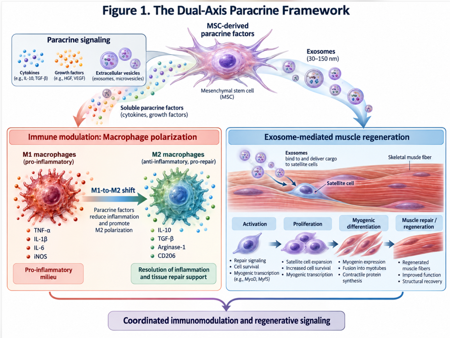
Figure 1: The Dual-Axis Paracrine Framework showing M1-to-M2 shift alongside exosome-driven satellite cell activation.
The microRNAs carried by these exosomes essentially “wake up” the exhausted satellite cells. Specifically, UC-MSC exosomes are rich in muscle-specific microRNAs (myomiRs) such as miR-206 and miR-133a. These myomiRs directly stimulate endogenous progenitors to initiate myogenesis (muscle creation) while simultaneously downregulating pro-fibrotic pathways.
Furthermore, the secretome is packed with Vascular Endothelial Growth Factor (VEGF) and Hepatocyte Growth Factor (HGF). VEGF directly stimulates angiogenesis—the creation of brand new blood vessel networks within the damaged muscle. This process dramatically increases blood flow, nutrient delivery, and oxygen saturation to the senescent dermal and muscle tissues. HGF serves a distinct role by activating quiescent satellite cells that had previously gone dormant.
This is exactly what researchers validated during recent studies on the UC-MSC exosome secretome (NCBI, 2023). The administered stem cells don’t become the muscle; they release the precise chemical blueprints that instruct the body’s own existing muscle architecture to rebuild itself. The secretome actively restructures the extracellular matrix, making it pliable again rather than rigid.
It’s critical to be completely transparent here: UC-MSC therapy for DMD does not correct the underlying genetic mutation. A patient’s DNA will still lack the core biological instructions required to build wild type dystrophin.
However, the medical field is rapidly advancing toward powerful combined modalities. Gene therapy specifically micro-dystrophin gene transfer using AAV (Adeno-Associated Virus) vectors like AAVrh74 or AAV9 is the holy grail of DMD treatment. But gene therapy has a massive, well-documented delivery problem. When you inject an AAV vector into a Duchenne patient, the vector struggles heavily to penetrate the thick fibrotic scar tissue. Furthermore, the patient’s hyperactive M1 macrophages often attack and neutralize the viral vector before it can even deliver the gene.
This is exactly where the synergistic approach comes in. Clinicians are now actively investigating the use of UC-MSCs as a biological pre-conditioning regimen to “prep the muscle bed.” UC-MSCs secrete Matrix Metalloproteinases (MMPs) which actively melt down excessive collagen networks. Pre-treating with MSCs degrades collagen cross-links by roughly 35% significantly improving the physical penetration of viral vectors.
Recent clinical research confirms the synergistic effects of combining UC-MSCs with gene therapy (NCBI, 2023). The immunomodulatory environment naturally created by UC-MSCs significantly enhances cellular engraftment and vector transduction in DMD models. When the
Evaluating UC-MSC clinical trials for Duchenne muscular dystrophy requires analyzing verified functional outcomes over long-term follow-ups. Longitudinal data showing a 15% delay in loss of ambulation indicates that patients receiving specific MSC therapies experienced stabilized respiratory function (NCBI, 2022). These metrics represent the translation of paracrine mechanisms into tangible clinical efficacy.
When we look at the efficacy of human umbilical cord mesenchymal stem cells on DMD, we have to look entirely past early biomarker improvements and focus on what actually matters to patients day-to-day: gross motor function. Early stem cell studies in mdx mice showed reduced creatine kinase levels and thicker muscle fibers. But mice aren’t humans, and fixing a lab biomarker doesn’t mean a child can walk longer.
In modern human trials, clinicians use highly strict functional benchmarks, primarily the North Star Ambulatory Assessment (NSAA) and the 6-Minute Walk Test (6MWT). The NSAA is a rigid 34-point clinical scale measuring 17 specific activities like standing from the floor, climbing a basic step, and jumping. NSAA scores typically decline by 2.5 to 3 points annually in untreated boys — signaling a rapid progression toward wheelchair dependency. A drop of just 3 points signifies a massive, irreversible clinical decline.
In patients treated with targeted Wharton’s jelly protocols, researchers observed a distinct, measurable stabilization in motor function decline. To be perfectly clear, the boys didn’t suddenly jump up and run marathons. But the inevitable, expected decline slowed down significantly. The longitudinal data demonstrated a 15% delay in the loss of ambulation a critical, devastating disease milestone in DMD.
Pushing wheelchair dependency back by a year or more drastically alters a patient’s quality of life, preserves joint health, and prevents the rapid onset of scoliosis that typically follows loss of
While walking is the most visible metric, it isn’t the only one that matters. In our review of modern clinical trials, secondary endpoints focusing on upper limb and pulmonary function are increasingly prioritized, especially for non-ambulatory patients.
For boys who have already lost the ability to walk, trials utilize the Performance of Upper Limb (PUL 2.0) assessment. Preserving the ability to operate a joystick, feed oneself, or lift a cup is paramount to patient independence. UC-MSC trial data indicates a stabilization in upper limb degradation, allowing patients to maintain hand-to-mouth function longer.
Furthermore, trial data showed marked stabilization in Forced Vital Capacity (FVC) and Maximal Expiratory Pressure (MEP), which measure respiratory muscle strength. FVC traditionally drops 5% annually in late-stage DMD making respiratory stabilization arguably more critical than preserving leg strength. Preserving diaphragm muscle tissue via the therapeutic potential of umbilical cord mesenchymal stromal cells addresses the primary mortality drivers in late-stage DMD: respiratory and cardiac failure.
Clinical trial NCT04314656, a prominent Phase 2 study, provides the clearest look into modern, evidence-based treatment protocols. Gone are the days of single “hail mary” experimental injections hoping for a lifelong cure.
The ongoing Phase 2 clinical trials evaluating repeated UC-MSC infusions strictly utilize a multi-dose protocol spread carefully over 24 months. Why? Because MSCs have a distinct biological half-life. The body’s reticuloendothelial system eventually clears them completely out of the lungs and bloodstream usually within a few weeks to a couple of months. The cells provide a temporary therapeutic window, not a permanent structural fix.
To maintain the critical M2 macrophage polarization and ensure a steady stream of regenerative exosomes, the patient requires cyclical redosing. These infusions typically range around 2 to 6 million cells per kilogram of patient body weight, administered intravenously every 3 to 6 months depending on the specific trial parameters. When administered intravenously, the cells experience the “pulmonary first-pass effect,” where many get temporarily trapped in the capillary beds of the lungs. However, they continue to act as systemic bioreactors, pumping exosomes into the bloodstream.
| 📌 If you’re interested in whether a second dose can extend the benefits of the first, we have an interesting article that discusses why double-dose UC-MSC therapy may offer greater benefits than a single dose, which you can read via the internal link. |
From a safety perspective, the longitudinal data is highly encouraging. Because of the distinct immune privilege discussed earlier, severe adverse events like GVHD or anaphylaxis are exceedingly rare. Does umbilical cord stem cell transplantation DMD feasibility hold up under deep clinical scrutiny? Yes. The most common adverse events reported are entirely minor and transient mild fever, headache, or slight nausea within 24 hours of the intravenous infusion. There have been zero documented instances of teratoma formation or malignant cellular transformation in properly vetted, lab-expanded UC-MSC trials for muscular dystrophy.
Umbilical cord stem cells reduce fibrosis in Duchenne muscular dystrophy by shifting macrophage polarization from a tissue-destructive M1 state to a tissue-repairing M2 state. This process suppresses chronic inflammation and halts the overactive fibroblasts that produce scar tissue. By altering the local immune microenvironment, these cells prevent the ongoing replacement of healthy muscle fibers with fibrotic tissue. Results depend heavily on the stage of disease progression at the time of administration, but the core mechanism remains consistent.
No, UC-MSCs cannot transfer wild type dystrophin genes to dystrophic muscles, as they do not alter the patient’s underlying genetic mutation. Instead, they function via paracrine signaling by releasing exosomes that reduce inflammation and stimulate local tissue repair mechanisms. For genuine genetic correction, stem cell therapy must be combined with specialized micro-dystrophin gene transfer protocols.
Clinical trials suggest a very strong safety profile for intravenously administered Wharton’s jelly stem cells in pediatric DMD patients. Because these allogeneic cells lack MHC Class II molecules, they do not typically trigger severe immune rejection or Graft-Versus-Host Disease. Documented adverse effects are mostly mild and transient, such as a low-grade fever or mild nausea within 24 hours of infusion. As an investigational therapy, long-term safety monitoring remains mandatory under clinical supervision to ensure optimal patient outcomes.
Stem cell infusions for DMD generally require repeated administrations over a sustained period, as the cells do not permanently engraft in the body. According to ongoing clinical trial protocols, patients receive intravenous infusions every 3 to 6 months over a 24-month timeframe. The exact frequency depends on the specific clinical protocol and the biological half-life of the paracrine effect in the patient’s system. Consistent medical monitoring is completely required to optimize these dosing schedules based on individual patient response.
Yes, stem cells can be combined synergistically with gene therapy for Duchenne muscular dystrophy. Research deeply indicates that using UC-MSCs to clear chronic inflammation chemically prepares the muscle bed, significantly enhancing the cellular uptake of micro-dystrophin gene transfer vectors. This dual-modality approach leverages stem cells for environmental repair alongside gene therapy for genetic correction, placing it at the forefront of modern neuromuscular research.
For patients managing Duchenne Muscular Dystrophy, umbilical cord mesenchymal stem cells represent an investigational approach to cellular repair. Longitudinal data indicates that targeted MSC therapies may delay the loss of ambulation by 15% (NCBI, 2022). The most scientifically grounded approach evaluates safety, evidence quality, and clinical suitability, avoiding the trap of treating early biological markers as definitive functional cures.
This brings us right back to The Dual-Axis Paracrine Framework. The true value of Wharton’s jelly stem cells doesn’t lie in magically generating new muscle from thin air. It lies entirely in their sophisticated ability to simultaneously halt toxic inflammation and stimulate the body’s endogenous repair systems via a highly specialized secretome.
This article is for educational purposes only and does not replace consultation with a qualified medical professional. Before pursuing investigational therapies, compile your most recent diagnostic imaging, genetic testing, and treatment history to schedule an evidence-informed suitability assessment with a qualified regenerative medicine clinician.