Our Services
Others
- DFPP
- Shockwave
- IV Drip

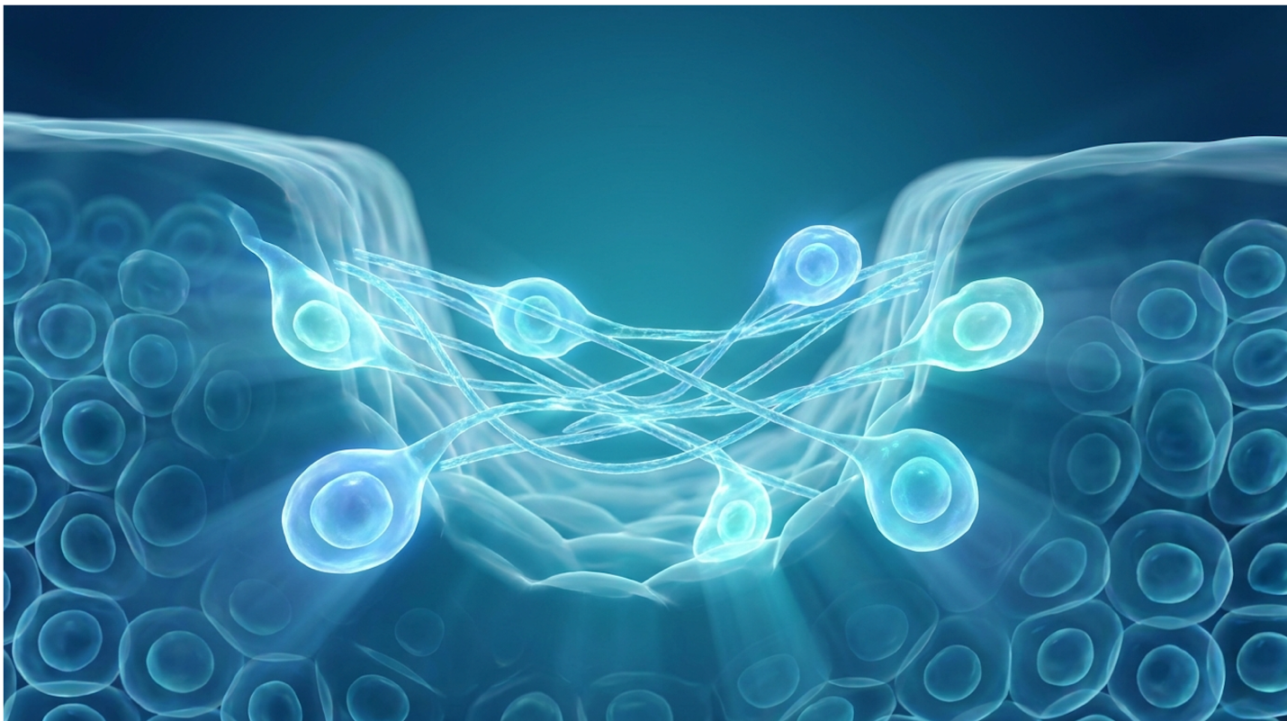
For patients evaluating advanced dermatological interventions for severe atrophic scarring, traditional modalities often provide limited structural repair. Chemical peels, ablative lasers, and mechanical resurfacing rely on a singular strategy: inflicting controlled tissue damage to force a healing response. But here’s the problem if your skin’s cellular infrastructure is already exhausted, demanding more from it yields diminishing returns.
Look, masking tissue deterioration doesn’t reverse the cellular senescence responsible for permanent dermal depressions. True recovery demands a biological intervention. Recent clinical data indicates that stem cell therapy for acne scars offers a fundamentally different mechanism for dermal regeneration. We aren’t talking about topical serums branded as “stem cell cosmetics.” We’re talking about living, laboratory-expanded cellular therapies administered in clinical environments.
Our clinical review indicates the gap between aesthetic marketing and biological reality is massive. In this guide, we analyze the clinical evidence behind UC-MSCs (Umbilical Cord Mesenchymal Stromal Cells) stem cell therapy for acne scars, evaluating its biological mechanisms, safety profile, and efficacy. We will review the paracrine signaling framework, compare targeted scar repair outcomes, and outline realistic expectations regarding clinical longevity and current cost.
UC-MSCs stem cell therapy for acne scars operates by promoting extracellular matrix synthesis and localized neovascularization rather than simply masking cosmetic defects.
Umbilical Cord Mesenchymal Stromal Cells (UC-MSCs) are multipotent progenitor cells that facilitate advanced cutaneous regeneration. The primary role of umbilical cord mesenchymal stromal cells in skin involves modulating local immune microenvironments and delivering vital nutrients to senescent dermal tissues. This makes them a focal point for modern tissue repair strategies.
Mesenchymal stromal cells accelerate cutaneous tissue repair primarily through paracrine signaling rather than direct cellular differentiation orchestrating localized healing pathways to radically increase native fibroblast activity (Nature research on MSCs, 2026).
When we discuss a legitimate stem cell scar treatment, precision matters. We have to separate the actual builders from the blood-replacers. In medicine, there are hematopoietic stem cells (HSCs) and mesenchymal stem cells (MSCs). Hematopoietic stem cells reside in bone marrow and cord blood; their entire job is to regenerate the blood and immune system. They are life-saving for leukemia, but they do virtually nothing to reconstruct skin.
Mesenchymal stem cells (MSCs) are the structural builders. Specifically, Umbilical Cord Mesenchymal Stromal Cells (UC-MSCs) are isolated from Wharton’s jelly the gelatinous connective tissue found within the umbilical cord. They are inherently multipotent. This means they possess the biological capacity to differentiate into various structural cell types, including osteoblasts, chondrocytes, and adipocytes.
| 📌 If you’re curious why umbilical cord cells are preferred over other stem cell sources, we have an interesting article that discusses why umbilical cord-derived UC-MSC stem cells are superior to other stem cell sources, which you can read via the internal link. |
But their real value in dermatology isn’t their ability to differentiate. It’s their immunoprivileged status. Because UC-MSCs lack the expression of HLA-DR antigens (the markers that typically trigger immune rejection), they can be utilized as an allogeneic treatment. Simply put, you can safely receive them from a donor without your immune system launching an aggressive attack. These cells are harvested entirely post-natally from discarded tissue, completely bypassing the ethical dilemmas associated with embryonic harvesting.
Clinical observations frequently reveal how pop-culture terminology confuses patients. Celebrities routinely boast about getting a “stem cell facial.” In reality? They are usually receiving Platelet-Rich Plasma (PRP) or, worse, plant-based apple stem cell extracts that have absolutely zero viability in human tissue. Which is better, stem cell or PRP? PRP relies entirely on your body’s existing, potentially aging platelets. Authentic UC-MSCs, cultivated in sterile, regulated laboratories, introduce young, highly active cellular machinery into the compromised tissue.
According to a comprehensive Nature research on MSCs, the viability and application of genuine MSCs drastically outperform basic blood derivatives in nearly every measurable metric. While understanding their origin is critical, their efficacy is entirely dependent on how they interact with existing skin structures.
As skin ages or suffers severe trauma from cystic acne, the extracellular matrix (ECM) deteriorates. The ECM is the physical scaffolding of your skin, primarily composed of collagen, elastin, and hyaluronic acid. When severe acne destroys this scaffolding, the overlying skin caves in, resulting in atrophic scarring.
UC-MSCs directly reconstruct this matrix by activating highly specific biochemical pathways. Through the ERK MAPK signaling pathway and the SIRT1 pathway, these cells protect existing structures from oxidative stress. ERK MAPK is heavily involved in regulating cellular proliferation, essentially acting as the biochemical alarm clock that tells sleeping dermal fibroblasts to wake up and get to work. Meanwhile, SIRT1 (a critical gene sequence associated with cellular longevity) shields the newly synthesized mitochondrial membranes from localized free radical damage.
By aggressively upregulating SIRT1, UC-MSCs actively delay the senescence (aging and functional death) of your native fibroblasts, giving them a biological second wind. This isn’t theoretical biology. It’s observable, quantifiable tissue repair. Once these pathways are fully activated, the synthesis of Type I collagen, fibronectin, and elastin skyrockets. Quiescent (sleeping) dermal fibroblasts wake up and begin furiously weaving new, healthy connective tissue to rebuild the missing scaffolding.
This mechanism is particularly effective at reversing cellular senescence the state where older cells refuse to divide but continue secreting toxic inflammatory markers. By clearing out these senescent cells and replacing them with a fresh ECM framework, the therapy prevents the excessive accumulation of melanin. This means it doesn’t just fill the physical depression; it actively reduces the dark, hyperpigmented shadows that often accompany deep scars.
Can stem cells heal acne scars? Current clinical data indicates that UC-MSCs offer an effective regenerative solution for skin repair by directly reconstructing damaged tissue. Rather than acting as a simple temporary dermal filler, mesenchymal stem cells address the underlying fibrotic tethering that causes atrophic depressions.
Stem cell therapies utilizing UC-MSCs directly reduce the depth of atrophic acne scars by stimulating Type 1 collagen synthesis establishing a permanent structural foundation rather than temporary volume (IFAAS clinical overview, 2026).
Not all acne scars are biologically identical, and treating them as such is a fundamental clinical error. We classify atrophic scars into three distinct morphological categories: icepick, boxcar, and rolling scars. Each presents a vastly unique structural deficit in the dermal architecture, and they respond differently to cellular therapies. Understanding these structural variations is the key to managing patient expectations.
Rolling scars feature undulating, tethered bands of fibrous tissue that anchor the epidermis deep into the subcutaneous layer and the SMAS (superficial musculoaponeurotic system). They create a wavy, uneven texture across the cheeks. UC-MSCs are incredibly effective here because they actively inhibit tissue-transforming growth factor (TGF-beta) signaling.
Why does this matter? Because TGF-beta is the primary driver that activates dermal myofibroblasts the specific cells responsible for pulling those fibrous bands tight. By interrupting this signal, UC-MSCs help release the tethering from the inside out. But let’s be honest about severe clinical tethering: mechanical subcision is often a required adjunct. A clinician uses a Nokor needle to mechanically slice those fibrous bands, creating a subdermal pocket. Flooding this newly created dead space with UC-MSCs prevents the bands from simply reattaching during the healing process, forcing the body to fill the void with new, healthy matrix instead.
Boxcar scars present broad depressions with sharply defined edges, while icepick scars are narrow, deep, V-shaped tracts extending deep into the dermis. The Paracrine Reset Framework addresses these volume deficits by flooding the base of the scar with localized growth factors, stimulating bottom-up cellular proliferation to push the depressed floor upward.
However, medical transparency is non-negotiable. While this biological intervention is powerful, it has distinct limitations. Is it a standalone treatment for ice pick acne scars of extreme depth? Not always. Extremely deep icepick tracts often possess highly epithelialized walls—meaning skin cells have grown down inside the scar tract, creating a tough lining that resists simple cellular remodeling. In these edge cases, targeted chemical destruction like TCA CROSS must frequently be paired with UC-MSC therapy to break down the scarred walls so the cells have a raw surface to rebuild upon.
A detailed IFAAS clinical overview underscores that while MSCs dramatically improve tissue volume, understanding morphological limitations ensures realistic patient expectations. You cannot ask a biological signal to override a physical obstruction without preparing the tissue first.
You cannot build new tissue without establishing supply lines. It’s biologically impossible. This is where neovascularization the formation of new blood vessels becomes the cornerstone of the best treatment for deep acne scars.
When UC-MSCs are introduced to scarred tissue, they immediately recognize the hypoxic (low-oxygen) environment characteristic of dense fibrosis. In response, they secrete massive amounts of Vascular Endothelial Growth Factor (VEGF). This specific molecule commands local endothelial cells to branch out, creating a dense micro-capillary network. This brand-new blood supply delivers the oxygen and raw materials required for sustained, long-term dermal repair.
With supply lines established, the focus shifts to the fibroblasts. Dermal fibroblasts are the factories that produce collagen. In scarred tissue, they are often dormant or producing disorganized, fibrotic scar tissue. The secretomes from the UC-MSCs stimulate these quiescent factories, forcing them to replace the disorganized scar tissue with healthy, beautifully aligned collagen networks.
Recently, clinical focus has rapidly expanded beyond the whole cell to the extracellular vesicles they produce: Exosomes. Exosomes are tiny, 30 to 150-nanometer extracellular vesicles. Because they lack a nucleus and native cellular hardware, they don’t trigger immune rejection and can rapidly cross tissue barriers. They are essentially biological envelopes packed with mRNA and microRNA that dictate fibroblast behavior.
Many advanced clinics now pair whole-cell UC-MSCs with purified exosomes to supercharge the signaling cascade. This dual-therapy combination yields a massive initial signaling spike from the exosomes to immediately wake up the fibroblasts, followed by sustained, long-term signaling from the engrafted live cells. Exosomes act as the immediate chemical messengers, while the UC-MSCs act as the sustained biological factories. An independent Ways2Well biological analysis highlights how this exact combination of neovascularization and exosomal signaling fundamentally rewrites the healing environment of damaged skin.
To appreciate the clinical shift UC-MSCs represent, we have to deeply contrast them with traditional dermatological staples: fractional CO2 lasers and microneedling.
Fractional lasers work via controlled thermal injury. They vaporize microscopic columns of skin, relying on acute inflammation to trigger a wound-healing cascade. Microneedling relies on mechanical injury to achieve a similar, though less aggressive, effect. Both methods operate on a simple principle: damage the skin to force it to rebuild itself.
This approach works beautifully for mild texture issues. But for severe atrophy, forcing already-exhausted senescent cells to work overtime through repeated thermal trauma often results in prolonged erythema (redness). In darker skin types (Fitzpatrick IV-VI), ablative thermal lasers carry a dangerously high risk of severe post-inflammatory hyperpigmentation (PIH). The intense heat from a CO2 laser can trigger melanocytes to overproduce pigment, leaving the patient with dark, permanent stamping marks.
UC-MSCs flip this model entirely. Instead of relying on acute inflammation to trigger healing, they actively suppress chronic inflammation while directly supplying the regenerative materials. You aren’t burning the tissue to stimulate it; you are feeding it. This makes the biological approach vastly superior for those seeking the best acne scar treatment for sensitive or melanin-rich skin.
| 📌 If you’re interested in how UC-MSCs calm chronic inflammation in damaged tissue, we have an interesting article that discusses mesenchymal stem cell therapy for immune modulation, which you can read via the internal link. |
When comparing stem cell therapy vs PRP for acne scars, the distinction becomes even sharper. PRP utilizes your own plasma, meaning the quality of the regenerative signal degrades directly in line with your chronological age and baseline systemic health. UC-MSCs provide a standardized, highly potent Day-Zero biological signal regardless of the patient’s age.
| Modality | Mechanism of Action | Inflammation Response | Typical Downtime |
| Fractional CO2 Laser | Controlled thermal ablation of epidermis/dermis | High acute inflammation, thermal stress | 7-14 days |
| Microneedling | Mechanical micro-injury to trigger native healing | Moderate acute inflammation | 3-5 days |
| PRP Injection | Autologous platelet degranulation | Mild, dependent on patient baseline | 1-3 days |
| UC-MSC Therapy | Biological paracrine signaling & ECM synthesis | Active suppression of inflammation | 1-2 days |
While the biological advantages are clear, patients must critically evaluate the practical timeline and financial commitment required for these advanced therapies.
Evaluating stem cell therapy cost requires analyzing both the initial financial investment and the mesenchymal stem cell therapy success rate. Because UC-MSCs induce fundamental biological changes rather than temporary cosmetic volumization, patients must plan for a progressive healing timeline and protocol-driven session frequencies to achieve optimal results.
The biological remodeling initiated by mesenchymal stem cell therapy typically requires 8 to 12 weeks to demonstrate measurable reductions in acne scar depth dictating patience over instant cosmetic fixes (Clinical protocols overview, 2026).
If you want instant results, get a hyaluronic acid dermal filler. It will perfectly mask your boxcar scar in thirty seconds and dissolve completely in eight months. Regenerative medicine does not work on a cosmetic timeline; it operates on a biological one.
The biological remodeling initiated by mesenchymal stem cell therapy typically requires 8 to 12 weeks to demonstrate measurable reductions in acne scar depth. Cellular regeneration and the synthesis of structurally sound Type I and Type III collagen take weeks, not hours.
During Weeks 1 to 2, the primary biological action is the rapid reduction of localized inflammation. The tissue is calming down, and macrophages are shifting into their M2 repair phase. You won’t see dramatic scar filling yet, though overall skin tone often brightens significantly as systemic inflammation drops.
From Weeks 3 to 6, neovascularization takes firm hold. Angiogenesis is actively building new capillaries, and fibroblast activity spikes. During this phase, clinicians closely monitor skin turgor and post-inflammatory erythema (PIE). Because robust capillary loops are actively forming, the skin might look intensely pink but this is a sign of healthy vascularization, not thermal damage. You might notice the skin feels thicker and substantially more resilient to the touch.
It is during Weeks 8 to 12 that the visible remodeling of atrophic scar depth becomes truly apparent. The newly synthesized extracellular matrix begins to firmly push up the floor of the dermal depressions. Beyond week 12, the tissue enters a prolonged collagen maturation phase where the new matrix fully stabilizes and cross-links for durability.
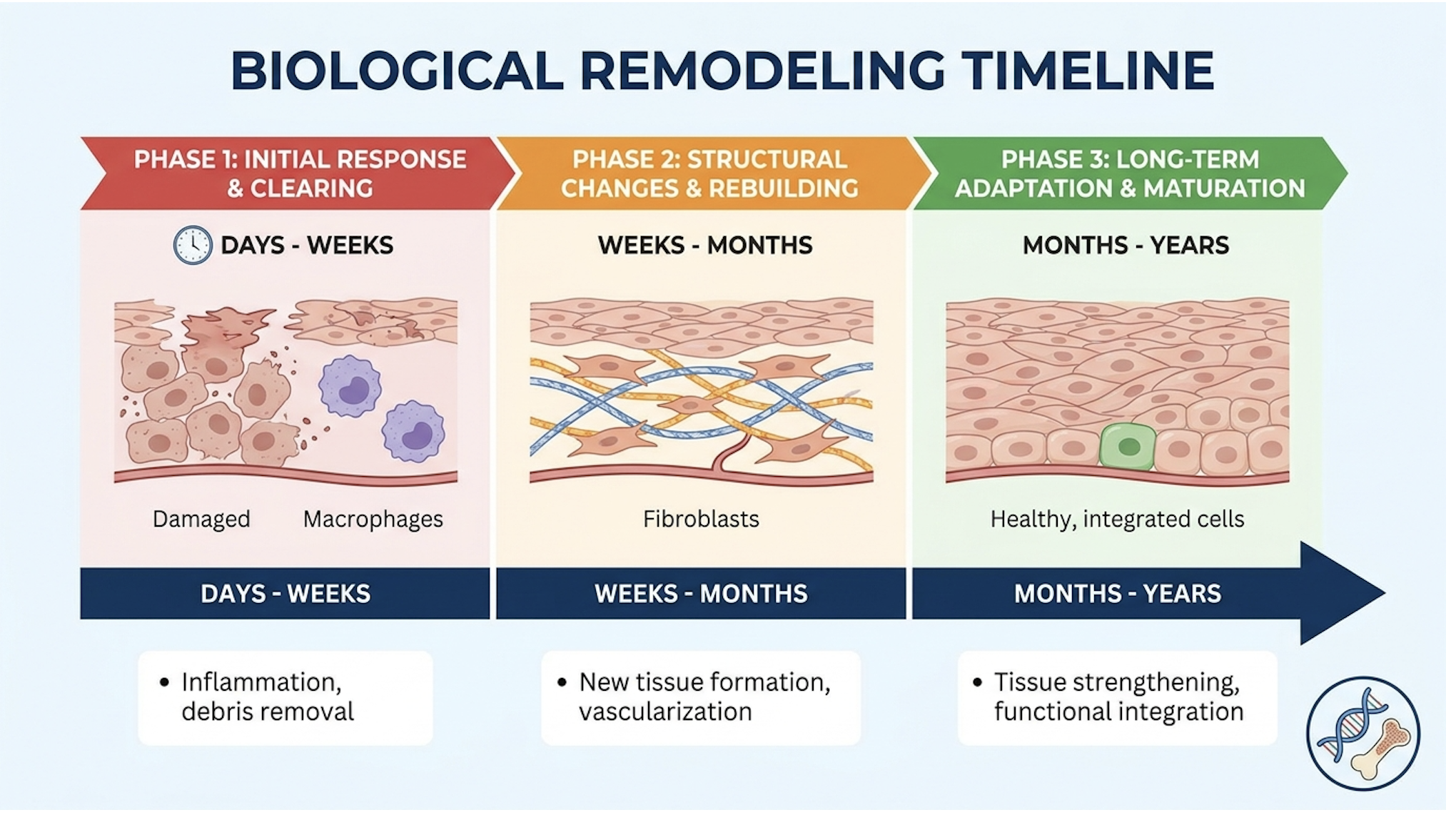
This progressive timeline stands in stark contrast to the instant, temporary swelling seen immediately after ablative laser treatments swelling that often tricks patients into thinking their scars have vanished, only for them to reappear exactly as before weeks later.
What does “success” actually mean in clinical dermatology? It rarely means a 100% flawless erasure of severe tissue damage. A medically accurate success rate for UC-MSCs refers to a measurable, volumetric reduction in scar depth and a statistical improvement on validated skin texture scoring systems.
Current clinical literature supports significant, quantifiable improvement. Patients routinely see a marked volumetric reduction in the depth of rolling and boxcar scars as the underlying matrix rebuilds. The skin’s tensile strength increases dramatically, and fibrotic tethering loosens, allowing the skin to drape more naturally over the underlying fat pads.
But how many sessions are needed for acne scar treatment? You simply cannot rebuild a ruined foundation in a single afternoon. Standardized clinical protocols generally prescribe 1 to 3 sessions, spaced approximately 3 to 6 months apart. This specific spacing allows the clinician to evaluate the full 12-week biological remodeling cycle before introducing a subsequent cellular load.
A stem cell acne scar treatment before and after portfolio from a reputable clinic should reflect this progressive timeline, showing gradual structural integration rather than overnight miracles. According to a clinical protocols overview, adhering to strictly timed sessions dramatically impacts long-term longevity and overall patient satisfaction.
Transparency regarding the financial commitment is mandatory. Stem cell therapy is expensive. This isn’t a simple topical cream mixed in a back room; it is a highly regulated, temperature-sensitive biological product.
The high cost is driven by incredibly rigorous laboratory standards. Cultivating UC-MSCs requires ISO-certified cleanroom environments, continuous cellular viability testing, pathogen screening, and specialized cold-chain logistics for transport. Live cellular products require cryogenic freezing and strict temperature monitoring during transit, contributing heavily to the final invoice. Furthermore, administration must be performed by a highly trained physician capable of targeted dermal placement.
| 📌 If you’re wondering why cell viability testing is so important for treatment quality, we have an interesting article that discusses the importance of cell viability in UC-MSC stem cell therapy, which you can read via the internal link. |
For patients looking at international pricing often a global hub for medical tourism legitimate therapies typically range from $2,500 to $6,000 per session in places like Bangkok or Latin America, while US-based investigational clinics can charge upwards of $5,000 to $10,000 per vial. This depends heavily on the exact cellular count (usually 20 million to 50 million cells per vial) and the clinic’s regulatory standing. If a clinic offers stem cells for a suspiciously low $300, you are almost certainly receiving unviable cellular debris or basic PRP, not laboratory-expanded UC-MSCs.
| 📌 If you’re interested in a detailed breakdown of treatment pricing in Bangkok, we have an interesting article that discusses stem cell therapy costs in Thailand for 2025, which you can read via the internal link. |
However, you must frame this cost against clinical longevity. Hyaluronic fillers require bi-annual top-ups indefinitely. Laser packages require weeks of cumulative downtime and lost wages. Because UC-MSC therapy repairs the underlying dermal matrix, the resulting collagen integration is permanent.
Understanding the mesenchymal stem cells controversy requires separating ethical debates surrounding embryonic cells from the clinical reality of umbilical cord derivatives. Furthermore, concerns regarding stem cell therapy cancer risk often stem from misunderstandings about cellular proliferation, making objective risk assessment essential for prospective patients.
Umbilical cord mesenchymal stromal cells exhibit a remarkably strong safety profile in clinical trials entirely lacking the teratoma-forming risks associated with pluripotent embryonic cells (Springer clinical review, 2026).
The term “stem cells” carries heavy historical baggage. Much of the ethical controversy centers entirely on embryonic stem cells, which require the destruction of an embryo to harvest.
Let’s clear the air immediately: UC-MSCs have absolutely nothing to do with embryonic harvesting. They are ethically sourced from discarded Wharton’s jelly within the umbilical cord following a healthy, full-term birth, with full, documented maternal consent. They bypass the ethical debates entirely.
However, the regulatory environment is intensely complex. In the United States, the FDA classifies extensively cultured, laboratory-expanded MSCs as a ‘351 biological product’ or drug. This categorization mandates a comprehensive Biologics License Application (BLA) and multi-phase clinical trials for approval. While this strict framework maximizes patient safety, it dramatically restricts domestic access to advanced cellular expansion, meaning UC-MSC applications in dermatology often remain investigational or are utilized off-label.
Conversely, regulatory bodies in international medical hubs like Japan, South Korea, and specific European jurisdictions have instituted accelerated, conditional approval pathways for regenerative medicine. This allows for earlier clinical access under rigorous monitoring. The real danger isn’t the cells themselves; it’s the unregulated clinics. A major downside to stem cell therapy is the proliferation of “bad actors” in the aesthetic market offering unverified treatments. Patients must demand transparent viability reports. If a clinic cannot provide a Certificate of Analysis (CoA) proving pathogen screening and exact cell counts, you are putting your health at risk.
Does stem cell therapy cause cancer? This is the most common, and most profoundly misunderstood, question in regenerative medicine. We must look at the exact biology of cellular proliferation to answer it accurately.
Pluripotent embryonic cells can turn into literally any tissue in the body. If injected haphazardly, they have a known biological risk of forming teratomas (benign tumors containing chaotic arrays of various tissue types). UC-MSCs are multipotent, not pluripotent. They have a highly restricted differentiation potential. They simply do not possess the biological programming to spiral into unchecked, chaotic growth.
Tumorigenesis the biological formation of a tumor requires a specific set of genetic mutations. Clinical reviews tracking thousands of patients over decades indicate that properly cultured, laboratory-verified UC-MSCs do not undergo malignant transformation in vivo. They arrive, signal the local fibroblasts, secrete their growth factors, and eventually undergo a strictly regulated process of natural apoptosis (programmed cellular death) after a few weeks, leaving behind the newly built collagen matrix.

So, what are the actual disadvantages of stem cells in this context? The risks are far more mundane. They include localized injection site reactions (mild bruising or swelling), the potential for infection if strict sterile protocols are violated during microneedling with stem cells, and severe financial loss if the therapy is administered to a poor candidate, yielding subpar results.
Regenerative medicine is not magic, and it is absolutely not for everyone. Identifying who is NOT a good candidate is just as critical as knowing who is. Some patients simply do not possess the biological environment necessary for UC-MSCs to thrive.
Here is the objective clinical exclusion criteria. You are not a candidate for stem cell therapy if you have:
Furthermore, patients seeking to treat active, cystic acne are terrible candidates. You must aggressively stabilize the acne first. If you inject expensive, pristine UC-MSCs into a dermis currently boiling with P. acnes bacteria and active inflammation, the toxic environment will destroy the secretomes before they can initiate tissue repair.
Finally, expectations matter. If a patient demands the 100% flawless removal of severe, tethered icepick scars without being willing to undergo adjunctive surgical therapies like subcision or TCA CROSS, they are a poor candidate. This strict patient selection underscores the broader clinical limitations inherent in regenerative aesthetics.
The aesthetic industry frequently prioritizes aggressive marketing over conservative medicine. A massive clinical pitfall is pursuing treatment at commercial “med-spas” lacking transparent laboratory standards. If a provider cannot show you the cold-chain transport logs and cellular viability reports from the day of your treatment, you risk receiving entirely dead cellular debris. Injecting dead cells results in zero biological remodeling it’s an incredibly expensive placebo.
| 📌 If you’re wondering how to choose a safe, properly regulated stem cell clinic, we have an interesting article that discusses whether stem cell therapy is safe in Thailand, which you can read via the internal link. |
Another frequent failure point is timing. Treating active acne rather than stabilized acne scars is a recipe for catastrophic clinical failure. Active P. acnes bacteria and high levels of existing acute inflammation create a hostile local microenvironment. This toxicity can completely neutralize the regenerative secretomes, essentially wasting the biological potential of the UC-MSCs. Tissue must be quiet and stabilized before we ask it to rebuild.
There are specific clinical scenarios where highly advanced cellular therapy is an unjustified overreaction. If your primary concern is purely superficial hyperpigmentation, macular erythema (red flat marks left after breakouts), or minor textural roughness, UC-MSCs are entirely unnecessary.
For these superficial issues, basic chemical peels (like TCA or Jessner’s) or targeted vascular lasers (like Vbeam) are far more cost-effective and clinically appropriate. Cellular therapy is designed exclusively for deep structural deficits the architectural collapses of the dermis. Deploying a premium biological intervention for a superficial epidermal issue is poor resource management. We frame this as informed decision-making: always match the intensity of the treatment to the severity of the biological deficit.
As noted in the medical disclaimer opening this guide, online educational content can never replace a hands-on, localized tissue assessment. Patients must actively seek a suitability assessment with a qualified, board-certified clinician at a reputable dermatology center.
A proper consultation involves extensively reviewing your comprehensive medical records, analyzing your prior treatment history (what lasers failed, what medications you take), and aligning on functional goals. A clinician needs to physically palpate your scars to check for underlying tethering. Do not skip the diagnostic phase in a rush to receive treatment.
What is the success rate of mesenchymal stem cell therapy for scars? Clinical success rates for mesenchymal stem cell therapy in treating acne scars are generally favorable, demonstrating measurable biological improvements in skin texture and scar depth. Success is typically defined as a volumetric reduction in atrophic depressions rather than complete scar erasure. Individual biological responses vary widely depending on age, scar morphology, and cellular viability. Patients must consult a dermatologist to establish realistic baseline expectations.
How long does mesenchymal stem cell therapy last? The structural improvements achieved through mesenchymal stem cell therapy are highly durable, often lasting several years. Because the therapy stimulates fundamental biological tissue remodeling and native collagen synthesis, the newly formed extracellular matrix integrates permanently into the dermal layers. Routine dermatological monitoring helps maintain long-term skin health.
Can stem cells turn cancerous after cosmetic treatments? Properly cultured multipotent umbilical cord mesenchymal stem cells (UC-MSCs) carry an exceedingly low risk of turning cancerous. Unlike pluripotent embryonic stem cells, which have the biological potential to form teratomas, UC-MSCs possess a restricted differentiation profile. Clinical data shows that UC-MSCs utilized in regulated therapeutic settings do not inherently promote malignant transformation. However, theoretical risks exist if stem cells are introduced to preexisting active malignancies, making rigorous medical screening mandatory prior to treatment.
Who is not a good candidate for stem cell therapy? Individuals with active systemic infections, severe unmanaged autoimmune disorders, or a history of active malignancies are generally not candidates for stem cell therapy. Additionally, patients with active inflammatory acne flare-ups must stabilize their skin condition before addressing the resulting atrophic scars. Treatment recommendations should only follow a thorough clinical review.
For patients facing severe atrophic acne scars, UC-MSCs stem cell therapy delivers targeted biological tissue remodeling. Stem cell therapies can significantly reduce scar depth by stimulating localized collagen synthesis. The most evidence-informed approach combines rigorous patient suitability assessments, laboratory-verified cellular viability, and adherence to progressive healing timelines. By stepping away from the endless cycle of thermal trauma and mechanical injury, patients can finally address the root architectural failure of their skin.
The efficacy of this treatment relies entirely on The Paracrine Reset Framework. By utilizing cellular secretomes to reprogram local immune responses and stimulate quiescent fibroblasts, patients can move beyond superficial cosmetic masking to achieve genuine structural repair of the dermal matrix. It is this precise biochemical cascade that separates verifiable medical interventions from the noise of the commercial beauty industry.
Because regenerative medicine is complex and highly individualized, the next step is securing a clinical assessment. Schedule a comprehensive consultation with a qualified medical specialist to evaluate your dermatological history, review your specific scar morphology, and determine if cellular therapy aligns with your functional goals. Do not let aesthetic frustration push you into unverified clinics; demand transparency, verify laboratory credentials, and prioritize your long-term biological safety.