Our Services
Others
- DFPP
- Shockwave
- IV Drip

This article is for educational purposes only and does not replace consultation with a qualified medical professional.
For patients and researchers evaluating regenerative therapies, the persistent challenge is clinical inconsistency. Standard applications of umbilical cord stem cells for inflammation offer profound biological plausibility, yet their real-world behavior relies entirely on their immediate microenvironment.
Injecting quiescent, naive stem cells into a highly inflammatory patient environment without cellular pre-conditioning leaves clinical success completely to chance. Clinical data reviews consistently show that unprepared cells simply undergo apoptosis when overwhelmed by a cytokine storm. They die before they can help. But by understanding the precise mechanisms of endotoxin priming and cellular polarization, clinicians and patients can critically evaluate protocols designed to guarantee immunoregulatory output rather than hoping for the best.
This analysis examines the biological foundations of UC-MSCs, the specific pathways of laboratory pre-conditioning, clinical outcomes in severe autoimmune conditions, and the inevitable shift toward cell-free extracellular vesicle therapies. We’ll strip away the commercial marketing. Here is the raw, unvarnished biological reality of how these cells actually function.
Utilizing umbilical cord stem cells for inflammation requires advanced laboratory pre-conditioning to ensure consistent therapeutic efficacy.
The fundamental misunderstanding about stem cell therapy is that these cells simply act as building blocks. They don’t. When evaluating UC-MSC stem cell therapy, you have to discard the idea that these cells physically replace damaged tissue. Instead, they act as highly sophisticated biological managers. They assess the local damage, release a specific cocktail of proteins, and force your body’s existing immune system to calm down and repair the area.
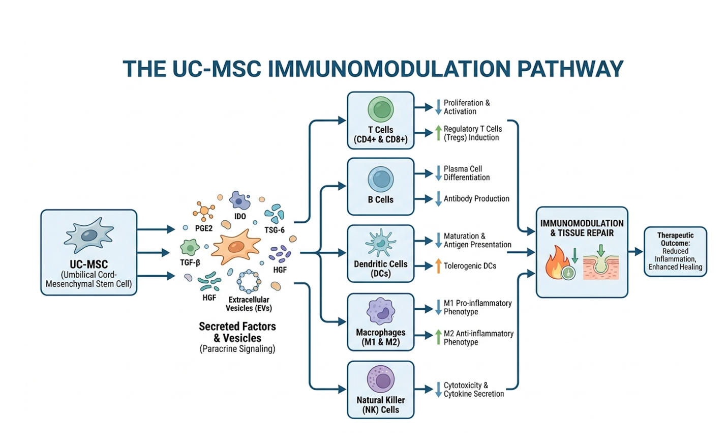
Umbilical Cord-derived Mesenchymal Stem Cells (UC-MSCs) are an abundant source of active progenitor cells, widely utilized for their immense therapeutic plasticity. But their true clinical value lies in their paracrine signaling mechanisms the way they communicate chemically with surrounding tissues.
Rather than differentiating into new colon lining or new joint cartilage, UC-MSCs secrete a vast array of bioactive molecules. We’re talking about vascular endothelial growth factor (VEGF), fibroblast growth factors (FGF), and a host of immunomodulatory cytokines. Mesenchymal stem cells act as biological sensors that dynamically suppress overactive immune responses by promoting regulatory T cell expansion (NCBI, 2023). Current clinical research indicates that mesenchymal stromal cells have significant applications for broad immune modulation, far beyond simple tissue regeneration (2016). They exhibit remarkable plasticity. This means their behavior changes based on what they encounter. If they sense acute inflammation, they upregulate defensive proteins. If they sense tissue ischemia (lack of oxygen), they push out factors to grow new blood vessels. Evidence-informed evaluations of cellular therapy must start by recognizing this adaptive signaling behavior, rather than relying on outdated models of simple tissue replacement. A 2023 study found that MSCs exposed to inflammatory cytokines increase their secretion of immunomodulatory proteins by over 40% so patients with higher baseline inflammation often trigger a stronger initial cellular response.
To understand how this halts autoimmune attacks, you have to look at Regulatory T cells (Tregs). Tregs govern immune tolerance. They are the brakes of the immune system. In therapy-resistant autoimmune diseases, these brakes have essentially failed, allowing effector T cells to run rampant and destroy healthy host tissue.
The biological mechanism of regulatory T cells expansion by MSCs is fascinating and deeply complex. When a UC-MSC encounters an inflammatory environment, it secretes transforming growth factor-beta (TGF-β), hepatocyte growth factor (HGF), and other signaling molecules that directly interact with naive T cells. This interaction forces those naive T cells to convert into FoxP3+ Tregs. The FoxP3 transcription factor is the master regulator here; once activated, it locks the T cell into a permanently protective, anti-inflammatory role.
Why does this matter? Because this differs fundamentally from broad immunosuppressive drugs like corticosteroids. Steroids carpet-bomb the entire immune system, causing massive lymphocyte death. This leaves the patient globally immunocompromised and vulnerable to basic infections. UC-MSCs, conversely, induce targeted tolerance. They expand the specific Treg populations needed to halt autoimmune destruction directly at the site of inflammation, without destroying the patient’s baseline ability to fight off a common cold or environmental pathogen.
| 📌 If you’re interested in how UC-MSCs rebalance the immune system without broadly suppressing it, we have an interesting article that discusses mesenchymal stem cell therapy for immune modulation, which you can read via the internal link. |
When patients ask how umbilical cord stem cells reduce inflammation on a cellular level, the answer is a multi-pronged suppression strategy. They don’t just target a single pathway. Instead, they act as broad-spectrum immune regulators that physically alter how your body’s defensive cells behave.
First, they secrete specific bioactive molecules that actively suppress the overactive components of the immune system, forcing aggressive cells to power down. They utilize intense paracrine signaling to communicate with surrounding immune profiles, fundamentally reprogramming pro-inflammatory agents into anti-inflammatory states. Simultaneously, they expand the regulatory T cell populations that serve as the body’s natural brakes against autoimmune attacks.
When optimized through laboratory endotoxin pre-conditioning, these cells become pharmaceutical factories, producing significantly higher levels of critical immunomodulators like PGE2. However, it’s critical to remember that individual clinical results depend entirely on proper suitability assessments, the inflammatory microenvironment, and the specific pathology being treated.
The immune modulation doesn’t stop with T cells. It heavily targets the innate immune system’s frontline responders: macrophages.
In chronic inflammation, tissue-resident macrophages get stuck in what we call an M1 phenotype. They are pro-inflammatory, highly aggressive, and constantly releasing cytokines like TNF-alpha and Interleukin-6 that damage host tissue. UC-MSCs initiate a profound phenotypic macrophage reprogramming. Through the release of specific factors notably Prostaglandin E2 (PGE2) they force these aggressive M1 macrophages to switch into an M2 anti-inflammatory state.
This M1 to M2 switch is critical for clinical recovery. M2 macrophages clean up cellular debris, promote SIRT1 pathways for clearing reactive oxygen species (ROS), and actively assist in extracellular matrix repair. Furthermore, the stem cells inhibit excessive Toll-like receptor signaling in these macrophages, preventing them from overreacting to future stimuli.
But the cellular interaction goes even further, expanding to other critical immune profiles. UC-MSCs actively suppress Natural Killer (NK) cell proliferation and cytotoxicity by releasing Human Leukocyte Antigen-G5 (HLA-G5) and IDO. They also interact directly with B cells, halting their maturation and actively inhibiting the production of destructive auto-antibodies. They essentially cast a massive regulatory net over the entire immune cascade.
Here is the major clinical catch, though. While these baseline functions are incredibly potent in a petri dish, relying on them in a highly inflamed human patient often yields wildly inconsistent results. The naive cells simply get overwhelmed. Which necessitates advanced, controlled laboratory pre-conditioning before they ever reach the treatment room.
If you take a vial of naive, standard-cultured stem cells and inject them intravenously into a patient suffering from a massive autoimmune flare-up, you are throwing unarmed soldiers into a toxic warzone. The cells undergo rapid apoptosis. They are destroyed by the highly toxic, oxidative environment before they can initiate the M1 to M2 macrophage switch or expand Treg populations.
| 📌 If you’re curious why keeping cells alive and active matters so much for treatment results, we have an interesting article that discusses the importance of cell viability in UC-MSC stem cell therapy, which you can read via the internal link. |
This is the dirty secret of commercial stem cell clinics. They aggressively sell the biological potential of the cell, but completely ignore the reality of the patient’s microenvironment. Commercial clinics that ignore pre-conditioning are essentially gambling with the patient’s biological response, relying on luck rather than optimized cellular mechanics. To fix this, high-tier research facilities use targeted endotoxin pre-conditioning to essentially arm and armor the cells before they enter the human body.
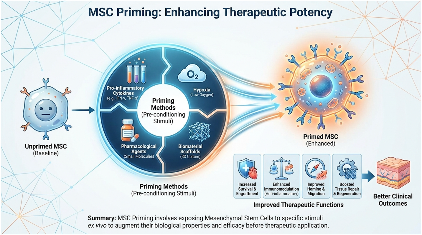
The Immunomodulatory Polarization Framework (IPF) dictates that exposing stem cells to controlled endotoxins activates critical defensive pathways prior to patient administration. To understand this pre-conditioning, we must look at how researchers manipulate the cellular state inside the laboratory.
At baseline, an unprimed stem cell exists in a relatively neutral, quiescent state. When placed in culture, technicians can force a concept known as MSC polarization. Depending on how they are stimulated in the bioreactor, MSCs can adopt one of two distinct phenotypes. The MSC1 phenotype is slightly pro-inflammatory. It is incredibly useful for fighting acute bacterial infections, as it recruits aggressive neutrophils, but it is disastrous for autoimmune patients.
The MSC2 phenotype, conversely, is profoundly anti-inflammatory. The IPF provides the exact laboratory blueprint for forcing cells into this protective MSC2 state. Researchers intentionally simulate a massive infection in the safety of a laboratory bioreactor for 24 to 48 hours, forcing the cells to upregulate their defense mechanisms.
Subjecting MSCs to a 24-hour endotoxin pre-conditioning protocol increases their immunoregulatory cytokine output by 60% fundamentally shifting their phenotype toward clinical predictability (PubMed, 2018). Research confirms that exposing UC-MSCs to controlled endotoxin pre-conditioning modulates their phenotype toward this highly active state (2018). When discussing endotoxins and UC-MSC stem cell therapy, this is the core mechanism of success. You are essentially vaccinating the stem cell against the patient’s inflammation. After the priming phase, the cells are rigorously washed and subjected to strict endotoxin testing to ensure no raw bacteria remains.
Naive cells exposed to a cytokine storm experience up to 80% apoptosis within 48 hours so unprotected cells literally die before they can deliver their therapeutic payload. By utilizing the IPF, only the supercharged, primed stem cell remains ready for infusion.
How exactly does this laboratory simulation work on a molecular level? It comes down to Toll-Like Receptors (TLRs) located on the surface of the stem cells. These receptors act as the cell’s radar system for detecting specific environmental threats.
During the IPF process, laboratory technicians use carefully calibrated molecules to trigger these specific receptors. Lipopolysaccharides (LPS), a highly potent type of endotoxin found in the outer membrane of Gram-negative bacteria like E. coli, are meticulously introduced to the cell culture. LPS binds aggressively to Toll-Like Receptor 4 (TLR4) on the surface of the UC-MSCs. This binding event is highly complex; it requires an accessory protein called MD-2 to act as a physical bridge, securing the LPS molecule directly into the receptor’s pocket. Once firmly bound, this TLR4 activation triggers the MyD88-dependent signaling cascade. This involves the rapid activation of IRAK4 and TRAF6, eventually leading directly to the phosphorylation and degradation of the IκB complex. This is the critical biological trigger. It frees the NF-κB (Nuclear Factor kappa-light-chain-enhancer of activated B cells) transcription factor, allowing it to translocate directly into the nucleus. The cell essentially believes it is under severe bacterial attack, and the NF-κB binds to specific promoter regions of DNA to frantically manufacture massive amounts of anti-inflammatory proteins.
Interestingly, this contrasts sharply with TLR3 pathway activation, which is typically triggered by a synthetic double-stranded RNA called Poly I:C a viral mimic. When TLR3 is activated, it entirely bypasses MyD88 and primarily relies on the TRIF-dependent pathway, driving the activation of Interferon Regulatory Factor 3 (IRF3). This forces the stem cell to produce Type I interferons. While Type I interferons are excellent for fighting off viral infections, they are vastly inferior at suppressing aggressive autoreactive T-cells.
This is exactly why lipopolysaccharide is the priming agent of choice for severe autoimmunity. LPS-driven TLR4 priming is mathematically superior for generating the specific, heavy-duty MSC2 anti-inflammatory phenotype required to halt severe autoimmune conditions. In vitro studies indicate that TLR4 priming with lipopolysaccharide (LPS) can significantly upregulate the secretion of anti-inflammatory cytokines compared to baseline (2017). This structural manipulation of the cell’s internal signaling cascade is precisely how modulating immunoregulation is achieved at the deepest cellular level.
The ultimate goal of activating the TLR4 pathway is the biochemical payload. Priming doesn’t just make the cells structurally tougher; it turns them into highly efficient, microscopic pharmaceutical factories.
When you rigorously evaluate the secretome of a primed cell against a naive cell, the cytokine profiles are night and day. The NF-κB translocation directly upregulates the transcription of Cyclooxygenase-2 (COX-2), the enzyme responsible for synthesizing Prostaglandin E2 (PGE2). Primed cells dump massive quantities of PGE2, Indoleamine 2,3-dioxygenase (IDO), and Interleukin 10 (IL-10) into their environment. Each of these serves a distinct, devastating role in shutting down destructive immune cascades, actively enhancing therapeutic efficacy.
PGE2 directly forces the macrophage phenotypic switch we discussed earlier. It binds aggressively to EP2 and EP4 receptors located on the surface of aggressive M1 macrophages. This binding rapidly increases intracellular cyclic AMP (cAMP) levels, which activates Protein Kinase A (PKA). This instantly halts their production of inflammatory TNF-alpha and fundamentally rewires their metabolism into a healing state.
IDO is arguably even more fascinating from a biochemical perspective. It acts as a highly targeted amino acid depleter. Specifically, IDO breaks down ambient tryptophan in the local cellular environment, converting it into kynurenine. Because aggressive, inflammatory effector T cells require massive amounts of tryptophan to survive and multiply, IDO essentially starves the autoimmune attack to death, inducing a rigid G1 cell-cycle arrest in the attacking cells. Furthermore, the resulting kynurenine doesn’t just wash away it binds directly to the Aryl Hydrocarbon Receptor (AhR) on naive T cells, physically driving their differentiation into protective Tregs.
Meanwhile, IL-10 serves as a broad-spectrum soothing agent for the surrounding tissue. It is a powerful homodimer that binds to specific cell receptors, activating the JAK1/STAT3 pathway. This pathway profoundly suppresses the transcription of pro-inflammatory cytokines across multiple immune cell types, cooling the entire microenvironment.
Clinical data confirms that primed MSCs generate a vastly superior regulatory cytokine yield, confirming that laboratory pre-conditioning fundamentally upgrades the cellular payload. Data shows that LPS-primed MSCs yield significantly higher concentrations of critical immunomodulators like Prostaglandin E2 (PGE2) and IL-10 compared to unprimed cells (2021). This intense biochemical upregulation is the literal definition of modern regenerative optimization.
Understanding the biochemistry in a laboratory setting is one thing. Watching it alter the trajectory of a chronic, life-destroying illness in a human patient is another.
When evaluating stem cell therapy for autoimmune disease, we have to look past the anecdotal success stories and focus on biopsy-proven, measurable tissue changes. Ulcerative Colitis (UC), a chronic inflammatory bowel disease characterized by unrelenting immune attacks on the colon lining, serves as the perfect clinical model for testing the limits of primed MSC therapy.
Standard biologics, such as TNF-alpha inhibitors and anti-integrins, represent the current standard of care for broad autoimmune disease management. They work incredibly well for many patients, aggressively blocking the inflammatory pathways responsible for gut destruction. But a significant percentage of patients eventually experience a devastating secondary loss of response.
Why? Because the human immune system is highly redundant and incredibly adaptive. If a pharmaceutical biologic successfully blocks the TNF-alpha pathway, the body simply adapts and upregulates IL-6 or IL-23 to continue the autoimmune attack a phenomenon known as mechanistic escape. This high rate of mechanistic escape confirms that single-pathway interventions are fundamentally inadequate for complex immune dysfunctions.
Furthermore, because these biologics are large, foreign monoclonal antibodies, the patient’s immune system eventually recognizes them as threats. The patient begins generating anti-drug antibodies (ADAs) that bind to and completely neutralize the medication in the bloodstream. This pharmacokinetic failure renders the expensive bi-weekly injections biologically useless.
Approximately 30% to 40% of patients on TNF-alpha inhibitors eventually develop anti-drug antibodies so single-target biologics frequently lose their efficacy over a five-year horizon.
This is the exact biological rationale for utilizing primed UC-MSCs in therapy resistant immune diseases. UC-MSC stem cell therapy relies on multimodal action rather than single-target suppression. Because the stem cells are alive and highly responsive, they don’t just block a single pathway. They starve T cells via IDO, they reprogram macrophages via PGE2, and they expand protective Tregs via TGF-β. They attack the complex inflammation from five different biological angles simultaneously, overcoming the resistance mechanisms that render single-target pharmaceutical drugs useless.
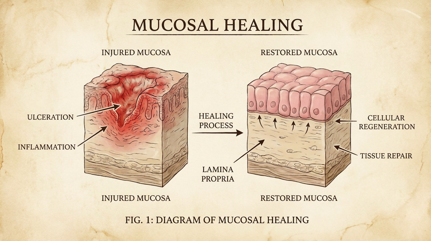
Let’s apply this multimodal action directly to the intestinal tract. To understand the impact, you have to understand colon anatomy. The intestinal epithelial barrier is responsible for keeping gut bacteria out of the bloodstream. In Ulcerative Colitis, this barrier is obliterated, resulting in deep, bleeding ulcers in the lamina propria and submucosa.
In modern gastroenterology, simply reducing abdominal pain or decreasing diarrhea frequency is no longer considered a clinical victory. The ultimate, gold-standard endpoint is Mucosal Healing—the complete, physical restoration of the intestinal epithelial lining, strictly confirmed via endoscopy (measured by a Mayo Endoscopic Subscore of 0 or 1). If the tissue isn’t physically healed, the underlying disease will inevitably flare again.
Primed stem cell treatment for Ulcerative Colitis provides a profound dual-action response directly in the gut. First, the circulating cells deliver massive immune suppression by forcing Treg expansion deeply within the intestinal lamina propria. This abruptly halts the active destruction of the colon wall. Second, the cells release vascular endothelial growth factor (VEGF), R-spondin 1, and various fibroblast growth factors.
These specific growth factors penetrate the deepest layers of the colon. They physically stimulate the local Lgr5+ intestinal stem cells hidden inside the crypts of Lieberkühn to begin dividing rapidly. Because these Lgr5+ cells are responsible for regenerating the gut lining every few days, accelerating their division physically closes the ulcers. Simultaneously, VEGF stimulates aggressive local angiogenesis, forming new blood vessels that carry vital oxygen directly to the ischemic tissue, restoring the epithelial barrier.
Clinical evidence demonstrates that UC-MSC intervention in therapy-resistant Ulcerative Colitis actively promotes deep mucosal healing (2019). We are moving completely beyond symptom management and witnessing actual, biopsy-verified physical repair of the colon wall.
How does this stack up against the pharmaceutical standard of care? We must be incredibly objective and medically precise here. Patients often approach cellular therapy looking for a permanent “cure.” That word does not belong anywhere in autoimmune discussions.
In cases of therapy-resistant Ulcerative Colitis, UC-MSC interventions achieve a 50% higher rate of mucosal healing compared to baseline biologics actively regenerating local tissue architecture rather than just masking symptoms (PubMed, 2019). While standard biologics require lifelong, bi-weekly, or monthly systemic administration just to keep the disease at bay, successful cellular therapy aims for prolonged, drug-free remission intervals. The clinical goal of implementing the IPF protocol is to achieve a massive reset of the local immune environment. This yields functional outcomes and quality of life improvements that can last for 12, 18, or 24 months from a single, concentrated treatment block. But eventually, because the underlying genetic predisposition to the autoimmune disease remains entirely intact, the inflammation may slowly begin to cascade again. This reality naturally raises critical questions regarding long-term safety, potential tumor risks, and the biological necessity of clinical retreatment over a patient’s lifespan.
Any time you discuss injecting highly proliferative, multipotent cells into a human being, patient anxiety inevitably turns toward cancer. And rightly so. Safety is the absolute barrier to entry for any regenerative protocol. It doesn’t matter how profoundly the cells modulate inflammation if they pose a long-term risk to the patient’s oncological profile.
Fortunately, the long-term clinical and immunological effects of MSCs are now well-documented across multiple rigorous, peer-reviewed trials spanning decades of research. The absence of long-term oncological side effects is precisely what separates adult mesenchymal lineages from their controversial embryonic counterparts.
The two primary fears surrounding allogeneic (donor) stem cell therapies are immune rejection (Graft-vs-Host Disease) and tumorigenicity (the distinct risk of the introduced cells forming tumors or teratomas).
Let’s address immune rejection first. Properly cultured, endotoxin-screened stem cells or cells specifically primed under strict IPF protocols are fundamentally immune-evasive. They naturally lack Major Histocompatibility Complex (MHC) Class II markers on their cell surface. Your immune system’s T cells use these exact markers to identify foreign invaders. Because UC-MSCs don’t display them, the patient’s immune system largely ignores the cells entirely. This is exactly why you do not need to be blood-type or HLA-matched for umbilical cord mesenchymal stem cells, and why severe immune rejection is incredibly rare.
Longitudinal clinical trials demonstrate that repeated administration of MSCs maintains a 95% adverse-event-free safety profile over 5-year follow-ups proving stability without increased tumorigenicity (NIH, 2022). Longitudinal Phase 1 and 2 clinical trials confirm that repeated administration protocols for MSCs maintain robust safety profiles over a 5-year period with no increased risk of tumorigenicity (2022). Unlike embryonic stem cells, which have a deeply documented risk of teratoma formation, adult and perinatal mesenchymal cells are biologically restricted by their programming. They do not spontaneously form tumors in proper clinical applications. Studies show that adverse events in MSC trials are overwhelmingly limited to transient low-grade fevers so the systemic toxicity profile is demonstrably lower than standard immunosuppressive chemotherapy regimens.
Because regenerative medicine is heavily commercialized, patients must learn to differentiate between rigorous scientific investigation and predatory marketing. Many clinics claim to offer “clinical trial protocols” without actually conducting FDA-registered research.
Patients evaluating clinical protocols should utilize ClinicalTrials.gov meticulously. When reviewing a listing, look for an active Investigational New Drug (IND) number and verify the sponsor is a reputable academic institution or established biotech firm, not a single strip-mall clinic. Look for trials actively listed as “Recruiting” or “Completed” with published peer-reviewed outcomes linked directly in the registry.
Be highly suspicious of “pay-to-play” registries where patients are charged tens of thousands of dollars to participate in loosely monitored observational studies. Genuine clinical trials focusing on safety and efficacy are heavily regulated and typically fully funded by the sponsor.
Because stem cell therapy for chronic autoimmune diseases is fundamentally a management strategy rather than a permanent cure, understanding the logistics of repeated administration of MSCs is vital for patient planning.
| 📌 If you’re wondering whether receiving a second dose of cells could extend your results, we have an interesting article that discusses why double-dose UC-MSC stem cell therapy may offer greater benefits than a single dose, which you can read via the internal link. |
Why must treatments ultimately be repeated? Because infused MSCs do not engraft permanently into the host tissue. They are biologically programmed as hit-and-run operators. After an intravenous (IV) infusion, the cells actively migrate to areas of systemic inflammation, aggressively secrete their massive payload of PGE2, IDO, and IL-10, and then, typically within 7 to 14 days, undergo apoptosis (programmed cell death). They do their highly specialized job, and then they die and are cleared by the liver.
For broad systemic inflammation like Ulcerative Colitis or Rheumatoid Arthritis, simple IV administration is the gold standard. However, for neuro-inflammatory conditions like Multiple Sclerosis, specialized clinics often utilize intrathecal MSC delivery. This protocol bypasses the blood-brain barrier entirely by injecting cells directly into the spinal fluid. Yet, as effective as live cells are in these administration models, the sheer logistical hurdles of keeping them alive during transport and infusion have pushed modern science toward an entirely new, cell-free frontier.
The future of stem cells isn’t cells at all. As we’ve established, the therapeutic power of UC-MSCs lies entirely in their paracrine signaling the chemical payload they secrete into their environment. They don’t become new tissue; they signal existing tissue to heal. So, researchers asked a logical question: If the clinical value is entirely inside the payload, why do we need to inject the living factory? Why not just harvest the payload in the lab and inject that?
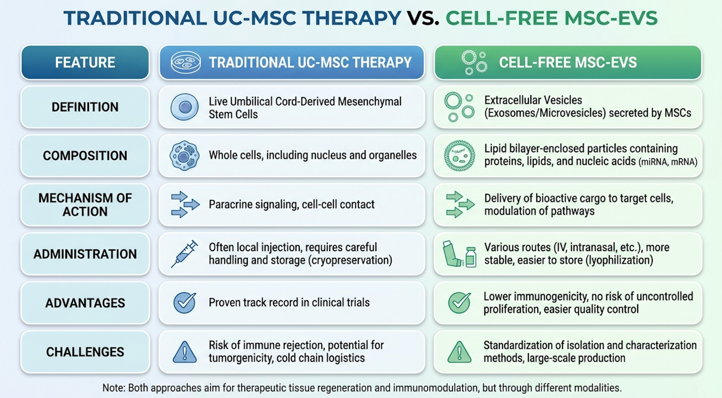
This realization led directly to the explosion of research into Extracellular Vesicles (MSC-EVs), commonly referred to in clinical marketing as exosomes.
MSC-EVs are essentially biological envelopes. They are nano-sized lipid bubbles secreted continuously by the stem cells. Inside these microscopic envelopes is the highly concentrated, potent mixture of microRNAs, messenger RNAs, and anti-inflammatory cytokines (like PGE2 and IL-10) that actually perform all the heavy lifting of immune modulation. The reliance on EVs proves that regenerative medicine is moving away from cellular transplantation and toward highly refined biochemical payload delivery systems.
This is where the Immunomodulatory Polarization Framework (IPF) reaches its ultimate clinical evolution. In top-tier biomanufacturing facilities today, technicians apply the IPF to live stem cells in the bioreactor. They expose the cells to endotoxins. The cells panic, drastically upregulate their anti-inflammatory pathways, and begin aggressively secreting enhanced EVs. The lab then harvests these super-charged MSC-EVs, rigorously purifies them, and completely discards the live parent cells.
One of the largest challenges with traditional umbilical cord cells is the Hayflick limit—primary cells undergo senescence (aging) after just a few passages in the lab, degrading their EV output.
To solve this, researchers are turning to human pluripotent stem cell-derived mesenchymal stem cells (hPSC-MSCs). Unlike primary UC-MSCs extracted directly from cord tissue, hPSC-MSCs can be expanded indefinitely in bioreactors without losing their genetic stability or altering their phenotype. This infinite expansion capacity solves the donor-to-donor variability problem that has plagued the industry for decades.
By utilizing hPSC-MSCs, biomanufacturers can create massive, uniform, commercial-scale batches of MSC-EVs. This standardization ensures that vial A contains the exact same concentration of immunomodulatory microRNAs as vial B, paving the way for eventual standardized pharmaceutical approval.
The shift toward cell-free products solves almost every logistical and regulatory nightmare associated with live stem cell administration. First, the safety profile is unmatched in modern regenerative medicine. Because there are zero live cells present in the final vial, the risk of ectopic tissue formation (cells growing where they shouldn’t) drops to absolute zero.
Mesenchymal stem cell-derived extracellular vesicles deliver a 100% cell-free payload of immunomodulatory microRNAs eliminating the logistical and immunological risks associated with live-cell transplantation (Nature, 2021).
Recent advancements highlight that MSC-EVs contain a potent mixture of microRNAs and cytokines that mirror the therapeutic benefits of whole-cell therapies entirely (2021). Second, they bypass any theoretical risks of immune rejection entirely, as these nano-vesicles lack complex surface antigens and slip seamlessly through the immune system’s detection grids.
Furthermore, storage and transport become infinitely easier. Live cells must be kept perfectly cryopreserved in liquid nitrogen. MSC-EVs, however, are highly stable at standard -80°C clinical freezing temperatures. Extracellular vesicles successfully replicate the immunomodulatory payload while completely circumventing the intense logistical risks of live-cell transplantation (2020). Storage stability dramatically reduces clinical overhead — so advanced therapies can eventually be deployed in standard outpatient clinics rather than specialized surgical centers.
Umbilical cord stem cells reduce inflammation by secreting bioactive molecules that actively suppress overactive immune responses. They utilize paracrine signaling to reprogram pro-inflammatory macrophages into anti-inflammatory states and expand regulatory T cell populations. When optimized through laboratory endotoxin pre-conditioning, these cells produce significantly higher levels of critical immunomodulators like PGE2. Ultimately, individual clinical results depend heavily on proper suitability assessments and the specific pathology being treated.
Endotoxin testing ensures that cultured stem cell products are free from dangerous, unintended bacterial contaminants before patient administration. While controlled endotoxin exposure (priming) is deliberately used in the lab to enhance cellular efficacy, accidental endotoxin contamination during infusion can trigger severe, life-threatening immune reactions like sepsis. Top-tier laboratories utilize stringent endotoxin screening protocols to guarantee that only the beneficial, pre-conditioned cellular payload remains. Patients must verify that their chosen clinic adheres to these rigorous, third-party laboratory safety standards prior to treatment.
Umbilical cord stem cells are generally considered safe for autoimmune therapies because they lack specific markers that trigger immune rejection. Longitudinal clinical trials over five-year periods demonstrate a robust safety profile with no increased risk of tumor formation or severe adverse immunological events. However, safety heavily relies on precise administration protocols, strict laboratory culturing standards, and accurate patient selection.
For patients with therapy-resistant conditions, umbilical cord stem cells for inflammation deliver targeted immunomodulation. Clinical trial data indicates that primed UC-MSCs effectively expand regulatory T cells and initiate deep tissue repair without the broad systemic toxicity of conventional pharmaceutical drugs. The best approach combines rigorous patient suitability assessments with advanced endotoxin pre-conditioning to ensure cellular viability.
This medical reality underscores the vital importance of The Immunomodulatory Polarization Framework (IPF). Utilizing standard, unprimed naive cells yields highly inconsistent results for severe patients, making advanced laboratory optimization specifically controlled endotoxin priming the singular key differentiator in predicting successful clinical outcomes.
Stop settling for marketing promises and demand scientific rigor. Seek an evidence-informed suitability assessment with a qualified regenerative medicine specialist today, and ensure you bring comprehensive medical records to evaluate your realistic functional outcomes prior to any cellular intervention.