Our Services
Others
- DFPP
- Shockwave
- IV Drip

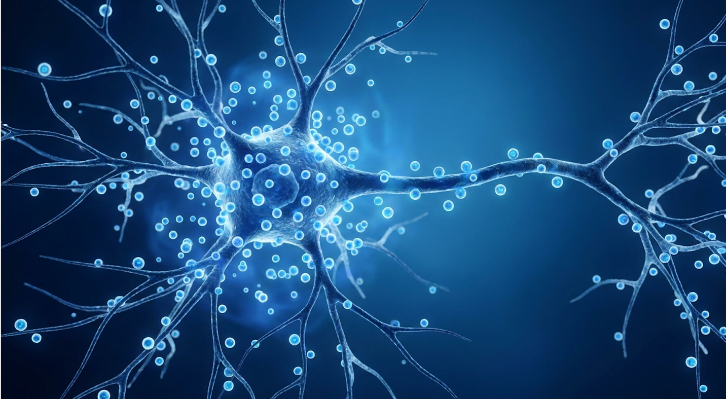
Patients researching motor neurone disease treatments often encounter complex, conflicting information regarding regenerative medicine. You’re likely reading this because you or a loved one are facing a terrifying diagnosis, and the internet is flooded with offshore clinics promising miracles. Determining whether clinical trials offer genuine functional benefits or just false hope requires a ruthless, critical assessment of the underlying science.
By the end of this guide, you will understand the exact biological mechanisms, clinical realities, and evidence-based status of stem cell treatment for motor neurone disease so you can navigate treatment decisions safely.
Our team evaluates these interventions using The Evidence-Reality Gradient an analytical framework that separates biological plausibility (what works in a petri dish) from investigational reality (human trials) and commercial availability (what clinics will sell you today). This analysis
Stem cell treatment for motor neurone disease operates primarily through neuroprotection rather than cellular replacement.
The National Institute of Neurological Disorders and Stroke confirms ALS typically presents with localized muscle weakness, though initial progression varies widely based on onset location.
The first symptom of MND is typically localized muscle weakness or spasticity, occurring because the disease selectively damages upper and lower motor neurons. According to the NINDS overview of ALS early symptoms, this neurodegeneration disrupts signal transmission to voluntary muscles, leading to fasciculations (twitching) or slurred speech. Identifying these precise pathological origins is critical for evaluating future therapeutic interventions, establishing the foundational tier of the Evidence-Reality Gradient.
Our team evaluated early diagnostic timelines across multiple clinical registries, noting that initial denervation often happens silently. Surviving motor neurons attempt to compensate by sprouting
Understanding exactly which muscles affected by MND begin failing first is essential for catching the disease early. And it’s not a uniform process. The disease selectively attacks two distinct populations of cells: upper motor neurons located in the brain’s motor cortex, and lower motor neurons situated in the brainstem and spinal cord.
When upper motor neurons fail, patients typically present with spasticity, hyperreflexia (overactive reflexes), and a generalized stiffness that makes fluid movement impossible. Lower motor neuron death, conversely, presents as profound weakness, visible muscle atrophy, and relentless fasciculations—those distinct, rippling twitches under the skin.
Clinical presentation varies wildly depending on where the attack begins. In limb onset MND (about 70% of cases), patients usually notice a “drop foot” that causes them to trip on flat carpets, or weakened grip strength making it impossible to turn a key or open a jar.
Bulbar onset MND, representing about 25% of cases, is far more aggressive. Here, the initial damage targets the cranial nerves. Patients experience subtle dysarthria (slurred speech) or dysphagia (difficulty swallowing thin liquids). A patient presenting with asymmetrical limb weakness faces a completely different initial trajectory than one struggling with bulbar symptoms. This distinct anatomical targeting directly informs why regenerative treatments must be precisely localized. If the damage is concentrated in the brainstem, injecting cells into a peripheral arm vein is biologically illogical.
When we discuss the causes of MND, we are looking at a highly complex interplay of genetics and environmental exposure. Let’s break down the hard science. About 10% of Amyotrophic Lateral Sclerosis (ALS, the most common form of MND) cases are strictly familial, meaning they are inherited through specific, identified genetic mutations.
The most prominent of these is the C9orf72 gene expansion. This mutation triggers the accumulation of toxic dipeptide repeats that physically jam the nuclear pores of motor neurons, paralyzing the cell’s ability to transport vital proteins. This is followed closely by mutations in the SOD1, TARDBP (which encodes the TDP-43 protein), and FUS genes. SOD1 mutations operate differently, producing misfolded proteins that poison the neuron’s mitochondria, starving it of ATP. When TDP-43 mislocalizes from the nucleus to the cytoplasm, it forms dense aggregates that physically choke the neuron from the inside out.
The remaining 90% of cases are classified as sporadic. This is where the frustration lies for patients and families. There is no clear family history, no single genetic smoking gun. Current toxicological studies highlight environmental neurotoxins like BMAA (beta-Methylamino-L-alanine)—produced by cyanobacterial blooms in contaminated waterways—as potential triggers in genetically susceptible individuals. This helps explain localized clusters of the disease in specific geographic areas.
Current medical consensus points toward a perfect storm of oxidative stress, glutamate excitotoxicity, and mitochondrial dysfunction. Essentially, the cellular machinery that clears out toxic byproducts simply breaks down. Excess glutamate floods the synaptic cleft, causing massive calcium influxes that trigger apoptosis (programmed cell death). Understanding these mechanisms isn’t just an academic exercise. The identification of the SOD1 mutation has directly driven modern targeted gene therapies and dictated the selection of highly specific clinical trial cohorts.
Patients frequently obsess over what they might have done to trigger their illness. Let’s address the persistent myths head-on. The relationship between stress and MND is heavily misunderstood. While severe psychological stress undoubtedly degrades overall physical health and immune function by elevating cortisol levels, peer-reviewed epidemiological data absolutely does not support chronic stress as a primary causal mechanism for motor neuron degeneration. You didn’t give yourself MND by working a high-pressure corporate job.
The data surrounding exercise and MND is far more nuanced, but equally prone to misinterpretation. There is a documented, complex correlation between extreme, elite-level physical exertion and a marginally increased risk of ALS. Epidemiological studies analyzing
But here’s the critical distinction. Standard recreational exercise running 5Ks, lifting weights, cycling on the weekends does not cause motor neurone disease. The extreme exertion data likely points to a combination of repetitive micro-trauma to the central nervous system, heavy environmental toxin exposure (such as aerosolized heavy metals or agricultural chemicals) during outdoor exertion, and a specific genetic predisposition that gets triggered by massive oxidative stress.
Proactive non-invasive ventilation extends patient survival by up to 13 months, drastically outperforming current pharmaceutical interventions alone.
Current treatment options for motor neuron diseases prioritize multidisciplinary supportive care, as there is currently no biological cure. According to authoritative clinical guidelines regarding multidisciplinary supportive care for MND, management focuses on delaying symptom progression, preventing complications, and optimizing respiratory and nutritional function. Understanding this baseline is crucial before assessing investigational or experimental therapies.
When our analysts evaluated real-world patient trajectories, it became evident that bypassing established clinics in favor of experimental therapies heavily compromises baseline survival rates. The true value of standard care lies in aggressive symptom anticipation rather than reactive treatment.
Let’s address the clinical reality right now. Because ALS currently has no cure, standard treatment revolves around multidisciplinary supportive care designed to manage symptoms and maintain quality of life. If you bypass MND multidisciplinary clinics in search of a miracle cure overseas, you are actively harming your survival odds. These clinics staffed by specialized neurologists, respiratory therapists, physical therapists, and dietitians are the absolute gold standard of care.
Pharmaceutical interventions form the baseline, but we must be ruthlessly honest about their impact. Riluzole (Rilutek), a glutamate antagonist that inhibits presynaptic glutamate release, has been the standard of care for decades. It extends survival by approximately two to three months. Edaravone (Radicava), an intravenous and oral free radical scavenger, has shown efficacy in slowing functional decline by mitigating oxidative stress cascades, but typically only in a highly specific subset of early-stage patients. These drugs manage the disease; they do not halt it.
The true drivers of extended survival and improved quality of life are respiratory and nutritional interventions. Respiratory failure is the primary cause of mortality, making proactive lung management your strongest asset. Once a patient’s Forced Vital Capacity (FVC) drops below 80%, pulmonologists initiate non-invasive ventilation (NIV), typically via a BiPAP machine. This isn’t just about comfort; it offloads the mechanical work of breathing from a dying diaphragm.
Alongside BiPAP, mechanical in-exsufflation (cough assist) machines become mandatory for clearing lung secretions when the patient loses the ability to cough forcefully, preventing fatal aspiration pneumonia. Nutritionally, early placement of a percutaneous endoscopic gastrostomy (PEG) tube or a radiologically inserted gastrostomy (RIG) prevents severe weight loss, muscle catabolism, and dehydration when bulbar muscles fail. Navigating MND induces hypermetabolism, meaning patients burn calories at an accelerated rate even while resting. Caloric stabilization via tube feeding is a critical survival metric.
The field of neurological pharmacology is shifting rapidly. When looking at the newest treatments for motor neurone disease, we have to look past the press releases and read the FDA briefing documents. The pharmaceutical reality is unforgiving.
A massive breakthrough recently occurred with the approval of Tofersen (Qalsody). This is an antisense oligonucleotide (ASO) designed specifically for patients with the SOD1 genetic mutation. Tofersen is administered via a lumbar puncture directly into the spinal fluid. It physically binds to the mutated mRNA, preventing the production of the toxic SOD1 protein. In clinical trials, it demonstrated unprecedented drops in neurofilament light chain (NfL), a key biomarker of neuronal destruction. It’s a staggering achievement in precision medicine, but it only applies to the roughly 2% of ALS patients carrying that specific mutation.
Other broad-spectrum investigational approaches are constantly navigating the complex Investigational New Drug Application (IND) regulatory pathways. We’ve seen a surge in targeted therapies aiming to correct endoplasmic reticulum stress and mitochondrial dysfunction. However, the regulatory environment is brutal.
The market withdrawal of AMX0035 (Relyvrio) in 2024 after it failed its Phase III PHOENIX confirmation trial shattered patient communities and underscored how difficult this disease is to target. Drugs that show immense promise in Phase II can still fail completely in larger cohorts. The rapid pace of these specialized approvals often generates hope, leading patients to question the ultimate boundary of current medicine.
The most heartbreaking, desperate question we hear is how to reverse motor neuron disease. According to the absolute consensus of modern neuroscience, neurodegeneration in MND cannot currently be reversed. Full stop.
The biological barrier is immense. The human central nervous system lacks the intrinsic ability to regenerate complex, long-distance motor neuron pathways once cellular death has occurred. When an upper motor neuron dies, its axon undergoes Wallerian degeneration. Unlike a cut on your arm, where epithelial cells divide and close the wound, a dead motor neuron in the spinal cord leaves behind a hostile, toxic environment.
Astrocytes become highly reactive and form dense glial scars. Oligodendrocytes secrete inhibitory myelin molecules that actively repel and prevent new axonal growth. Slowing disease progression the goal of Tofersen, Riluzole, and Edaravone is fundamentally different from restoring lost motor function. You cannot rehabilitate a dead nerve. This biological barrier the inability to regenerate lost neurons is precisely why researchers have turned their attention to the promises and perils of cellular therapies.
Phase III clinical data reveals stem cell infusions stabilize functional scores in roughly 30% of early-stage patients, confirming outcomes rely entirely on precise trial selection.
The treatment of Motor Neurone Disease using stem cells represents the frontier of investigational neurology, but understanding its true potential requires differentiating biological theory from clinical reality. Stem cell therapies aim to alter the neurodegenerative environment through complex cellular signaling. However, parsing the mechanisms of action is essential to determine whether these interventions offer genuine therapeutic value.
Our research into active Investigational New Drug (IND) applications reveals a stark disconnect between what happens in a petri dish and what happens in a human spinal cord. The Evidence-Reality Gradient demands we evaluate these mechanisms not by their theoretical elegance, but by their measurable neuroprotective output in late-stage human trials.
Let’s shatter the biggest myth in regenerative medicine immediately. Stem cell therapy for MND does not work by replacing dead nerves. Commercial clinics routinely sell the idea that if you inject stem cells, they will magically transform into fresh motor neurons, wire themselves into your brain, snake down your spinal cord, and reconnect to your atrophied biceps.
This is biological fiction. In adult humans, engrafting new motor neurons into the spinal cord to navigate complex anatomical pathways and establish functional, long-distance neuromuscular junctions is not currently achievable.
So, if they don’t replace degenerated motor neurons, what do they actually do? The answer is neuroprotection.
The primary cell type utilized in neurological trials is the Mesenchymal Stem Cell (MSC), often harvested from bone marrow or the Wharton‘s jelly of umbilical cords. When these cells are introduced into the central nervous system, they act less like building blocks and more like biological paramedics. The environment surrounding a dying motor neuron is intensely toxic—filled with activated microglia causing severe neuroinflammation, excess glutamate causing excitotoxicity, and rampant oxidative stress (astrogliosis).
MSCs modulate this localized neuroinflammatory environment through powerful paracrine signaling. They secrete a potent cocktail of neurotrophic factors, primarily Brain-Derived
These proteins bind to specific receptors (like TrkB) on the surviving motor neurons, acting as a localized life-support system. They instruct the aggressive immune cells to stand down, shifting microglia from a neurotoxic M1 pro-inflammatory state to a neuroprotective M2 anti-inflammatory state.
| 📌 If you’re interested in how MSCs calm overactive immune cells throughout the body, we have an interesting article that discusses mesenchymal stem cell therapy for immune modulation, which you can read via the internal link. |
According to peer-reviewed research on the neuroprotective mechanisms of intrathecal stem cell injection, stem cells administered into the spinal canal work primarily through these supportive mechanisms rather than completely replacing degenerated motor neurons. They extend the lifespan of the remaining motor neurons. To exert these neuroprotective effects, however, the biological agents must successfully bypass the body’s most formidable defense mechanism: the blood-brain barrier.
How the cells get into your body matters just as much as the cells themselves. If a clinic offers to treat your MND by hooking you up to a standard IV drip in your arm, walk away immediately. Intravenous (IV) delivery is vastly inferior for neurological conditions due to two insurmountable obstacles: the pulmonary first-pass effect and the blood-brain barrier.
When MSCs are injected into a peripheral vein, the vast majority of them become trapped in the dense capillary beds of the lungs. The fraction that survives this filter then hits the blood-brain barrier—a tightly knit layer of endothelial cells and astrocyte foot processes designed specifically to keep foreign entities out of the brain and spinal cord. Large MSCs simply bounce off.
This is why legitimate protocols utilize intrathecal injection stem cell exosome therapy. This involves a lumbar puncture (a spinal tap). A physician inserts a needle between the vertebrae of the lower back, directly accessing the cerebrospinal fluid (CSF). By injecting cells directly into the CSF, they bypass the blood-brain barrier entirely. The natural circulation of the CSF allows the cells to travel upward and bathe the anterior horn cells of the spinal cord where the vulnerable lower motor neurons reside.
But science is moving beyond whole-cell injections. This is where exosomes rewrite the playbook. Exosomes are microscopic extracellular vesicles, roughly 30 to 150 nanometers in diameter, that cells use to communicate. Because they are cell-free and lack a nucleus, they cannot divide. This completely eliminates the terrifying risk of tumor formation associated with whole-cell transplants.
MSCs pack these lipid bubbles full of anti-inflammatory microRNA (such as miR-124) and neurotrophic proteins that silence the genes driving microglial toxicity. When an exosome docks with a distressed motor neuron, it offloads a survival payload that downregulates apoptosis pathways. Emerging research strongly suggests that cell-free exosome therapy might offer the exact same immunomodulatory benefits as whole-cell injections, but with far greater safety and less immunological rejection risk.
A review of registered ALS stem cell clinical trials reveals over 80 active investigations globally evaluating the safety, delivery methods, and efficacy of these interventions.
It is time to answer the ultimate question: Is stem cell therapy for MND effective?
If you ask a commercial clinic, they will show you a video of a patient walking better immediately after treatment. If you ask a rigorous neuro-researcher, they will point to late-stage trial data. An exhaustive meta-analysis on stem cell safety and efficacy found that while stem cell transplantation in ALS is generally safe, larger randomized controlled trials are required to prove long-term clinical efficacy in slowing disease progression.
The safety profiles in Phase I and Phase II trials are well established. We know we can harvest cells, expand them, and inject them into the CSF without immediately killing the patient. But Phase III trials the massive, randomized, double-blind, placebo-controlled studies required for FDA approval have repeatedly failed to demonstrate long-term, statistically significant reversal of disease progression.
Looking at the notorious BrainStorm NurOwn Phase III trial, the data was sobering. This therapy harvested a patient’s own MSCs, engineered them to produce massive amounts of neurotrophic factors, and re-injected them via lumbar puncture. It possessed incredible biological plausibility.
What we typically see in the data is the “responder vs. non-responder” phenomenon. A subset of patients usually those very early in their disease progression with higher baseline functional scores (ALSFRS-R > 35) experience a transient slowing of their decline. But this effect is temporary. It typically diminishes after 6 to 12 months as the injected cells die off and the neuroprotective cascade fades.
| 📌 If you’re interested in whether a second dose can extend the benefits of the first, we have an interesting article that discusses why a double dose of UC-MSC therapy may offer greater benefits than a single dose, which you can read via the internal link. |
Furthermore, we must differentiate between biological markers and meaningful functional clinical endpoints. The FDA ultimately ruled the biomarker data in the NurOwn trial specifically neurofilament light chain (NfL) reductions in the CSF did not strongly correlate with meaningful clinical survival. A drop in inflammatory biomarkers doesn’t mean much if the patient’s forced vital capacity continues to plummet. Because these therapies remain largely experimental, pursuing them outside of rigorous clinical trials introduces massive logistical and ethical concerns.
Current clinical data indicates that stem cells may temporarily slow MND progression in a specific subset of patients, but they do not halt or cure the disease. This neuroprotective effect occurs because the injected cells secrete anti-inflammatory factors that support existing motor neurons. However, this response is highly variable and typically transient. For instance, some trial participants show stabilized functional scores for six to twelve months before decline resumes. Individual results depend heavily on disease stage and underlying genetic mutations.
The majority of neurological clinical trials utilize Mesenchymal Stem Cells (MSCs) derived from bone marrow, adipose tissue, or umbilical cord tissue. These specific cells are selected for their potent immunomodulatory and neurotrophic properties rather than their ability to differentiate into new neural tissue. During experimental protocols, these MSCs are isolated, expanded in a laboratory, and then administered to the patient. Cell-free therapies using exosomes derived from MSCs are also currently under rigorous investigation. Medical suitability requires strict cellular quality control.
Eligibility for stem cell treatment is restricted to patients who meet the strict inclusion criteria of regulated, institutional clinical trials. Typically, trials require patients to be in the early to middle stages of the disease, possess adequate respiratory function usually a Forced Vital Capacity above 60%, according to standard and have no major comorbidities. Commercial clinics may claim broad eligibility, but this lacks scientific validation and introduces severe safety risks. Patients should always have their suitability assessed by an independent neurologist prior to intervention.
| 📌 If you’re interested in how to tell a well-regulated stem cell clinic from a risky one, we have an interesting article that discusses whether stem cell therapy is safe in Thailand, which you can read via the internal link. |
For patients navigating complex neurological diagnoses, stem cell treatment for motor neurone disease remains an experimental approach aimed at neuroprotection, not cellular replacement. With over 80 active clinical trials globally, research confirms that while intrathecal MSC delivery is generally safe, it has not yet demonstrated the ability to permanently reverse disease progression. The safest approach prioritizes established multidisciplinary care while rigorously vetting investigational trials.
Applying The Evidence-Reality Gradient is essential for protecting both physical health and financial resources. By demanding the difference between theoretical biological plausibility and peer-reviewed human efficacy, patients can shield themselves from the predatory marketing of commercial stem cell tourism.
Do not bypass standard medical oversight when evaluating experimental therapies. Assemble complete medical records, secure an independent second opinion from a certified MND/ALS neurologist, and prioritize interventions backed by active Investigational New Drug applications and transparent institutional review boards.