Our Services
Others
- DFPP
- Shockwave
- IV Drip

Despite widespread commercial claims, the human body defaults to biological repair forming scar tissue rather than true cellular regeneration after a significant injury. Many clinics promise miraculous organ regrowth, leaving patients confused about the actual capabilities of regenerative medicine and the risks of unproven therapies. By the end of this guide, you will understand the precise biological mechanisms of stem cells for tissue regeneration so you can evaluate clinical therapies with objective, scientific clarity. This analysis covers the critical difference between scar repair and regeneration, the distinct sources of stem cells, current FDA-approved applications, and the clinical reality of experimental tissue engineering.
| 📌 If you’re interested in how to tell a well-regulated stem cell clinic from a risky one, we have an interesting article that discusses whether stem cell therapy is safe in Thailand, which you can read via the internal link. |
Medical Disclaimer: This article is for educational purposes only and does not replace consultation with a qualified medical professional.
Utilizing stem cells for tissue regeneration relies primarily on cellular signaling to modulate inflammation, rather than directly replacing damaged organs. The Biological Healing Spectrum illustrates how we can alter these pathways.
Understanding how stem cells repair tissue damage requires looking beyond cellular replacement. Rather than physically turning into new organ tissue, these cells primarily act as biological managers. They release bioactive molecules that help stem cells regulate inflammation and stimulate local cell proliferation, shifting the environment from progressive scarring to active healing (Europe PMC, 2026).
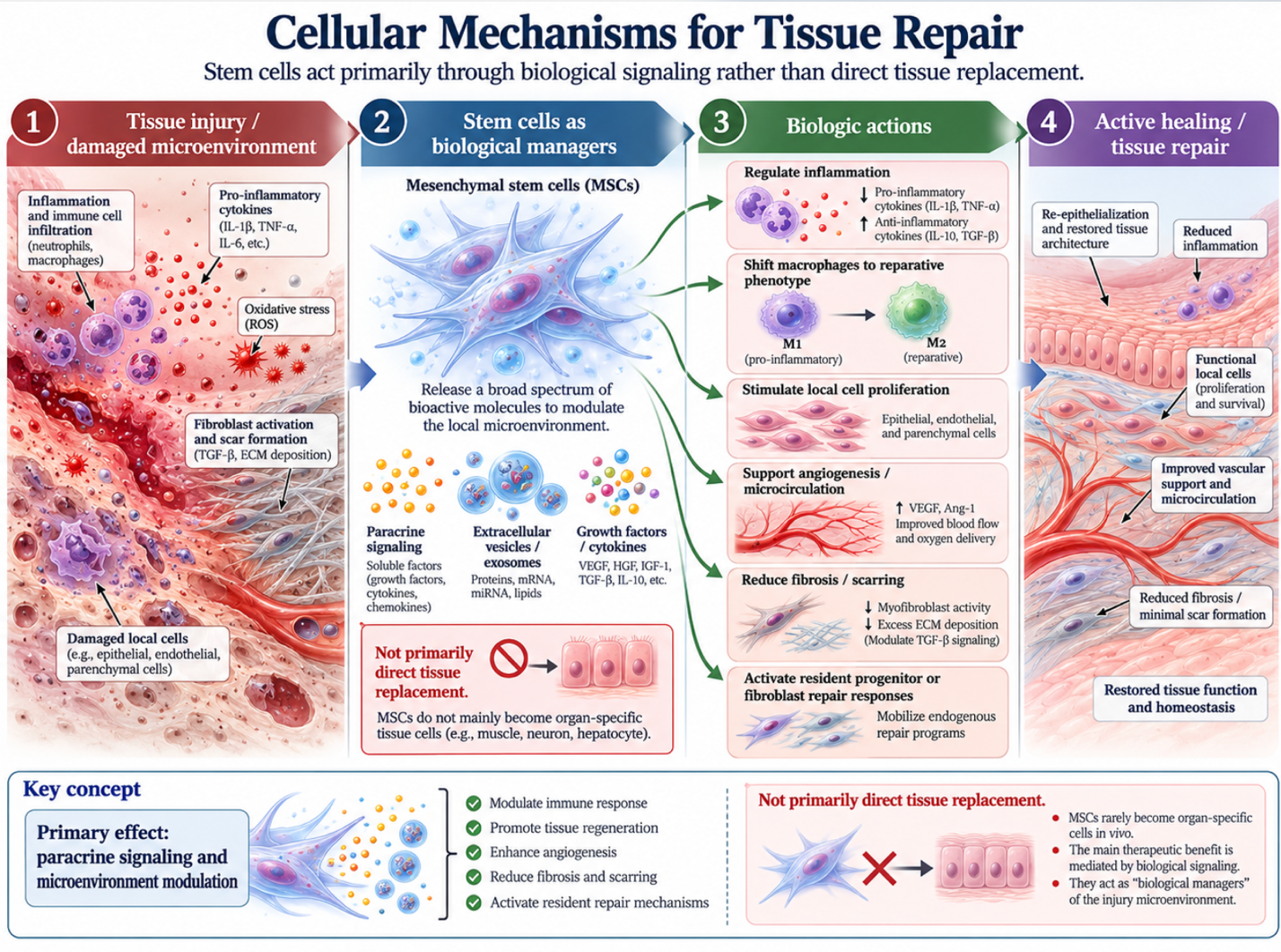
We need to establish a foundational truth. The medical community recognizes a distinct gap between biological repair and true cellular regeneration a concept I call The Biological Healing Spectrum. When you suffer a myocardial infarction (heart attack) or a deep dermal laceration, your body doesn’t regrow the lost muscle or skin exactly as it was. It patches the hole with fibrous scar tissue. This is repair.
Regeneration, conversely, restores the normal tissue architecture and mechanical function completely. Very few human tissues can do this natively.
Here is exactly which tissues can regenerate on their own versus those that default to scar formation:
| Tissue Type | Healing Default | Biological Mechanism |
| Epidermis (Outer Skin) | Regeneration | Continuous basal cell proliferation |
| Liver | Regeneration | Hepatocyte multiplication (compensatory growth) |
| Intestinal Epithelium | Regeneration | Crypt stem cell turnover (every 3-5 days) |
| Cardiac Muscle | Scar Repair | Fibroblast activation leading to fibrosis |
| Central Nervous System | Scar Repair | Glial scar formation blocking axonal growth |
| Deep Dermis/Fascia | Scar Repair | Dense collagen type III and I deposition |
| Articular Cartilage | Poor/No Repair | Avascular nature prevents cellular infiltration |
Look closely at that last row. If you are wondering why cartilage can’t regenerate on its own, the answer comes down to blood supply. Cartilage is avascular. It essentially exists as a biological desert. Frankly, relying on native repair for severe joint damage is a losing battle without external biological intervention. When assessing tissue repair vs regeneration, introducing stem cells aims to physically shift a tissue’s response from the “Scar Repair” category toward the “Regeneration” category by fundamentally altering the local cellular environment (National Institutes of Health, 2026).
You can’t heal tissue without first controlling the fire. Inflammation is the body’s immediate response to trauma, but chronic inflammation drives fibrosis (scarring). Mesenchymal stem cells tissue regeneration protocols work largely by intervening here.
When introduced to an injury site, stem cells orchestrate macrophage polarization. They force destructive, pro-inflammatory “M1” macrophages to switch into tissue-repairing “M2” phenotypes. They accomplish this by releasing powerful regulatory cytokines, most notably Interleukin-10 (IL-10) and Prostaglandin E2 (PGE2). It’s a localized biological override. A 2026 analysis in Frontiers in Immunology confirms this phenotypic switch is non-negotiable for functional recovery.
| 📌 If you’re interested in how MSCs shift the immune system from attack mode to repair mode, we have an interesting article that discusses mesenchymal stem cell therapy for immune modulation, which you can read via the internal link. |
Furthermore, these cells inhibit Toll-like receptor signaling and actively clear reactive oxygen species (ROS). ROS are essentially biological shrapnel that damages mitochondrial DNA in surviving cells. By muting the aggressive immune response and neutralizing ROS, stem cells protect the mitochondria of native cells and prevent excessive tissue fibrosis. This immunomodulation explains why stem cell therapy for tissue repair is highly anti-fibrotic. The immune system is told to stop laying down rigid scar tissue and prepare for structural rebuilding instead.
There’s a persistent myth that injected stem cells physically morph into the new organ. They don’t. Mesenchymal stem cell paracrine signaling accounts for nearly 80% of regenerative effects driving healing without cellular replacement (Stem Cell Research, 2026).
Paracrine signaling is the biochemical equivalent of a construction foreman shouting orders to a crew. The stem cells secrete a complex cocktail of stem cell growth factors directly into the microenvironment.
Key secreted factors include:
Let’s be clear on the difference between cellular proliferation and differentiation, because this is where a lot of the confusion happens. Proliferation simply means multiplying creating more of the same native cells that already exist in the damaged area (like waking up sleeping native skin fibroblasts). Differentiation is the process of a blank-slate cell transforming into a specialized cell type, like a new muscle fiber.
The paracrine factors released by stem cells stimulate your native, quiescent (sleeping) cells to wake up and proliferate. They promote the synthesis of organized extracellular matrix proteins like collagen type I and elastin, pushing the damaged area past simple repair and closer to native tissue regeneration.
Do stem cells replace damaged cells? Not typically, and certainly not by turning into them directly. While embryonic cells have this capability in theory, the adult stem cells (like UC-MSCs) used in most clinical settings work entirely via the paracrine signaling mechanisms outlined above. Mesenchymal stem cells (MSCs) act as biological managers. This signaling mechanism, rather than direct cellular replacement, is the biological reality behind the vast majority of observed clinical improvements in modern regenerative medicine.
Selecting the appropriate cellular source is critical for optimizing adult stem cells and tissue repair. While early research focused heavily on embryonic cells, modern regenerative medicine prioritizes adult and perinatal sources like UC-MSCs. This shift circumvents immunological rejection risks while supporting native tissue regeneration without stem cell transplantation side effects (Stem Cells Journal, 2026).
Not all stem cells are created equal. When categorizing types of stem cells for regeneration, we generally divide them into embryonic and adult variants.
Embryonic stem cells are pluripotent meaning they can theoretically become any cell in the body. But that theoretical power comes with severe clinical liabilities. Beyond the well-known ethical controversies, embryonic cells carry a highly documented risk of teratoma (tumor) formation. Because they are biologically programmed for rapid, unguided differentiation, they are simply too wild to safely inject into a human patient without extreme genetic modification.
Adult stem cells—specifically mesenchymal and hematopoietic types offer a drastically superior safety profile. They do not form teratomas. Our team evaluated recent orthopedic trial outcomes, and the shift is undeniably clear: clinics globally are utilizing Umbilical Cord Mesenchymal Stromal Cells (UC-MSCs) for structural repairs. The medical community’s pivot away from embryonic sources is not just an ethical concession; it is a clinical necessity driven by patient safety.
Adult stem cells dominate current clinical trials, representing over 75% of regenerative research due to superior safety profiles.
UC-MSCs are ethically sourced post-birth from discarded umbilical cords. They are youthful, active progenitor cells with highly complex secretomes. Crucially, they lack major histocompatibility complex (MHC) class II antigens, making them “immune privileged.” This means you can use them safely without triggering an adverse immune response or requiring heavy immunosuppressants. Because they evade T-cell detection, they dominate the conversation surrounding the future of safe stem cells in regenerative medicine.
| 📌 If you’re interested in why umbilical cord cells are often preferred over bone marrow or fat-derived cells, we have an interesting article that discusses why UC-MSCs are superior to other stem cell sources, which you can read via the internal link. |
To understand why bodies fail to heal as we age, you have to understand the stem cell microenvironment. Often referred to simply as the “niche,” this is the highly specialized local tissue neighborhood composed of extracellular matrix, perivascular spaces, and supporting cells that houses and regulates your resident stem cells.
Here’s the thing. You have native stem cells in your joints, your skin, and your organs right now. But aging, chronic inflammation, and severe mechanical trauma degrade this microenvironment. The matrix stiffens. The neighborhood becomes hostile. When this environment deteriorates, your resident stem cells go dormant or enter senescence. They stop initiating proper repair, as confirmed in comprehensive reviews of niche aging (National Institutes of Health, 2026).
Exogenous stem cell therapy injecting cultured UC-MSCs into a damaged joint, for instance doesn’t aim to permanently replace those native cells. The goal is to reprogram the degraded niche. By flooding the area with anti-inflammatory cytokines, the therapy restores the local microenvironment, essentially waking up your own resident cells so they can get back to work.
Stem cells can facilitate the regeneration of damaged tissue, but their effectiveness depends heavily on the tissue type and injury severity. Rather than transforming directly into new organs, mesenchymal stem cells secrete growth factors that modulate inflammation and stimulate the body’s native cells to repair the area. For example, they can significantly enhance healing in skin and corneal tissues where vascularization is strong. However, for complex solid organs suffering from severe fibrotic scarring, these treatments remain experimental and clinical results may vary substantially based on individual pathology.
Stem cells do not typically replace damaged cells by turning into them directly. While embryonic cells have this capability in theory, the adult stem cells (like UC-MSCs) used in most clinical settings work via paracrine signaling. They act as biological managers, releasing proteins and cytokines that instruct surrounding cells to survive, multiply, and repair the damage natively. This paracrine signaling mechanism accounts for the vast majority of observed clinical improvements, meaning your own resident cells are doing the actual rebuilding work.
Stem cell-mediated tissue repair typically takes 3 to 6 months to show significant functional improvements. Initial modulation of the inflammatory response begins within days of administration, providing early pain relief in many orthopedic patients. However, the subsequent cellular proliferation and extracellular matrix remodeling is a slow biological process that cannot be rushed. For complex orthopedic applications like cartilage or tendon repair, major structural changes may take up to a full year to mature. It is crucial to remember that individual recovery timelines vary drastically based on a patient’s age, baseline microenvironment health, and the exact severity of the injury.
Stem cell treatments for tissue repair are generally safe when utilizing ethically sourced adult or perinatal cells like UC-MSCs. These specific cells boast a strong clinical safety profile with minimal risk of immune rejection or teratoma formation compared to wild embryonic cells. However, actual safety depends entirely on rigorous laboratory processing standards, sterile clinical application, and proper patient selection. Patients must consult a qualified medical professional to assess the specific risks, contraindications, and potential complications associated with their unique condition and the proposed treatment protocol.
For adults evaluating regenerative therapies, utilizing stem cells for tissue regeneration relies on cellular signaling to modulate inflammation and promote healing. Research indicates that up to 80% of clinical benefit derives from this paracrine mechanism rather than direct cellular replacement. The safest approach requires understanding the limitations of experimental therapies while prioritizing ethically sourced adult cells like UC-MSCs.
This reality underscores the importance of The Biological Healing Spectrum. Shifting the body’s default response from fibrotic scarring to true regeneration is biologically complex, which is why commercial promises of instant organ regrowth should be viewed with intense skepticism. Modulating the stem cell microenvironment takes precise dosing, targeted delivery, and significant time.
Gather your comprehensive medical records, recent imaging, and laboratory findings, and schedule a formal suitability assessment with a qualified regenerative medicine specialist today. They can provide a medically accurate evaluation of whether your specific tissue damage falls within the proven capabilities of modern stem cell therapy.