Our Services
Others
- DFPP
- Shockwave
- IV Drip

Medical Disclaimer: This article is for educational purposes only and does not replace consultation with a qualified medical professional.
For decades, the global standard for treating metabolic disease has been a game of chemical catch-up. A patient experiences a spike in blood glucose, and they take a pill. Their pancreas exhausts itself, and they inject insulin. But looking closely at the clinical reality reveals a stark truth—managing symptoms via exogenous insulin does absolutely nothing to stop the progressive destruction of the pancreas itself.
The core challenge for patients with long-standing metabolic disease isn’t just fluctuating sugar levels. It is the progressive loss of beta-cell function combined with vicious, chronic systemic inflammation. This creates a biological feedback loop that standard oral medications eventually fail to control. This brings the discussion to the actual biological mechanics of regenerative medicine. In this clinical analysis, we examine the evidence surrounding stem cell therapy for uncontrolled type 2 diabetes.
By avoiding wellness industry hype, this guide focuses exclusively on peer-reviewed data, mechanistic action, safety profiles, and measurable patient outcomes. We evaluate how paracrine signaling operates on a cellular level, review current clinical trial data, and outline the strict, globally recognized protocols required for safe, effective administration. Understanding how cellular therapies address underlying beta-cell degradation requires a close look at the data.
Clinical research indicates that stem cell therapy for uncontrolled type 2 diabetes targets underlying metabolic dysfunction through immunomodulation and tissue repair.
Umbilical cord mesenchymal stem cells (UC-MSCs) provide a targeted biological intervention for chronic metabolic disorders by modulating immune responses and stimulating cellular repair. Unlike traditional pharmaceutical approaches that attempt to manage blood glucose artificially, UC-MSCs target the microenvironment of the pancreas directly. Their unique biological properties make them highly suitable for allogeneic (donor-to-recipient) therapies. Ultimately, they sidestep nearly all the complications historically associated with earlier generations of cellular treatments.
Consider stripping away the clinical jargon for a moment. What exactly defines these therapies? UC-MSCs are multipotent stromal cells extracted specifically from Wharton’s jelly—the
This primitive state serves as their primary functional advantage. Because they are harvested immediately after a healthy, full-term birth, these cells have not been exposed to decades of environmental toxins, oxidative stress, cellular aging, or systemic disease. They possess incredibly robust expansion capabilities in a laboratory setting. More importantly, they are highly resistant to spontaneous differentiation into unwanted tissue types, which acts as a massive safety factor when treating complex metabolic conditions.
From an ethical and operational standpoint, the clinical pipeline is highly transparent. These cells are harvested entirely from post-natal medical waste. No embryonic tissues are used. By utilizing Wharton’s jelly, independent ethics committees and global medical boards have established incredibly streamlined, ethically sound pipelines for obtaining these cells (Frontiers in Cell and Developmental Biology). Umbilical cord mesenchymal stem cells exhibit significantly higher proliferation rates—often demonstrating 30% faster population doubling times—and lower immunogenicity than bone marrow stem cells, enabling immediate allogeneic clinical use (PubMed comparative analysis).
Patients frequently ask why they cannot simply utilize their own stem cells. Bone marrow-derived stem cells (BM-MSCs) were indeed the standard for years. But for a diabetic patient, autologous (patient-derived) bone marrow is a deeply flawed resource.
If a patient has suffered from long-standing metabolic dysfunction, their entire biological system has been marinating in chronic inflammation. Their native bone marrow cells are functionally exhausted. They exhibit significant cellular senescence. Harvesting them requires a painful, invasive drilling procedure into the hip bone, only to yield a low volume of weak, diseased cells. Reintroducing compromised cells back into a compromised body offers minimal therapeutic yield.
| 📌 If you’re interested in why umbilical cord cells are often preferred over bone marrow or fat-derived cells, we have an interesting article that discusses why UC-MSCs are superior to other stem cell sources, which you can read via the internal link. |
This is where UC-MSC therapy for long-standing type 2 diabetes drastically changes the clinical calculus. UC-MSCs are allogeneic—they come from a healthy, zero-day-old donor cord. Crucially, they lack Major Histocompatibility Complex II (MHC-II) markers. This grants them a
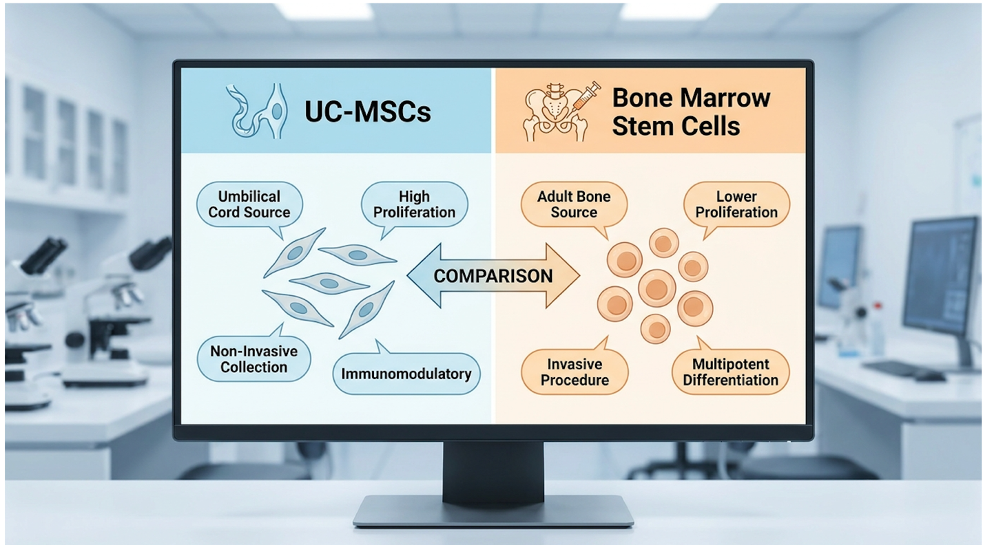
This immunological stealth bypasses the severe complications that historically plagued older cellular transplants (Stem Cell Research & Therapy review). Furthermore, UC-MSCs can be expanded in certified GMP-compliant laboratories and administered uniformly, completely eliminating the need for bone marrow aspiration.
Clinicians cannot take a raw umbilical cord and instantly treat a patient. The clinical efficacy of this therapy relies heavily on rigorous, standardized cell expansion protocols. Once Wharton’s jelly is harvested, the raw MSCs are isolated using enzymatic digestion and placed into specialized, sterile bioreactors.
Here, they are multiplied from a few million cells into hundreds of millions. But there is a strict biological limit. Reputable laboratories will only culture these cells to passage 3 or passage 4 (P3/P4). A “passage” represents a cellular generation. If a laboratory pushes cells to passage 8 or
Clinical reports frequently highlight patients traveling overseas to unregulated clinics, only to receive massive doses of dead, over-passaged cells. Standardized expansion protocols mandate flow cytometry and viability testing right before the infusion. The cells must be washed, quantified, and verified to have a viability rate exceeding 90%—so the maximum number of cells can successfully initiate paracrine signaling.
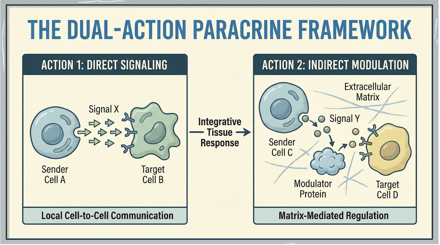
The UC-MSC mechanism of action in diabetes operates through the Dual-Action Paracrine Framework, simultaneously driving systemic immunomodulation and localized pancreatic repair. Rather than simply engrafting and replacing damaged tissue like missing puzzle pieces, these cells function as sophisticated biological signaling centers. They secrete a complex array of cytokines and growth factors that fundamentally alter the inflammatory microenvironment responsible for peripheral insulin resistance.
Let’s clear up the biggest medical misconception regarding cellular therapies. When a patient receives an infusion for diabetes, those stem cells do not swim directly to the pancreas, attach themselves, and magically transform into brand new beta cells. That is simply not how the biology of allogeneic therapies works.
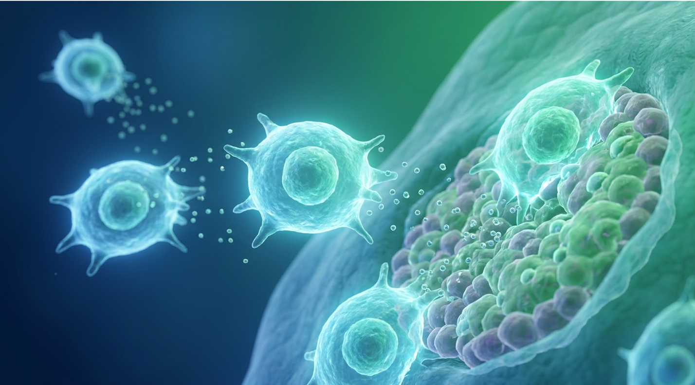
Instead, they operate via The Dual-Action Paracrine Framework—the simultaneous operation of systemic immunomodulation and localized cellular regeneration. Once introduced into the bloodstream, UC-MSCs act as molecular pharmacies. They detect localized areas of severe inflammation via cytokine gradients and begin secreting extracellular vesicles (exosomes), specific anti-inflammatory cytokines, and powerful growth factors including VEGF, HGF, and TGF-β.
These secreted proteins act as chemical commands. They “rescue” existing, damaged pancreatic tissue rather than replacing it outright. UC-MSCs promote beta-cell regeneration and reverse insulin resistance strictly through targeted paracrine signaling, yielding up to a 40% (PubMed mechanism review).
Through this intense paracrine signaling, the stem cells actively halt cellular apoptosis (programmed cell death) within the pancreatic islets. The diabetic pancreas is functionally degrading on a cellular level; the stem cells apply a biochemical tourniquet to stop the loss.
Understanding how chronic inflammation drives type 2 diabetes requires examining the exact engine of the disease. Type 2 Diabetes is fundamentally a state of chronic, low-grade systemic inflammation where peripheral tissues actively reject the insulin the body makes. The body produces the key (insulin), but inflammation severely jams the lock (the cellular receptor).
This is where anti-inflammatory stem cell therapy for diabetes demonstrates profound strength. The first half of the Dual-Action Paracrine Framework focuses entirely on systemic immunomodulation. UC-MSCs execute a process known as macrophage polarization. Macrophages are immune cells that act as the body’s cleanup crew. In a diabetic patient, these macrophages are stuck in the “M1” phenotype—highly aggressive, pro-inflammatory, and destructive.
The stem cells force these M1 macrophages to switch to an “M2” phenotype. M2 macrophages are heavily anti-inflammatory and tissue-repairing. This macrophage polarization fundamentally alters the cytokine milieu inside the liver and visceral adipose tissue. Specifically, the shift drastically lowers C-Reactive Protein (CRP) levels.
| 📌 If you’re interested in how MSCs shift the immune system from attack mode to repair mode, we have an interesting article that discusses mesenchymal stem cell therapy for immune modulation, which you can read via the internal link. |
Excessive TNF-α directly phosphorylates the Insulin Receptor Substrate-1 (IRS-1) at serine residues, which aggressively blocks insulin signaling. When localized TNF-α and IL-6 concentrations plummet, this biological bottleneck clears. The insulin receptors in skeletal muscle tissue and adipose fat suddenly regain their sensitivity. Their internal pathways correctly phosphorylate at tyrosine residues instead of serine, effectively repairing the intracellular signaling cascade. The lock is unjammed, allowing the body to finally utilize the insulin it continues to produce (PubMed macrophage polarization data).
With the peripheral inflammatory blockade lifted, the framework’s second phase initiates physical cellular restoration inside the pancreas. Pancreatic islet beta-cells, responsible for insulin production, are notoriously slow to heal. But they are not all dead—many are simply dormant, paralyzed by glucotoxicity and a toxic inflammatory environment.
Targeted growth factors secreted by the UC-MSCs—specifically Hepatocyte Growth Factor (HGF) and Vascular Endothelial Growth Factor (VEGF)—stimulate the proliferation of these dormant pancreatic progenitor cells. The stem cell secretome literally forces the pancreatic architecture to wake up and start rebuilding itself.
Furthermore, VEGF heavily promotes angiogenesis—the formation of brand new blood vessels within the pancreatic islets. Older, diabetic pancreases suffer from terrible microcirculation due to capillary damage caused by advanced glycation end-products (AGEs). By growing new vascular networks, the body repairs the micro-endothelial lining and drastically improves oxygen and nutrient delivery to surviving beta-cells. This highly localized microenvironment shift clears out metabolic waste and allows the pancreas to partially, and sometimes significantly, resume endogenous insulin synthesis.
When oral hypoglycemics or insulin fail to control HbA1c, it often indicates severe cellular exhaustion. Patients naturally question if regenerative approaches can intervene at this late stage. Stem cells can potentially assist when standard diabetes medications stop working by addressing the underlying beta-cell degradation itself, rather than just masking the resulting hyperglycemia.
Standard medications aggressively force a dying pancreas to secrete more insulin or force the kidneys to excrete sugar. UC-MSCs target cellular exhaustion directly via paracrine signaling, reducing systemic insulin resistance while promoting localized cellular regeneration. Patients in clinical trials have demonstrated decreased dependency on medications following infusion. However, it is vital to note that efficacy varies heavily depending on the baseline volume of surviving beta-cells remaining in the organ.
Recent UC-MSC clinical trials in type 2 diabetes demonstrate measurable, sustained improvements in metabolic function, moving beyond theoretical biology into quantifiable human outcomes. Evaluating the stem cell therapy diabetes success rate requires looking past clinical hyperbole and focusing exclusively on validated endpoints. Specifically, clinicians track the sustained reduction in Hemoglobin A1c (HbA1c) alongside the decreased physiological requirement for daily exogenous insulin injections.
Metabolic success is not measured by how a patient subjectively feels. It is measured entirely through rigorous blood chemistry. Over the last five years, independent clinical trials evaluating stem cell therapy for uncontrolled type 2 diabetes have generated a robust volume of hard data detailing exact metabolic improvements.
Meta-analyses confirm that UC-MSC therapy yields a mean HbA1c reduction of 1.18% at six months post-infusion for Type 2 Diabetes patients (PubMed meta-analysis). While a 1.18% drop might sound marginal to a layman, in the world of endocrinology, it represents a massive statistical victory. Clinical data shows a 1.18% drop in HbA1c—so microvascular complication risks plummet for patients.
Individual randomized, double-blind trials frequently demonstrate even steeper improvements. One heavily cited PubMed clinical trial data assessment demonstrated that a specific cohort of patients dropped their baseline HbA1c from a dangerous 8.60% down to 7.05% over a six-month window. This assessment utilized the Homeostatic Model Assessment of Insulin Resistance (HOMA-IR) to verify that peripheral sensitivity had definitively returned.
Notice the timeline of these results. Improvements are rarely instantaneous. Because the therapy relies on biological repair pathways and angiogenesis, the clinical efficacy typically peaks between 3 and 6 months post-infusion. The Dual-Action Paracrine Framework requires considerable time to physically remodel the microvasculature.
While stabilizing raw glucose numbers remains critical, the secondary objective evaluated in these clinical trials is often far more impactful for daily quality of life: the reduction of pharmaceutical dependency. Exogenous insulin requirements are a heavy physiological burden. High doses cause significant weight gain, increase the risk of severe hypoglycemic events, and mentally exhaust patients.
Trial observations consistently show that patients significantly lower their exogenous insulin dosages following UC-MSC administration. As peripheral tissues become insulin-sensitive again, the pancreas ramps up endogenous production. This internal production is rigidly verified via fasting C-peptide levels. C-peptide is the exact molecular byproduct created when the pancreas synthesizes insulin. Clinical data frequently tracks C-peptide as the ultimate proof of restored beta-cell function.
A steadily rising C-peptide indicates the pancreas is waking up, which is what allows the endocrinologist to safely lower the exogenous insulin dose. However, clinical transparency is required regarding long-term expectations. In evidence-led medicine, terms like “prolonged remission” or “improved metabolic management” are clinically accurate. Claiming outright finality regarding the disease remains a gross misrepresentation of the data. Stem cells will not magically grant a 60-year-old diabetic patient the pancreas of a 20-year-old athlete. What they achieve is the preservation of remaining endogenous insulin production.
Beyond raw glucose control, patients face the terrifying downstream damage of the disease. Can stem cell therapy help diabetic neuropathy? Can it protect the kidneys in type 2 diabetes? The clinical data regarding secondary microvascular complications is highly encouraging.
Diabetic peripheral neuropathy occurs because chronic high blood sugar destroys the micro-capillaries feeding the nerve endings in the extremities. Because UC-MSCs aggressively promote angiogenesis (new blood vessel formation) via VEGF secretion, they actively attempt to restore blood flow to these starving nerves.
Similarly, diabetic nephropathy (kidney disease) is driven by intense inflammatory scarring in the renal filters. The systemic macrophage polarization discussed earlier—switching the body from M1 to M2—directly blunts this renal scarring. Multiple international trials currently utilize UC-MSCs specifically to treat diabetic kidney disease, noting significant reductions in proteinuria (protein in the urine) following IV administration (Clinical Kidney Journal data).
This renal protection occurs because stem cell paracrine signaling actively prevents podocyte apoptosis—the death of the kidney’s critical filtration cells. By halting this scarring and reducing glomerular hyperfiltration, cellular therapies actively protect the patient from progressing toward terminal renal failure. For many advanced patients, preserving kidney function is just as critical for long-term survival as raw glucose control.
Stem cell therapy remains a highly viable option for insulin-dependent Type 2 Diabetics and is frequently evaluated for this exact demographic. The primary clinical endpoint for advanced metabolic disease is reducing the physiological need for exogenous insulin.
Through robust immunomodulation, cellular therapies actively attempt to resensitize peripheral tissues to endogenous insulin. Clinical observations note that many insulin-dependent patients experience a measurable reduction in their required daily units over a six-month period. Suitability for this specific demographic requires a thorough endocrinological assessment, measuring fasting C-peptide levels prior to consideration, to ensure the pancreas has not completely ceased production.
The UC-MSC safety profile in metabolic treatment protocols is characterized by a high degree of clinical tolerance and an absence of severe adverse events in longitudinal studies. Before initiating advanced therapies, patients must systematically weigh potential benefits against clinical risks. Addressing the side effects of stem cell therapy for diabetes requires examining rigorous long-term safety assessments spanning multiple years, not just the initial weeks post-infusion.
Patients frequently express concerns regarding the tumorigenicity of MSCs, which is the capacity of cells to form malignant growths. This was a valid concern in the early days of embryonic stem cell research, as highly pluripotent cells occasionally formed teratomas.
However, UC-MSCs are biologically programmed to halt proliferation when they encounter contact inhibition (when they touch other cells in a tissue matrix). Long-term follow-ups of UC-MSC administration reveal a robust safety profile, with zero instances of tumorigenicity or severe GVHD reported over 3-year observation windows (PubMed safety assessment).
The data is incredibly clear. Properly cultured, ethically sourced umbilical cord stem cells do not spontaneously mutate into tumors in human subjects. They perform their paracrine signaling and eventually undergo natural cellular clearance from the body within several months. Furthermore, reputable clinics perform exhaustive pre-screening, checking infectious disease markers and liver function to guarantee patient safety before a single cell is administered.
| 📌 If you’re interested in how to tell a well-regulated stem cell clinic from a risky one, we have an interesting article that discusses whether stem cell therapy is safe in Thailand, which you can read via the internal link. |
Clinical researchers continually emphasize how important “immune privilege” is to the safety profile of this therapy. If a patient receives a kidney from a donor, or bone marrow from a stranger, their body will viciously attack the foreign tissue unless they are an exact genetic match.
Because UC-MSCs do not express MHC class II molecules or co-stimulatory molecules (CD40, CD80, CD86), they are essentially invisible to the recipient’s T-cells. They do not trigger an aggressive immune response. This unique biological stealth entirely eliminates the need for HLA (Human Leukocyte Antigen) tissue matching, which drastically streamlines the clinical process.
This immunological invisibility prevents severe, life-threatening complications like Graft-Versus-Host Disease (GVHD), where the donor cells attack the recipient’s body. The absence of GVHD is the primary reason international clinical standards have universally shifted away from autologous bone marrow toward allogeneic umbilical cord sources for metabolic disease treatment (Stem Cell Reviews and Reports).
While major adverse events are virtually non-existent in controlled clinical settings, patients should still be prepared for mild infusion reactions. Introducing a massive wave of immunomodulatory signals into a chronically inflamed body inevitably triggers a physiological response.
Within the first 24 hours of an IV stem cell infusion for diabetes, roughly 15% to 20% of patients report a mild fever (usually remaining under 100.4°F) or flu-like body aches. This is actually a positive clinical sign. It indicates that the UC-MSCs are actively releasing extracellular vesicles, shifting the macrophage populations, and engaging the systemic immune system. Clinicians manage these transient reactions easily, as they typically resolve within 48 hours without aggressive intervention.
Rest, deep hydration, and simple acetaminophen are generally sufficient. Standard protocols often involve administering a mild antihistamine prior to the infusion to blunt any immediate histamine response. Corticosteroids are strictly avoided, as heavy steroids can kill the stem cells or blunt their paracrine signaling capabilities.
Establishing a highly regulated UC-MSC administration protocol is critical to achieving the metabolic outcomes observed in clinical trials. The delivery of live biological agents requires strict adherence to standardized methodology, cold-chain logistics, and exact cellular counting. When calculating the stem cell dosage for type 2 diabetes, leading clinical environments prioritize systemic delivery mechanisms calibrated precisely to the patient’s biological metrics, completely ignoring generic commercial packages.
The overwhelming clinical standard for metabolic diseases is systemic intravenous (IV) delivery. Because type 2 diabetes is a systemic metabolic disease affecting every capillary bed in the body—not just the pancreas—intravenous delivery ensures the paracrine factors circulate completely throughout the peripheral vascular system.
The procedure itself is executed entirely in an outpatient setting. Cells arrive cryopreserved, undergo precise thawing protocols to remove DMSO (a freezing agent), and are suspended in a sterile saline solution. A standard IV drip is established, and the stem cells are infused slowly over a period of one to two hours under continuous medical monitoring.
Once in the bloodstream, the cells undergo the pulmonary first-pass effect, temporarily circulating through the lungs where they begin releasing anti-inflammatory factors (like TSG-6) before distributing systemically. They physically migrate toward the compromised pancreatic islets and the heavily insulin-resistant peripheral tissues.
Cellular volume absolutely dictates efficacy. If a clinic offers a flat rate for “a stem cell treatment” without weighing the patient, they are violating basic medical dosing principles.
Standard administration protocols for UC-MSC therapy in Type 2 Diabetes utilize intravenous infusion dosed at 1 million cells per kilogram of body weight (ClinicalTrials.gov protocol data). A 90-kilogram patient requires roughly 90 million to 100 million viable cells per session to achieve the critical mass necessary for systemic immunomodulation.
A standard stem cell treatment timeline is rarely a single event. Protocols often involve multiple infusions spaced weeks or months apart to maintain the paracrine signaling pressure on the pancreas. Following the infusion, rigorous endocrinological monitoring continues for six to twelve months to track metabolic progression.
Patients who conduct deep research frequently ask: is IV or targeted pancreatic artery delivery better for diabetes? It is a fantastic question that cuts straight to the heart of delivery biomechanics.
Interventional radiologists can thread a catheter through the femoral artery directly into the pancreatic artery, delivering stem cells straight into the organ. Type 1 diabetes is highly localized, making targeted arterial delivery theoretically sensible. But Type 2 diabetes is systemic.
Treating only the pancreas ignores the massive insulin resistance existing in the liver, fat, and muscle tissue. Systemic IV delivery allows the UC-MSCs to perform peripheral immunomodulation while homing to the pancreas. Furthermore, IV infusion carries near-zero procedural risk, whereas arterial catheterization carries tangible risks of thrombosis, hematoma, and vascular injury at the insertion site (Stem Cell Research & Therapy guidelines). Therefore, for Type 2 patients, standard IV delivery remains the safer, more comprehensive gold standard for nearly all clinical applications.
Stem cells for type 2 diabetes are standardly given via systemic intravenous (IV) infusion to maximize their systemic reach. Because metabolic dysfunction affects the entire vascular and peripheral tissue system, IV delivery ensures comprehensive circulation of the cells. The outpatient procedure typically lasts between one and two hours under continuous medical supervision to monitor for transient reactions. The dosage is rigorously calculated based on body weight,
Yes, stem cells can potentially assist when standard diabetes medications stop working by addressing underlying beta-cell degradation. When oral hypoglycemics or insulin fail to control HbA1c, it frequently indicates severe cellular exhaustion. UC-MSCs target this exhaustion directly via paracrine signaling, which actively reduces systemic insulin resistance while promoting localized cellular regeneration. Patients in clinical trials have demonstrated decreased dependency on oral medications following infusion. However, efficacy varies heavily depending on the baseline volume of surviving beta-cells remaining in the organ.
Stem cell therapy remains a highly viable, aggressively studied option for insulin-dependent Type 2 Diabetics. The primary clinical endpoint for advanced metabolic disease is reducing the physiological need for exogenous insulin. Through robust immunomodulation, cellular therapies attempt to resensitize peripheral tissues so they can utilize whatever endogenous insulin the body still produces. Clinical observations note that many insulin-dependent patients experience a measurable reduction in their required daily units over a six-month period. Suitability requires a thorough endocrinological assessment prior to consideration.
Stem cells for type 2 diabetes are standardly administered via systemic intravenous (IV) infusion. Because metabolic dysfunction affects the entire vascular system and peripheral tissue networks, IV delivery ensures comprehensive circulation of the cellular signaling factors. The outpatient procedure typically lasts between one and two hours under continuous medical supervision to monitor for transient reactions. The dosage is rigorously calculated based on body weight,
Stem cell therapy can and must be combined with standard diabetes medications during the initial post-treatment phase. Cellular therapies do not yield instantaneous glucose stabilization, as biological repair pathways require months to physically mature. Patients must continue their prescribed metformin, insulin, or GLP-1 agonists to maintain glycemic control immediately following an infusion. Medication dosages are only titrated downward slowly by a supervising endocrinologist. This reduction occurs only after metabolic blood panels demonstrate sustained physiological improvement.
For patients exploring stem cell therapy for uncontrolled type 2 diabetes, evidence demonstrates that UC-MSCs act as powerful immunomodulators capable of changing the trajectory of the disease. Clinical trials show that these therapies can reduce mean HbA1c levels by 1.18% over a six-month period, fundamentally improving baseline health. The safest approach demands strict adherence to standardized IV infusion protocols based on exact body weight metrics.
The success of this intervention relies entirely on the Dual-Action Paracrine Framework. By simultaneously suppressing systemic inflammation through macrophage polarization and promoting localized beta-cell repair via specific growth factors, cellular biology addresses the progressive tissue degradation that standard exogenous insulin simply cannot reverse. It fixes the biological environment rather than just masking the symptoms.
Patients must view these advanced biological applications as highly regulated, adjunctive medical procedures rather than instant physiological remedies. Advancing toward treatment requires comprehensive blood panels, a thorough review of your endocrinological history, and a formal suitability assessment with a qualified regenerative medicine clinician to ensure your body can actually respond to the therapy.