Our Services
Others
- DFPP
- Shockwave
- IV Drip

Globally, up to 500,000 people suffer a spinal cord injury annually, prompting intense scientific investigation into regenerative medicine (WHO, 2023). But here’s the reality check. Stem cell therapy for spinal cord injury represents a highly complex, emerging medical frontier not a guaranteed cure. If you’ve been searching for answers online, you’ve likely encountered a massive disconnect between peer-reviewed clinical biology and aggressive medical tourism marketing.
Navigating the transition from acute trauma to chronic management often exposes patients to conflicting, sometimes predatory information regarding investigational treatments. This is especially true regarding overseas medical hubs like Thailand, where regulatory frameworks allow for faster clinical translation of regenerative protocols. Patients and their families are routinely bombarded with promises of total recovery that simply aren’t supported by the current literature.
By the end of this guide, you will understand the exact biological mechanisms of cellular repair, current clinical trial outcomes, and the logistical realities of seeking specialized care abroad. We’re
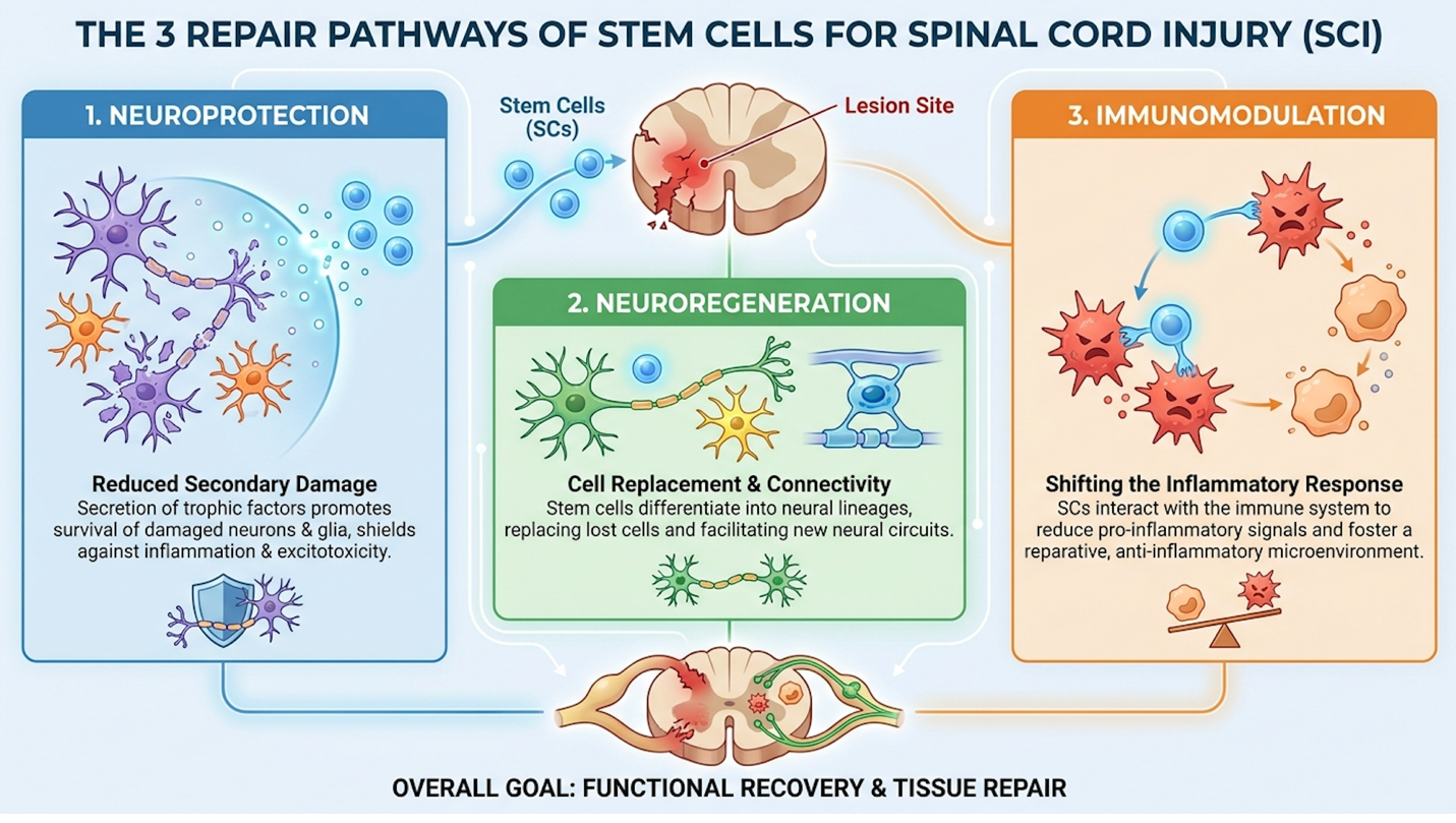
Investigational stem cell therapy for spinal cord injury relies on The Triphasic Neural Repair Model a biological framework targeting specific neurological deficits through immunomodulation, axon regeneration, and vascular restoration.
Stem cell therapy for spinal cord injury repair pathways operate through The Triphasic Neural Repair Model, a biological framework targeting specific neurological deficits. Mesenchymal stem cells act through immunomodulation and neuroprotection, significantly reducing secondary inflammation and facilitating nerve axon regeneration (NIH, 2019). This complex cellular interaction addresses both immediate tissue damage and long-term neural pathway restoration, treating the local lesion site as a dynamic environment rather than a static scar. From an editorial standpoint, the fundamental misrepresentation of these biologics as simple structural replacements rather than complex signaling directors is what drives so much dangerous medical misinformation online.
The initial mechanical trauma of a spinal cord injury isn’t what causes the most catastrophic tissue loss. The real damage happens hours and days later. When the cord is crushed or severed, the highly specialized blood-spinal cord barrier (BSCB) physically ruptures. This is a physiological
These rogue immune cells release neurotoxins, free radicals, and excitatory amino acids that rapidly destroy surrounding, otherwise healthy nerve tissue. This secondary inflammatory cascade effectively doubles the size of the initial physical lesion if left unchecked.
This is exactly where the first phase of The Triphasic Neural Repair Model intervenes. Stem cells don’t magically morph into new spinal cord right away. Instead, they operate like localized pharmaceutical factories. When administered intrathecally, mesenchymal stem cells secrete highly specialized bioactive molecules that fundamentally alter the local immune microenvironment before irreversible damage cascades throughout the cord.
Research evaluating the mechanisms of mesenchymal stem cell immunomodulation highlights a fascinating biological pivot (2019). The introduced cells force local macrophage populations to shift from a destructive, pro-inflammatory phenotype (M1) to a tissue-repairing phenotype (M2). They aggressively downregulate Tumor Necrosis Factor-alpha (TNF-α) while upregulating anti-inflammatory cytokines like IL-10. This halts the secondary wave of cell death.
| 📌 If you’re interested in how stem cells calm the secondary inflammation that follows a spinal injury, we have an interesting article that discusses mesenchymal stem cell therapy for immune modulation, which you can read via the internal link. |
While mitigating initial inflammation stops the lesion from expanding, restoring motor function requires direct cellular bridging via nerve axon regeneration.
Nerves in the central nervous system are notoriously stubborn. Unlike peripheral nerves in your arm or leg, spinal cord axons inherently resist regrowing after severe trauma. Axon regeneration involves forcing these severed communication lines to sprout new extensions across the site of the injury. You are essentially attempting to re-establish disrupted sensory and motor pathways across a biological chasm.
Cellular therapy tackles this resistance through the targeted upregulation of neurotrophic factors. Think of these factors as highly potent biological fertilizers. Introduced stem cells actively secrete Brain-Derived Neurotrophic Factor (BDNF), Glial Cell Line-Derived Neurotrophic Factor (GDNF), and Nerve Growth Factor (NGF). A consensus report on neurotrophic factor secretion in spinal cord repair confirms that these proteins act directly on surviving host neurons (NIH, 2020).
To understand why this is so revolutionary, look at the hostile environment of the extracellular matrix (ECM) inside the injured cord. Normally, severed axons encounter chondroitin sulfate proteoglycans (CSPGs) chemical barriers that force the nerve endings to curl back into “retraction bulbs.” The axons just give up. But when BDNF and GDNF flood the zone, they act as powerful chemical attractants.
They prevent the neurons from undergoing apoptosis (programmed cell death) and stimulate quiescent dermal and neural fibroblasts. These fibroblasts then deposit favorable matrix proteins like fibronectin and laminin. The stem cells essentially build a temporary biological scaffold, coaxing the patient’s own native axons to extend specialized growth cones directly across the damage zone. Rebuilding these neural pathways, however, is intensely metabolically demanding. These newly sprouting nerve extensions will immediately die without adequate vascular restoration.
You simply cannot rebuild neurological tissue in an ischemic, oxygen-starved environment. A severe spinal lesion obliterates the local capillary networks, creating an ischemic dead zone. Hypoxia sets in rapidly, triggering the death of the very neural pathways the therapy aims to salvage. Angiogenesis the physiological process through which new blood vessels form from pre-existing vessels is an absolute prerequisite for any permanent tissue restoration in the spinal cord.
During the third phase of the repair model, the transplanted cells secrete massive amounts of Vascular Endothelial Growth Factor (VEGF). This specific protein acts as a distress signal, responding to hypoxia-inducible factor 1-alpha (HIF-1α) pathways in the damaged tissue. VEGF stimulates local endothelial cells to aggressively proliferate and construct new, highly organized vascular networks directly into the necrotic spinal tissue.
This enhanced local microcirculation is what delivers the vital oxygen, glucose, and systemic immune cells required by those metabolically hungry, regenerating axons we discussed earlier. Furthermore, studies on vascular regeneration in SCI demonstrate that these newly formed capillaries help flush out toxic residual cellular debris (Nature, 2021). This physically clears the path for nerve growth.
Executing this triphasic repair model effectively requires selecting the precise cellular derivative tailored to the specific neurological deficit.
One of the most dangerous marketing oversimplifications is treating all “stem cells” as identical products. They aren’t. Understanding the biological distinction between Mesenchymal Stem Cells (MSCs) and Neural Stem Cells is non-negotiable if you are evaluating treatment protocols. Many commercial clinics deliberately blur these lines to sell less complex MSC treatments as complete cellular replacements.
Mesenchymal stem cells are multipotent cells typically harvested from Wharton’s jelly (umbilical cord tissue), adipose (fat) tissue, or adult bone marrow. Their primary superpower is not replacing dead tissue. Instead, their dominance lies in systemic anti-inflammatory (paracrine) signaling. When a clinic discusses mesenchymal stem cell therapy SCI protocols, they are primarily targeting the immunomodulation and vascular regeneration phases of repair. MSCs act as biochemical directors, telling the host’s body how to heal itself.
Neural stem cells are entirely different. These are lineage-specific progenitor cells. They are biologically pre-programmed with a specific destiny: differentiating directly into neurons, astrocyte glia, and critical oligodendrocyte precursor cells (OPCs).
OPCs are incredibly vital for structural repair. They are responsible for producing myelin the insulating lipid sheath that wraps around naked nerve axons. Without myelin, electrical signals leak out, and the nerve cannot conduct impulses efficiently (a process called saltatory conduction). Current neural stem cell phase I/II clinical trials heavily focus on evaluating human neural stem cell transplantation safety and immunological integration (2021).
The clinical application diverges sharply based on these traits. Doctors use MSCs to forcefully alter a hostile, inflammatory immune response, whereas they investigate Neural stem cells for direct, structural cellular replacement in the damaged cord. Understanding these distinct cellular mechanisms provides the necessary biological context to interpret recent clinical trial outcomes and recovery data accurately.
The newest breakthrough in spinal cord therapy emphasizes targeted functional improvements rather than absolute systemic cures. A comprehensive review of recent clinical trials indicates that stem cell therapies for spinal cord injury successfully promoted measurable motor recovery in 45% of patients with incomplete injuries (NIH, 2022). Evaluating these outcomes requires analyzing specific neurological benchmarks rather than anecdotal internet claims. From a clinical perspective, we must transition patient expectations away from total physiological reversal and toward the incremental, life-altering gains in physical independence that modern protocols can actually deliver.
When clinical literature cites a 45% motor recovery rate, we need to unpack what that actually means for a patient’s daily life. It rarely means getting out of a wheelchair and sprinting.
The distinction between complete and incomplete injuries dictates this data. Neurologists classify SCI using the ASIA Impairment Scale (AIS). Incomplete spinal cord injuries where some sensory or motor pathways remain intact across the lesion (ASIA grades B, C, or D) show vastly higher responsiveness to cellular therapy. The intact native axons provide a physical, surviving bridge that the stem cells can amplify and remyelinate.
Complete anatomical transections (ASIA grade A), where the cord is entirely severed with zero motor or sensory function below the lesion, currently demonstrate negligible structural regeneration with biologics alone.
In these clinical trials, doctors measure recovery by testing specific myotomes (muscle groups) on a 0 to 5 grading scale. Meaningful functional benefit translates to concrete, life-altering improvements. A patient moving from a C5 to a C6 functional level on the ASIA scale might regain wrist extension, granting them the critical ability to feed themselves or operate a manual wheelchair independently.
These broader statistical reviews of recent clinical trial reviews are further corroborated by localized, intensive academic trials evaluating precise sensory feedback (2022).
Search the internet for five minutes, and you’ll inevitably encounter the query: “Is there a cure for spinal cord injury in 2026?” Let’s be unequivocally clear. While therapeutic applications are advancing at a blistering pace, a universal biological cure that completely reverses paralysis for all patients is medically inaccurate and ethically irresponsible.
Viral videos showing a “paralyzed man walk again after stem cell” therapy require massive contextual caveats. What these viral clips omit is the clinical reality behind the scenes. These profound recovery cases almost exclusively involve patients with incomplete injuries who are undergoing grueling, daily physical rehabilitation regimes.
Furthermore, the true 2026 frontier involves pairing cellular biologics with advanced Epidural Electrical Stimulation (EES) implants. Developed by researchers at institutions like EPFL, EES
The stem cells provide the biological substrate, actively modulating inflammation and promoting axon sprouting. Concurrently, the EES implant provides the electrical spark to force those new pathways to communicate. The stem cells alone did not make the patient walk. Clinics that guarantee specific motor outcomes or promise 100% resolution of paralysis with standalone injections are exploiting desperate families. Valid neurosurgical consensus on SCI cure timelines indicates we are currently optimizing marginal gains in independence (Journal of Neurosurgery, 2023). Setting realistic baseline expectations also requires understanding the biological half-life of these cellular interventions.
A pervasive misconception is that injected stem cells take up permanent residence in the spinal cord, living there forever to maintain your mobility. That simply isn’t how the biology works.
When discussing the longevity of stem cell therapy, we have to look at the “hit and run” hypothesis of paracrine signaling. The introduced cells typically survive in the host tissue for only a matter of weeks, occasionally a few months, before the body naturally clears them via apoptosis. Their primary function during that brief window is to permanently alter the host’s tissue microenvironment. They secrete exosomes and cytokines that jumpstart the native repair mechanisms we discussed in the Triphasic model.
Because the biologics eventually clear, the necessity of ongoing physical therapy becomes paramount. Neurological recovery is fundamentally activity-dependent, so patients must engage in aggressive locomotor training to cement their functional gains. You have to force the nervous system to use the newly established, fragile neural pathways, or they will degrade.
Achieving any degree of sustained functional outcome depends entirely on intervening during the correct biological window.
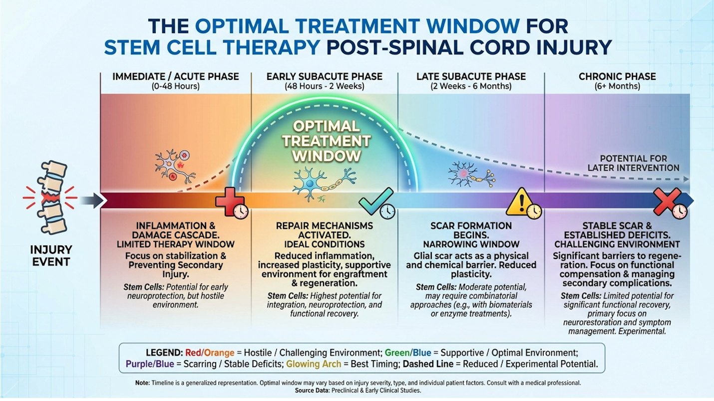
The spinal cord injury treatment window dictates the biological viability of cellular intervention. Research indicates the most optimal treatment window for stem cell transplantation occurs during the subacute phase, 7 to 14 days post-injury, before impenetrable glial scarring forms (NIH, 2023). Missing this critical chronological benchmark severely diminishes the efficacy of regenerative protocols. Medically speaking, treating chronological eligibility as a flexible guideline rather than a strict biological imperative is a massive failure in clinical judgment.
Timing is the single most ruthless variable in neuro-regeneration. The biological environment of the spinal cord changes radically week by week following the initial trauma.
Why not inject stem cells immediately on day one? Immediate acute intervention (0 to 3 days post-injury) is heavily contraindicated in most clinical protocols. During this acute phase, the lesion site is a warzone of extreme localized swelling, necrotic tissue, and severe hemodynamic instability. Introducing fragile biologics into this highly toxic, actively dying tissue environment ensures they will be destroyed almost immediately by the host’s surging immune response.
However, research pointing to the subacute phase as optimal window demonstrates a biological sweet spot (2023). Around the 7 to 14-day mark, the extreme acute inflammation begins to subside, but the body has not yet permanently sealed off the injury site. This offers the optimal biological environment for introduced cells to survive, integrate, and initiate the paracrine signaling required to salvage surrounding threatened neurons.
Once this subacute window closes, the body initiates a protective but ultimately prohibitive biological response.
The central nervous system prioritizes containment over repair. When the spinal cord sustains damage, specialized cells called astrocytes rush to the lesion border. They rapidly multiply and intertwine, forming a dense, fibrous wall known as a glial scar (astrogliosis).
The body does this to prevent the toxic inflammatory cascade from spreading up and down the spinal cord. It’s a life-saving mechanism in the short term. But in the long term, this glial scar
This brings us to the harsh reality of stem cell therapy for chronic spinal cord injury (5+ years). For patients injured years ago, this glial scar is fully mature and practically impenetrable. The efficacy of cellular therapy for these chronic cases drops precipitously. While a chronic patient might still experience some mild immunomodulation or a reduction in neuropathic pain, structural repair and significant motor recovery are highly unlikely without an experimental enzyme to dissolve the scar first.
This immutable biological barrier informs the rigorous medical guidelines determining who qualifies for advanced therapies.
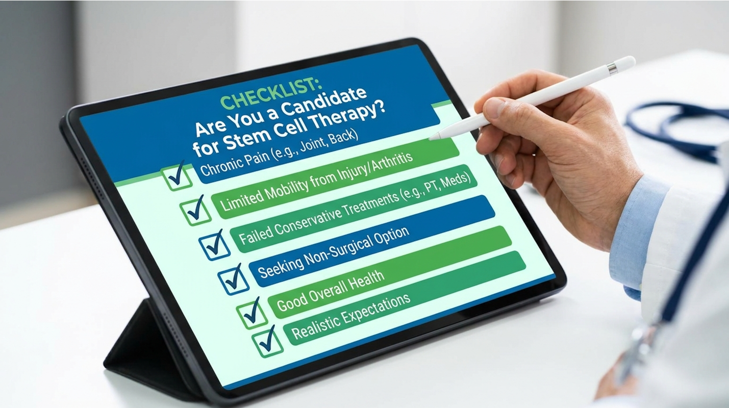
Not everyone is a candidate for regenerative intervention. In fact, reputable clinics will reject a significant percentage of applicants following a detailed neurological review. Understanding who is not a good candidate for stem cell therapy saves patients tremendous financial and emotional distress.
Explicit medical contraindications include patients with active systemic infections, uncontrolled autoimmune disorders, or a history of specific malignancies (as growth factors could theoretically accelerate dormant tumor growth). Patients suffering from extreme hemodynamic instability or those requiring ongoing intensive care for secondary organ failure are immediately disqualified.
Crucially, patients with a complete anatomical cord transection where neuro-imaging confirms the spinal cord has been physically cut in half with massive physical separation are not viable candidates for biologics alone.
The WHO guidelines on regenerative medicine contraindications reiterate the absolute necessity of a personalized clinical suitability assessment (2023). Never commit financially to an overseas clinic before a board-certified neurologist has reviewed your recent MRI.
Beyond strict medical eligibility, patients must also navigate the broader regulatory and ethical complexities surrounding these treatments.
When families begin researching options, they inevitably encounter fierce debates. Understanding why are people against stem cell therapy requires separating historical baggage from modern clinical realities.
Much of the public hesitation stems from outdated ethical debates regarding embryonic stem cells. Today, virtually all reputable offshore facilities utilize ethically sourced adult MSCs derived from donated umbilical cord tissue (Wharton’s jelly) following healthy, full-term cesarean births. The embryonic debate is largely irrelevant to modern SCI protocols.
| 📌 If you’re curious why umbilical cord tissue is favored over fat or bone marrow as a cell source, we have an interesting article that discusses why umbilical cord-derived UC-MSC stem cells are superior to other stem cell sources, which you can read via the internal link. |
The valid, contemporary controversies revolve entirely around patient safety and clinical regulation. Medical professionals strongly oppose unregulated clinics that utilize unproven, uncharacterized cell lines. Without stringent laboratory protocols, there is a legitimate risk of severe central nervous system infection or in cases involving poorly manipulated induced pluripotent cells (iPSCs) the terrifying risk of tumor formation (teratomas) within the spinal cord.
| 📌 If you’re wondering how patient safety is protected at regulated clinics, we have an interesting article that discusses whether stem cell therapy is safe in Thailand, which you can read via the internal link. |
For eligible candidates seeking ethically sourced, evidence-based care, Southeast Asia has emerged as a primary logistical hub for specialized treatment facilities.
The stem cell therapy cost in Thailand for spinal cord injury interventions establishes a baseline starting around $15,100 USD (approximately ฿513,400 THB). This baseline covers standard cellular processing and administration, though comprehensive regenerative packages often require extended physical rehabilitation and multidisciplinary care, heavily altering the final financial investment. Ultimately, navigating overseas medical pricing requires radical transparency from providers; any clinic dodging direct cost inquiries should be disqualified immediately.
| 📌 If you’re interested in a broader look at treatment pricing, we have an interesting article that discusses stem cell therapy costs in Thailand for 2025, which you can read via the internal link. |
When you see a starting price of $15,100, you need to know precisely what that capital procures. Stem cell therapy for spinal cord injury cost structures in Bangkok are significantly lower than experimental cash-pay trials in the West, but they remain a massive out-of-pocket investment.
This base tier typically covers the core biological deliverables. It pays for the extensive laboratory cell expansion—culturing millions of highly viable MSCs in a sterile, ISO-certified cleanroom environment. It covers the mandatory pre-operative medical screening, blood panels, and the initial surgical administration (usually an intrathecal lumbar puncture or localized injection by an interventional radiologist).
Medical tourism data tracking average price points for regenerative SCI care confirms this $15,100 baseline is standard among top-tier, regulated providers in the region (ISCT, 2023). Costs fluctuate, but a clinic offering treatment for $3,000 is cutting terrifying corners on lab sterilization, while a clinic charging $60,000 for a basic MSC infusion is drastically overcharging.
Securing this level of care requires selecting a facility that prioritizes stringent laboratory standards over aggressive marketing.
The base price gets you in the door. The actual final medical bill depends heavily on the specific medical protocol tailored to the severity of your lesion.
Cell dosage is the primary cost multiplier. An administration of 100 million MSCs is vastly cheaper than a high-dose protocol requiring 200 to 300 million cells spaced over several weeks. Furthermore, the method of administration alters fees. A simple intravenous (IV) drip carries lower facility fees than an intrathecal injection requiring fluoroscopic guidance in a sterile surgical theater.
| 📌 If you’re wondering whether a higher cell dose leads to better results, we have an interesting article that discusses why double-dose UC-MSC stem cell therapy may offer greater benefits than a single dose, which you can read via the internal link. |
Finally, do not underestimate the financial weight of post-procedure rehabilitation. Stem treatment for paralysis is largely ineffective in a vacuum. Cells alone cannot rebuild motor function without the targeted mechanical stimulation provided by months of specialized physiotherapy.
While evaluating these logistical hurdles, patients and their families must remain vigilant against the inherent risks associated with unregulated medical tourism. Following rigorous CDC guidelines for medical travel ensures that you are asking the right questions regarding laboratory certifications, contingency care, and infectious disease screening before crossing borders.
Any regenerative treatment plan involving intrathecal administration or a significant, five-figure financial investment demands extreme medical scrutiny. Do not rely solely on the clinical assessment of the doctor selling you the treatment.
The American Academy of Neurology guidelines suggest that patients pursuing experimental overseas therapies must secure a secondary evaluation (2023). Take the proposed treatment protocol from the Thai clinic to a board-certified, independent neurologist in your home country. They can objectively assess your individual physiological risks, identify potential medication interactions, and confirm whether your specific lesion morphology makes you a viable candidate.
Stem cell therapy for spinal cord injury demonstrates measurable motor improvements in up to 45% of patients with incomplete lesions during clinical trials. Success relies heavily on intervening during the critical subacute phase, typically 7 to 14 days post-injury. Patients with complete anatomical transections or dense chronic scarring rarely experience significant structural regeneration. For example, recent Phase 1 academic trials show promising sensory gains, but outright cures remain scientifically unsupported. Individual outcomes vary extensively based on lesion severity and subsequent physical rehabilitation protocols.
The primary side effects of intrathecal stem cell administration include mild localized pain, transient fever, and temporary headaches. When utilizing ethically sourced, rigorously screened mesenchymal stem cells, severe adverse immunological rejections remain statistically rare. However, utilizing unregulated clinics carries profound risks, including severe central nervous system infections and potential tumor formations. Patients should meticulously verify the clinic’s laboratory processing standards to mitigate contamination risks. Always consult an independent neurologist to assess individual physiological risks prior to proceeding.
For patients navigating the crushing complexities of spinal cord injuries, stem cell therapy in Thailand offers advanced regenerative options, provided treatment occurs within the critical subacute biological window (NIH, 2023). The data is clear: the most effective approach requires combining rigorous, unbiased medical eligibility assessments with comprehensive, months-long post-procedure physical rehabilitation. It is a grueling process of marginal gains, not a magic switch.
The science driving these outcomes relies fundamentally on The Triphasic Neural Repair Model. By systematically targeting immunomodulation, coaxing axon regeneration through neurotrophic factors, and forcing vascular restoration, we can address the specific biological deficits of an SCI. However, this framework only translates to real-world mobility when executed transparently by evidence-based clinical facilities.
Take the next step with clinical objectivity. Gather your complete medical records, recent high-resolution MRIs, and your comprehensive treatment history to schedule a formal, independent clinical suitability assessment with a qualified regenerative medical specialist before booking any international travel.