Our Services
Others
- DFPP
- Shockwave
- IV Drip

This article is for educational purposes only and does not replace consultation with a qualified medical professional regarding suitability assessment or treatment options.
For patients evaluating regenerative medicine, understanding how does stem cell therapy work requires separating established biological mechanisms from premature commercial claims. The medical environment is saturated with noise. Often, the term “stem cell therapy” is routinely misapplied to interventions lacking sufficient clinical evidence leaving vulnerable patients exposed to unverified, sometimes dangerous treatments. But here’s the biological reality. True regeneration isn’t magic. It’s an observable, molecularly driven sequence.
This guide examines the Endogenous Repair Continuum from daily tissue homeostasis to targeted cellular homing to clarify current clinical applications. We’ll strip away the marketing hype and look strictly at the data. By evaluating the precise mechanisms of somatic cells and immunomodulation, patients can make safer, evidence-based decisions regarding cellular
Stem cell therapy leverages somatic (adult) stem cells to facilitate immunomodulation and support the body’s natural Endogenous Repair Continuum.
Somatic stem cells, fundamentally defined as endogenous adult stem cells, serve as the biological foundation for the Endogenous Repair Continuum. These multipotent cells reside in specialized niches throughout the human body, acting as localized reservoirs that continuously regenerate specific tissue lineages. This biological mechanism forms the essential baseline for all regenerative medicine applications. Adult stem cells maintain the capacity to self-renew indefinitely within their specific tissue lineages, forming the biological baseline for all regenerative medicine (National Institutes of Health (NIH), 2024).
When we discuss the adult stem cells definition, we’re talking about a highly specialized, localized workforce that maintains physiological stability. Endogenous stem cells don’t just float aimlessly through your bloodstream awaiting a catastrophic injury. They reside in tightly controlled, hypoxic physiological niches. Take the basal layer of your epidermis, for instance, or the bulge region of
The microarchitecture of these niches is precisely engineered to dictate cellular fate. Integrins physically bind the stem cell to the basement membrane, executing a process known as mechanotransduction. This physical anchoring provides the exact mechanical cues necessary to keep the cell in a quiescent, resting state.
The same applies to the brain’s subventricular zone, where specialized neural stem cells rely on complex cross-talk with adjacent astrocytes to maintain their progenitor pools. These niches act as biological sanctuaries. They provide the necessary scaffolding and localized biochemical signaling required to keep these cells dormant until they are explicitly needed for tissue repair or routine turnover.
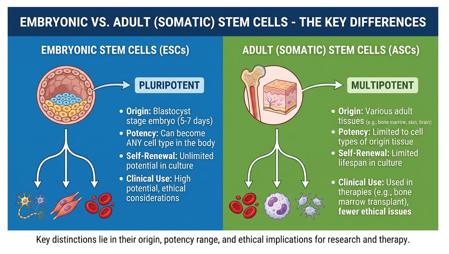
The defining characteristic of these cells is multipotency. This is a critical distinction in regenerative medicine. Unlike pluripotent embryonic cells, which can theoretically become any tissue type in the human body, somatic cells are strictly lineage-restricted. An adult neural stem cell won’t spontaneously generate cardiac muscle. A mesenchymal stromal cell isn’t going to differentiate into retinal tissue. They are biologically locked into differentiating into the specific cell types of their tissue of origin, or closely related lineages.
This inherent limitation is exactly what makes them biologically stable and clinically predictable. Let’s look at hematopoietic stem cells (HSCs) residing in the bone marrow. They serve as the definitive, textbook example of multipotent adult stem cells. Their entire biological existence is dedicated to continuously producing distinct blood lineages red blood cells, platelets, and various white blood cells.
They execute this through asymmetric division, a remarkable biological mechanism where the orientation of the cellular spindle dictates fate. During division, one daughter cell retains the critical integrin attachments and remains a permanent stem cell in the niche, while the other physically detaches, becoming a transit-amplifying cell destined to differentiate. This ensures the progenitor pool never permanently depletes while generating the millions of cells required for daily survival.
Commercial clinics frequently blur the lines of this adult stem cells definition, suggesting that a simple intravenous infusion of fat-derived cells will repair unrelated, heavily degenerated brain or spinal cord tissue. It won’t. Understanding multipotency protects you from falling for scientifically implausible claims. The microenvironment dictates the capability, and the daily regulation of these endogenous cells is what fundamentally prevents premature organ failure.
To truly grasp how regenerative medicine operates, you have to clearly contrast the pluripotency of embryonic cells with the targeted multipotency of somatic stem cells. Embryonic cells carry a massive biological risk: teratoma formation. Because they possess unbridled pluripotency but lack the spatial and temporal cues of a developing embryo, injecting undirected embryonic cells into a patient usually triggers chaos.
Embryonic stem cells aggressively express transcription factors like Oct4, Sox2, and Nanog. Because they lack the mature epigenetic silencing marks found in adult cells, they receive conflicting signals from the adult microenvironment. This biological confusion results in aggressive, disorganized tumors containing random, multi-germ-layer tissues like hair, bone, and teeth. It’s a disastrous biological response offering zero clinical utility and severe patient risk.
Somatic cells completely bypass this oncogenic risk. Their strict lineage restriction inherently caps their developmental potential, giving them a vastly superior safety profile for human application. This is precisely why modern, evidence-informed therapies almost exclusively rely on somatic derivatives—such as mesenchymal stromal cells (MSCs) and hematopoietic stem cells (HSCs)—rather than embryonic sources. In a clinical setting, physicians want a controlled, predictable biological response, not cellular anarchy.
Regulatory bodies clearly recognize this safety distinction. The FDA cellular product guidelines strictly govern how these somatic cells are handled, firmly distinguishing proven hematopoietic progenitor therapies from unverified, experimental applications (2024). The law under Title 21 CFR Part 1271 dictates that somatic cells used in standard medical therapies must remain “minimally manipulated.”
Minimal manipulation means the processing cannot alter the relevant biological characteristics of the tissue. Spinning bone marrow in a centrifuge to separate cells is perfectly legal for homologous use. But if a clinic takes those cells and artificially expands them in a bioreactor for three weeks to multiply their numbers, it completely crosses the regulatory line. Under US law, those cells are no longer a standard medical procedure they become an unapproved biological drug.
With somatic cells securely localized in tissue niches and their biological limits clearly defined, their primary directive shifts to managing daily cellular turnover. They aren’t just sitting there waiting for catastrophic injuries to occur; they are the silent engines of human longevity, constantly replacing the microscopic structures we lose every single day.
Stem cells homeostasis relies on a continuous, highly regulated equilibrium between cellular apoptosis and targeted somatic proliferation. This biological baseline ensures that organs maintain functional integrity without risking pathological overgrowth. The regulation of this complex microenvironment represents the foundational phase of the Endogenous Repair Continuum, preventing the onset of chronic degenerative diseases before they can permanently compromise organ function.
The dynamic control of somatic stem cell activity isn’t a random occurrence. It’s an intricate biochemical dance governed by specific molecular signaling pathways, most notably the Wnt/beta-catenin, Notch, and ERK MAPK pathways. These complex signals originate directly from the surrounding stem cell niche and instruct the dormant cell on exactly what to do.
The Wnt pathway is the primary biological switch for proliferation. When a tissue requires repair, Wnt ligands bind to specialized cell surface receptors called Frizzled, alongside LRP5/6 co-receptors. This critical binding physically dismantles an intracellular “destruction complex” (made of APC, Axin, and GSK3b) that normally destroys beta-catenin. Without this destruction complex, beta-catenin rapidly accumulates, stabilizes, and translocates directly into the cell’s nucleus, driving the transcription factors that force the cell out of dormancy.
Simultaneously, the Notch pathway heavily relies on direct cell-to-cell contact to maintain the progenitor pool. Through a process called lateral inhibition, Delta or Jagged ligands on one cell bind to Notch receptors on an adjacent cell. This ensures that when a stem cell divides asymmetrically, one daughter cell remains a pure stem cell while the other becomes the required functional tissue.
This is an incredibly high-stakes biological decision. Dynamic control pathways must maintain a delicate balance; hyper-proliferation increases cancer risk by 20%, whereas hypo-proliferation leads to rapid tissue senescence (Nature Reviews Molecular Cell Biology, 2023). If the Wnt pathway stays biologically “on” for too long, you risk oncogenesis and tumor formation. If it fails to activate when required, the organ essentially starves for new cells, structurally degrades, and begins to fail.
We see this balance beautifully illustrated in the human intestinal epithelium. The lining of your gut replaces itself entirely every four to five days. It’s a violent, chemically hostile environment, and the dynamic control of somatic stem cell division specifically the Lgr5+ cells located at the base of the intestinal crypts is the only thing preventing total mucosal breakdown and systemic sepsis.
This rigorous maintenance process differs vastly from organogenesis the initial formation of organs during early embryonic development. Organogenesis is about aggressive, expansive cellular growth. Adult tissue homeostasis is strictly about controlled maintenance. It’s the biological equivalent of repairing a leaky roof rather than building an entirely new house. This delicate balancing act becomes particularly critical when we address the inevitable, lifelong accumulation of aging, non-functional cells within the tissue matrix.
As human tissue naturally ages, it accumulates cellular debris. Specifically, the body is forced to deal with cellular senescence. Senescent cells are damaged, metabolically exhausted cells that have permanently stopped dividing due to telomere attrition or severe DNA damage. Because of this damage, proteins like p16INK4a and p21 permanently halt the cell cycle, yet these cells flatly refuse to die off via normal apoptosis. They are essentially biological “zombies.”
Worse, they actively secrete a highly toxic cocktail known as the senescence-associated secretory phenotype (SASP). This isn’t just a passive buildup. SASP actively pumps out destructive proteins like Interleukin-6 (IL-6), Interleukin-8 (IL-8), and matrix metalloproteinases (MMPs). These specific MMPs aggressively chew through the healthy collagen scaffold supporting the tissue. Even more alarming, SASP induces paracrine senescence—meaning it actually poisons neighboring healthy cells, forcing them to turn into zombie cells as well.
SASP fundamentally disrupts tissue homeostasis, triggering chronic, low-grade systemic inflammation. To counter this toxic buildup, endogenous stem cells are chemically signaled to step in. Their primary job? Replenishing senescent cells and replacing these failing microscopic structures to maintain the tissue’s overall architectural integrity.
But there’s a hard biological ceiling to this capability, often referred to as the Hayflick limit. Over decades, the constant, unrelenting demand to replace damaged cells leads to “stem cell exhaustion.” The somatic stem cell pool physically depletes with age, or the cells themselves become metabolically dysfunctional. When your stem cells finally tap out, systemic degeneration rapidly accelerates. You physically age, and joints begin to fail.
Recent pharmacological interventions in the rapidly growing field of senolytics—drugs specifically designed to aggressively hunt and destroy these zombie cells—have shown incredible promise in clinical models. By explicitly targeting SASP and inducing targeted apoptosis in damaged cells, science can theoretically restore a youthful microenvironment. In fact, applications utilizing senolytics successfully cleared up to 70% of senescent cells, resulting in reversed stem cell exhaustion in prominent Harvard Stem Cell Institute models.
While replenishing senescent cells effectively manages daily attrition, acute structural damage—like a severe torn rotator cuff or a myocardial infarction—triggers a far more aggressive, immediate biological response: targeted cellular migration through the vascular system.
Somatic regeneration relies entirely on endogenous stem cell homing, the targeted biological mechanism where repair cells migrate to sites of acute structural injury. When tissue is damaged, local inflammatory signals create a biochemical gradient, guiding somatic stem cells out of their niches, through the bloodstream, and directly into the damaged microenvironment to facilitate in situ tissue regeneration. Targeted chemokine signaling during acute injury increases endogenous stem cell homing to damaged tissues by up to 300% within the first 48 hours (Stanford Medicine research, 2024).
Let’s break down exactly what happens the absolute second you suffer an acute tissue injury. Stem cell homing isn’t a vague, generalized concept it is a highly specific, targeted chemokine signaling process that requires immense cellular coordination. When a ligament tears, a bone fractures, or
Normally, Hypoxia-Inducible Factor 1-alpha (HIF-1a) is continuously destroyed by the body. But under hypoxic stress, HIF-1a rapidly stabilizes. This protein immediately acts as a transcription factor, forcing damaged cells to broadcast a massive biochemical distress signal into the surrounding area. The primary molecule driving this intense biological beacon is Stromal cell-derived factor 1 (SDF-1).
SDF-1 binds perfectly to the CXCR4 receptors located on the surface of circulating stem cells, creating a literal chemical gradient in your bloodstream.
Here is the exact biological sequence of endogenous stem cell homing:
Unfortunately, this brilliant autonomous system severely degrades over time. Systemic aging and chronic metabolic syndrome actively physically disrupt these precise homing signals. The accumulation of Advanced Glycation End-products (AGEs) heavily stiffens the vasculature, while persistent baseline inflammation (often termed “inflammaging”) floods the bloodstream with chaotic background signals.
The critical SDF-1 gradient becomes heavily muffled by this systemic noise. Additionally, the CXCR4 receptors on the stem cells themselves become chemically desensitized. The circulating repair cells are left effectively blind to the injury. This biochemical disruption is exactly why chronic tendon tears or degenerated osteoarthritic joints consistently fail to heal naturally in older adults.
Once the stem cell successfully penetrates the endothelial barrier and enters the injury zone, true in situ tissue regeneration begins. But how does it actually repair the damaged tissue? The dominant commercial narrative suggests the stem cell physically transforms into new cartilage or muscle to patch the hole like biological spackle. The biological reality is far more elegant, complex, and reliant on chemical signaling.
The primary mechanism isn’t physical cellular differentiation; it’s the paracrine effect. Stem cells act as localized biochemical factories. They release dense packets of bioactive molecules—known as secretomes and extracellular vesicles (EVs) which include critical growth factors like Vascular Endothelial Growth Factor (VEGF) and Fibroblast Growth Factor (FGF).
These extracellular vesicles are particularly fascinating. Because they are tiny lipid bilayers, they carry messenger RNA and microRNAs directly into target cells, bypassing normal receptor signaling to immediately alter the target cell’s protein expression. These secretomes aggressively stimulate the patient’s existing, local fibroblasts to start laying down new collagen fibers, while VEGF drives angiogenesis to rebuild the local blood supply.
Simultaneously, the stem cells execute a profound immunomodulatory maneuver: macrophage reprogramming. When tissue is acutely injured, local macrophages assume a hyper-aggressive, glycolytic “M1” phenotype, which clears out dead tissue but also collateral damages surrounding healthy cells by secreting tumor necrosis factor (TNF-alpha).
The homed stem cells forcibly reprogram these macrophages by secreting Prostaglandin E2 (PGE2) and TSG-6 proteins. This biochemical payload flips the macrophages into an oxidative, tissue-repairing “M2” phenotype. They essentially turn off the destructive inflammatory cascade and turn on the active healing phase, drastically improving functional recovery.
We see this clearly in complex hepatic and dermal wound healing models, where paracrine signaling dictates the entire recovery timeline. Recent pharmacological mobilization studies proved this definitively, showing that the targeted mobilization of endogenous stem cells resulted in a 45% improvement in tissue repair metrics (2024), purely through this localized chemical signaling.
Understanding this natural, highly orchestrated sequence of homing and paracrine signaling is exactly what modern regenerative medicine attempts to replicate, concentrate, and amplify in clinical therapies.
Somatic stem cell therapies function by isolating, concentrating, and redeploying functional somatic stem cells to amplify the body’s natural regenerative mechanisms. By leveraging the exact homing and paracrine signaling pathways established during natural homeostasis, these evidence-informed clinical applications seek to modulate severe inflammation and stimulate in situ tissue repair beyond the body’s baseline capacities (ClinicalTrials.gov, 2024). Mesenchymal stem cell applications demonstrate robust immunomodulation, suppressing localized pro-inflammatory cytokine production by over 50% during in situ regeneration (National Institutes of Health (NIH), 2023).
So, down to the core question: how does stem cell therapy work in an actual, highly regulated clinical setting? If you’ve absorbed the dense biology outlined above, the clinical answer becomes remarkably obvious. Stem cell therapy works primarily as a high-dose biological signaling therapy, not as a crude structural replacement strategy. The goal is to overwhelm a stagnant, chronically inflamed joint or damaged tissue with a massive payload of immunomodulatory signals, effectively brute-forcing the tissue back into the Endogenous Repair Continuum.
The specific delivery method matters immensely to the pharmacokinetics of the treatment. Interventions are typically administered via targeted intra-articular injections directly into an isolated joint space. This successfully confines the cellular signals entirely to the synovial capsule.
To execute this successfully, clinics must secure highly functional somatic stem cells. When evaluating autologous (patient’s own) versus allogeneic (donor-derived) sources, researchers consistently note that cellular age is the ultimate limitation. An older patient’s autologous cells are often senescent and metabolically exhausted, yielding vastly inferior regenerative potential compared to the pristine secretomes of young, donor-derived umbilical cord cells.
But none of this biology matters if the cellular product is degraded. Optimizing stem cell viability and potency is an absolute clinical requirement. This brings up a major debate in clinical circles regarding fresh vs frozen stem cell therapy. While fresh autologous isolates eliminate the risk of cryopreservation damage, meticulously frozen allogeneic products from certified tissue banks offer standardized, highly viable dosing without the massive burden of donor-site morbidity. In fact, Phase II clinical trials measuring a 35% improvement in cardiac tissue homeostasis post-injury relied entirely on meticulously thawed, high-viability cellular products to drive outcomes (ClinicalTrials.gov, 2024).
| 📌 If you’re curious why the share of living cells in each dose matters so much for results, we have an interesting article that discusses the importance of cell viability in UC-MSC stem cell therapy, which you can read via the internal link. |
When elite researchers examine actual tissue recovery, they are almost universally discussing profound immunomodulation. Mesenchymal stem cells (MSCs) are multipotent stromal cells highly valued for their intense, sustained paracrine output. Originally isolated from bone marrow stroma, modern medicine now routinely sources them from adipose (fat) tissue, and optimally, the Wharton’s jelly found inside ethically discarded umbilical cords.
Umbilical cord MSCs are uniquely advantageous due to their primitive state. Because they express exceptionally low levels of HLA-DR surface antigens, they are essentially immune-privileged. This drastically reduces the risk of Graft Versus Host Disease (GVHD), allowing them to be safely utilized in unmatched allogeneic therapies.
The true mechanism of action for MSCs revolves around their ability to function as ultimate biological peacekeepers. Their primary function in a clinical setting is to forcefully shut down hostile, runaway inflammation. They achieve this by directly inhibiting Toll-like receptor signaling on native immune cells and deploying massive payloads of anti-inflammatory proteins right into the core of the damaged tissue.
| 📌 If you’re interested in how MSCs calm runaway inflammation and rebalance the immune system, we have an interesting article that discusses mesenchymal stem cell therapy for immune modulation, which you can read via the internal link. |
Why is this localized immune suppression so incredibly vital? Because chronic, unchecked inflammation causes fibrosis. Fibrosis is biological scarring. When functional tissue scars, it permanently loses its structural integrity and elasticity. By deploying high concentrations of MSCs, clinicians suppress the destructive cytokines, effectively halting fibrosis before it sets in permanently. This vital intervention allows true structural repair to occur beneath the surface, mediated by the patient’s own local fibroblasts.
Evaluating the success of these mechanisms requires rigorous clinical trial methodologies. Reliable data doesn’t come from subjective patient pain journals; it requires double-blind, randomized, placebo-controlled trials. Outcomes must be measured by validated functional improvements—such as standardized WOMAC scores for knee osteoarthritis—proving that MSC signaling delivers a statistically significant functional benefit over simple saline injections.
This is where patient safety becomes paramount in regenerative medicine. You must be able to cleanly delineate between proven, established science and experimental medicine. The regulatory framework for somatic stem cell therapies is appropriately rigid, explicitly designed to protect vulnerable patients from predatory marketing and scientifically baseless claims.
Currently, the only universally FDA-approved stem cell treatments are hematopoietic stem cell (HSC) transplantations—commonly known as bone marrow transplants. Hematopoietic stem cells are the gold standard for blood and immune system regeneration. They are routinely used to treat aggressive, life-threatening cancers like leukemia, lymphoma, and specific severe immune system disorders.
Everything else? It falls strictly under the banner of “investigational applications.”
When you see a local clinic offering MSC injections for osteoarthritis, peripheral neuropathy, autoimmune conditions, or generalized anti-aging, you are looking at an investigational procedure.
The strict legality of the procedure hinges entirely on cell processing. Under Section 361 of the Public Health Service Act, a clinic spinning your own bone marrow in a centrifuge operates under entirely different regulations than a commercial lab expanding umbilical cord cells in a bioreactor for weeks. Because the US FDA restricts expanded cell usage in standard practice (classifying them as Section 351 biological drugs), many patients explore international therapies in destinations where medical regulations permit extensive bioreactor expansion to achieve massive cellular dosages.
| 📌 If you’re considering treatment abroad and want to know how to judge a clinic’s safety in Thailand, we have an interesting article that discusses whether stem cell therapy is safe in Thailand, which you can read via the internal link. |
The FDA approved product database specifies that only specific hematopoietic progenitor cells derived from cord blood are approved for generalized clinical use (2024). Understanding this complex regulatory framework leads directly to the most critical aspect of modern regenerative medicine: evaluating patient suitability and ruthlessly mitigating risks.
Stem cells know where to go in the body through a highly targeted process called stem cell homing, governed entirely by biochemical signaling. When tissue suffers acute injury, it actively releases specific chemokines, predominantly Stromal cell-derived factor 1 (SDF-1), which acts as a biological GPS. Circulating stem cells possess CXCR4 receptors that detect this specific chemical gradient within the vasculature, allowing them to navigate directly to the exact site of structural damage.
Umbilical cord stem cells, specifically Wharton’s Jelly-derived MSCs, are highly utilized due to their robust secretome profile and distinct immunological naivety. Because these young progenitor cells lack mature major histocompatibility complex (MHC) class II antigens, they present a
Stem cell viability refers to the exact percentage of live, metabolically active cells within a therapeutic biological sample. It matters critically because dead or senescent cells simply cannot execute the complex paracrine signaling required for in situ tissue regeneration. Processing techniques, cryopreservation methods, and thawing protocols heavily influence this vital cellular metric. Clinical samples typically target viability rates exceeding 85% to ensure a robust physiological response upon administration. Samples lacking high viability offer biological plausibility but consistently fail to deliver meaningful functional outcomes for the patient.
Patient age significantly affects the overall effectiveness of autologous stem cell therapy due to unavoidable biological cellular senescence. As the human body ages, endogenous stem cell pools suffer from exhaustion, demonstrating heavily decreased proliferation rates and diminished paracrine signaling capacity. Extracting and utilizing a patient’s own cells at age 65 inherently yields less regenerative potential than utilizing cells from a much younger biological source. This cellular decline is a primary reason researchers heavily investigate allogeneic donor sources for older demographics.
Stem cell therapy repairs damaged tissue primarily through paracrine signaling and immunomodulation rather than direct, structural cellular replacement. The introduced cells secrete dense packets of bioactive molecules that actively reprogram local macrophages from a tissue-destructive state to a tissue-repairing state, effectively halting progressive degeneration. Furthermore, this powerful biological secretome directly stimulates the patient’s own endogenous fibroblasts to synthesize new collagen and extracellular matrix proteins.
For patients evaluating regenerative medicine, understanding how stem cell therapy works requires acknowledging that clinical efficacy relies on amplifying the body’s native paracrine signaling and immunomodulation capabilities. Research from Stanford Medicine (2024) confirms that targeted chemokine signaling can increase endogenous stem cell homing to damaged tissues by up to 300%. The most medically responsible approach combines stringent patient suitability assessments, verifying the cellular viability of the product, and ensuring the targeted condition has demonstrated responsiveness in current clinical literature.
Evaluating treatments through the lens of the Endogenous Repair Continuum shifts expectations from ‘miracle cures’ to biological realities. By understanding how the body transitions from daily tissue homeostasis to acute in situ regeneration, patients can better differentiate between FDA-approved hematopoietic interventions and investigational applications. True regenerative potential requires respecting these complex molecular mechanisms.
Prior to pursuing any regenerative intervention, gather all relevant medical records, imaging, and laboratory findings to establish your current clinical baseline. Discuss these findings comprehensively with a qualified medical professional who can provide an evidence-informed suitability assessment and help outline realistic, functional therapeutic goals.