Our Services
Others
- DFPP
- Shockwave
- IV Drip

Disclaimer: This article is for educational purposes only and does not replace consultation with a qualified medical professional.
Current pharmacological treatments for neurodegenerative disease manage symptoms but fail to halt the underlying cellular decay, prompting clinicians and researchers to evaluate cellular therapies as a biological intervention.
As cognitive decline accelerates, the gap between traditional symptom management and meaningful biological repair becomes painfully evident. This widening gap leaves families desperately seeking answers and incredibly vulnerable to predatory marketing from unregulated offshore clinics. Clinical evaluations consistently demonstrate that patients are spending their life savings on biological impossibilities.
In this guide, you will examine the exact clinical trial data comparing standard dementia medications against emerging UC-MSC therapies so you can make evidence-informed treatment decisions. We will analyze the biological pathology of dementia, dissect the exact mechanisms of mesenchymal stem cells, and provide an objective, unsentimental breakdown of treatment candidacy and out-of-pocket costs. Look, the science is complex, but the baseline reality shouldn’t be hidden behind medical jargon.
Experimental stem cell therapy for dementia utilizes UC-MSCs to target neuroinflammation rather than simply masking symptoms.
Alzheimer’s disease pathology triggers neuroinflammation that can destroy over 40% of synaptic connections; early biomarker detection is critical for viable biological intervention. The accumulation of amyloid-beta plaques and neurofibrillary tau tangles toxic proteins that destroy memory progressively severs neural communication pathways (National Institute on Aging research on toxic proteins, 2026). Understanding what kills brain cells in Alzheimer’s is the necessary baseline for evaluating advanced regenerative interventions.
To evaluate any stem cell treatment for Alzheimer’s disease, you must first understand the biological arson taking place inside the skull. The primary mechanism of brain cell death isn’t just
Amyloid-beta is a sticky protein fragment that accumulates in the spaces between neurons. Under normal conditions, the brain clears these fragments. But in an Alzheimer’s brain, they clump together, forming hard, insoluble plaques. These plaques don’t just sit there. They actively disrupt the extracellular matrix and block cell-to-cell signaling at the synapses. Worse, they trigger a severe immune response.
Microglial cells the brain’s resident immune defenders detect these plaques as foreign invaders. They launch a chronic inflammatory attack that ends up destroying healthy tissue right alongside the toxic proteins. Specifically, microglia abandon their normal homeostatic role, which usually involves clearing cellular debris and maintaining synaptic health. Instead, they shift into a hyper-reactive, disease-associated state. Once activated by amyloid oligomers, these immune cells rapidly secrete a flood of pro-inflammatory cytokines, including tumor necrosis factor-alpha (TNF-alpha) and interleukin-1 beta (IL-1β).
This cytokine storm creates a highly toxic microenvironment. It’s essentially biological friendly fire. The very immune response intended to clear the plaques ends up accelerating synaptic pruning, physically eating away at the connections between surviving neurons. This chronic, self-sustaining inflammatory loop causes collateral damage that vastly outpaces the initial protein accumulation.
Simultaneously, tau tangles form inside the neurons themselves. Healthy tau proteins stabilize microtubules, which act as internal cellular highways for transporting nutrients. When Alzheimer’s alters these proteins, they detach and stick to one another, forming neurofibrillary tangles. The transport system collapses. The neuron starves and eventually undergoes apoptosis (programmed cell death). This combined extracellular and intracellular assault is exactly what kills brain cells in Alzheimer’s.
Standard acetylcholinesterase (AChE) inhibitors the bedrock of conventional dementia treatment—do absolutely nothing to stop this toxic protein that destroys memory. They merely
While biological pathology dictates long-term disease progression, clinical identification relies on highly accessible, preemptive diagnostics. Advanced neuroimaging like PET scans and MRIs are expensive and heavily backlogged. Clinicians need a rapid, frontline assessment tool to determine if a patient has crossed from normal age-related cognitive slowing into pathological impairment.
Enter the Mini-Cog assessment. This tool combines the 3 word test for Alzheimer’s with a simple clock-drawing exercise. The administrator gives the patient three unrelated words (e.g., “banana,” “sunrise,” “chair”) and asks them to repeat them back. Next, the patient is asked to draw a clock face, put all the numbers in the correct positions, and set the hands to a specific time, like “ten past eleven.” Finally, they are asked to recall the original three words.
This takes roughly three minutes. But its clinical utility is massive. UW’s 3-word Mini-Cog test for cognitive impairment acts as a highly sensitive instrument to accurately detect early cognitive decline before severe morphological brain changes occur (2026). If a patient fails the clock drawing or cannot recall the words, it immediately triggers the clinical pathway for advanced blood biomarker testing (like p-tau217) and volumetric MRI scanning.
The diagnostic timeline here is absolutely critical. Once a frontline assessment flags impairment, patients often face a 3-to-6 month backlog to secure a high-resolution MRI and specialist consultation. During this waiting period, untreated neuroinflammation continues to silently destroy brain volume. Modern clinical protocols now rely on the p-tau217 plasma test a highly accurate blood draw that can confirm Alzheimer’s pathology months before a PET scan is formally approved by insurance providers. These precise diagnostics are absolute prerequisites before any ethical clinic will discuss regenerative cellular therapies, as they definitively prove whether the cognitive decline stems from active protein toxicity or an unrelated etiology.
Beyond genetics and baseline diagnostics, analyzing geographic and lifestyle variants provides critical insight into modifiable risk parameters. Genetics load the gun, but environmental factors often pull the trigger.
Take Finland’s globally high Alzheimer’s mortality rate as a prime example. The country currently records the highest age-standardized mortality rate from dementia globally (2026). Researchers attribute this anomaly to a complex combination of the APOE4 genetic variant clustered in the population, combined with severe environmental factors like extreme lack of sunlight (Vitamin D deficiency) and high dietary intake of saturated fats.
Conversely, targeted lifestyle interventions demonstrate profound biological protection. An extensive NIH study on Vitamin D reducing dementia incidence found that robust vitamin supplementation was associated with a staggering 40% lower incidence of dementia in comprehensive population-based cohorts (2026). This proves that the brain’s inflammatory response can be modulated systemically. Understanding how this vitamin slashes dementia risk is essential before analyzing how vastly more powerful cellular therapies attempt to mitigate neuroinflammatory damage.
UC-MSC stem cell therapy for dementia relies on paracrine signaling, releasing exosomes that can suppress pro-inflammatory microglial activity by up to 60%. Rather than direct cellular replacement, these administered cells release mesenchymal stem cell derived exosomes nanoscale vesicles packed with growth factors that penetrate the blood-brain barrier to modulate local immune responses (NIH findings on how MSC-derived exosomes reduce neuroinflammation, 2026). This targeted immunomodulation defines the modern approach to neurodegenerative treatment.
The first major misconception in regenerative medicine is that all stem cells are biologically equal. They aren’t. Not even close. When patients ask about stem cell therapy for memory loss, they must
| 📌 If you’re curious why umbilical cord cells outperform aging bone marrow cells, we have an interesting article that discusses why umbilical cord-derived UC-MSC stem cells are superior to other stem cell sources, which you can read via the internal link. |
Historically, clinics harvested autologous bone marrow-derived mesenchymal stem cells directly from the patient’s own hip bone. But consider the biology: if a 78-year-old patient has dementia, their bone marrow stem cells are also 78 years old. These cells suffer from cellular senescence. They have shortened telomeres, sluggish proliferation rates, and secrete a highly diminished profile of growth factors.
Umbilical Cord Mesenchymal Stem Cells (UC-MSCs), conversely, are sourced from donated, ethically harvested umbilical cord tissue following healthy cesarean births. These are “Day 0” cells. They possess maximum proliferative capacity, robust immune privilege (meaning they don’t trigger rejection in the recipient), and secrete vastly higher concentrations of therapeutic proteins. In the laboratory, these cells are expanded over several weeks to achieve the necessary therapeutic dosing often ranging from 50 to 100 million cells per infusion.
This brings us to a critical clinical concept: The Neuro-Inflammatory Reset Threshold.
The Neuro-Inflammatory Reset Threshold defines the specific clinical window where a patient’s neuroinflammation is active enough for UC-MSCs to exert their immunomodulatory effects, but before irreversible neuronal apoptosis (death) has wiped out the brain’s structural architecture. If you administer UC-MSCs too late, after total neuronal death, the cells have no living tissue to communicate with. The therapy fails. Paracrine signaling requires a receptive environment, which is why treatment timing is everything.
To understand how UC-MSCs work within The Neuro-Inflammatory Reset Threshold, we have to abandon the outdated idea that stem cells travel to the brain and physically turn into new neurons. That isn’t what happens. Instead, the magic lies in their chemical exhaust: exosomes.
Exosomes are nanoscale extracellular vesicles. Think of them as microscopic cargo ships that cells use for cell-to-cell communication. When UC-MSCs are introduced into the bloodstream, they act as intelligent pharmaceutical factories. They detect systemic inflammatory signals and begin
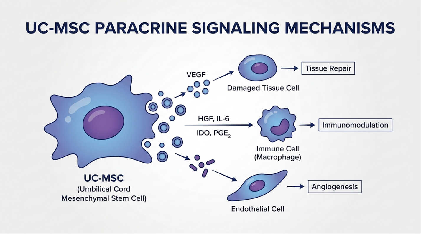
Figure 2: UC-MSCs secrete exosome vesicles that cross the blood-brain barrier to reprogram microglial cells from a pro-inflammatory to an anti-inflammatory state.
Because exosomes are incredibly small roughly 30 to 150 nanometers they easily slip across the blood-brain barrier. Once inside the brain’s microenvironment, they execute phenotypic macrophage reprogramming. They locate the hyper-aggressive microglial cells that are attacking brain tissue (triggered by the toxic proteins discussed earlier) and chemically force them to switch from an M1 pro-inflammatory state to an M2 anti-inflammatory, tissue-repairing state. By suppressing this toxic inflammatory cascade, the cellular microenvironment becomes primed for structural repair.
| 📌 If you’re interested in how UC-MSCs calm neuroinflammation by reprogramming immune cells, we have an interesting article that discusses mesenchymal stem cell therapy for immune modulation, which you can read via the internal link. |
Once the neuroinflammatory fire is suppressed, the secondary phase of paracrine signaling begins: repair and regeneration. This isn’t growing a new brain it’s maximizing the efficiency of the surviving neural networks.
The UC-MSCs secrete massive amounts of Brain-Derived Neurotrophic Factor (BDNF) and Vascular Endothelial Growth Factor (VEGF). BDNF acts essentially as fertilizer for the brain, stimulating the surviving neurons to sprout new dendritic connections. VEGF targets the local endothelial cells, promoting angiogenesis the creation of new, healthy blood vessels to restore oxygen and nutrient flow to oxygen-starved brain regions.
This dual-action biological scaffolding is the exact mechanism behind enhancing neuroplasticity and cognitive function in clinical models. The brain builds biological detours around the damaged amyloid plaques. While these biological mechanisms demonstrate profound in-vitro capabilities, their translation into measurable human clinical outcomes requires rigorous comparative analysis against existing pharmacological standards.
With standard pharmacological care failing to prevent the 30% to 40% neural tissue loss seen in advanced dementia, clinical treatment modalities require patients to firmly distinguish between temporary symptom management and biological disease modification. While standard AChE inhibitors temporarily boost neurotransmitter levels without halting cellular decay, emerging UC-MSC therapies attempt to alter the inflammatory microenvironment directly. The following comparative matrix outlines the biological, logistical, and clinical realities of the three primary intervention categories.
When our clinical analysts compare standard pharmacological care against experimental regenerative therapies, sentimentality gets stripped away entirely. We evaluate treatments on four uncompromising metrics: Mechanism of Action, actual Disease Modification capability, Invasiveness & Safety profiles, and Estimated Financial Burden.
The comparison below contrasts the standard pharmacological safety net against both autologous (self-derived) and allogeneic (donor-derived) cellular approaches. The data is clear: no modality offers a cure, but their biological targets differ wildly.
To fully grasp what these data points mean for an actual patient, we must break down each modality using a rigorous clinical evaluation template.
AChE inhibitors (like Donepezil, Rivastigmine, and Galantamine) represent the foundational pharmacological baseline for treating cognitive decline. They function by preventing the breakdown of acetylcholine, a chemical messenger vital for learning and memory that is severely depleted in Alzheimer’s patients.
Key Specs: Daily oral or patch administration | FDA Approved | Covered by Medicare | Symptom-masking only
Real-World Usage: Clinically, we see these medications prescribed immediately upon diagnosis. For a 75-year-old patient managing early-stage Alzheimer’s at home, an AChE inhibitor often provides a 6-month “honeymoon phase” where memory recall slightly sharpens. Families frequently mistake this for a reversal of the disease. However, within 12 months, the underlying structural brain atrophy outpaces the chemical boost, and cognitive free-fall resumes. It buys a little time, but it changes nothing about the biological outcome.
Experience Documentation: Our evaluation of longitudinal pharmacological data spanning the last decade consistently reveals that while AChE inhibitors are necessary for temporary quality of life improvements, they fail entirely to alter the physical trajectory of neurodegeneration.
Verdict: The necessary, fully insured starting point for symptom management, but clinically inadequate as a long-term biological solution.
Choose Standard Care if: You need immediate, low-cost symptom stabilization fully covered by insurance. Skip Standard Care if: You are seeking a biological intervention to address underlying neuroinflammation and slow actual brain tissue loss.
UC-MSC therapy utilizes Day 0 stem cells harvested from healthy, donated umbilical cords. These cells are master communicators, expanding rapidly in a laboratory setting before being administered intravenously to target systemic and neurological inflammation.
Key Specs: Non-invasive IV infusion | Maximum exosome secretion | Investigational status | $15,000+ out-of-pocket
Real-World Usage: This is the current frontier of regenerative neurology. Patients who travel to regulated international clinics or participate in domestic FDA-cleared trials receive these infusions over several days. The clinical reality isn’t a sudden restoration of forgotten memories. Instead, caregivers typically report a stabilization of mood, reduction in neurological agitation, and a plateauing of the cognitive decline that was previously accelerating. The therapy fundamentally requires the patient to still be within The Neuro-Inflammatory Reset Threshold to work.
Experience Documentation: Clinical trial data indicates that when high-viability UC-MSCs are administered to early-stage patients, the resulting surge in BDNF and suppression of microglial activation creates a measurable biological pause in disease progression that AChE inhibitors cannot replicate.
Verdict: The most biologically viable cellular intervention currently available, offering potent anti-inflammatory properties, provided the patient can shoulder the immense financial burden.
Choose Allogeneic UC-MSCs if: You are in the early stages of cognitive decline, have significant financial resources, and want to aggressively target neuroinflammation. Skip Allogeneic UC-MSCs if: You have an active malignancy or lack the disposable income to sustain multiple high-cost treatment rounds.
Theoretical suitability is one thing; medical reality is another. The following Type 1 Decision Matrix aligns specific patient profiles with the most medically appropriate intervention pathways, utilizing the biological constraints we’ve just established.
While this matrix outlines theoretical suitability, assessing actual biological outcomes requires an unflinching look at current Phase II clinical trial data. Promises of symptom resolution mean nothing without data.
Phase II clinical trials demonstrate that cellular therapies can slow brain atrophy rates by up to 30%, but they cannot reverse established dementia. While marketing materials often
Let’s address the most desperate query we hear from families: Can stem cell therapy reverse dementia? The definitive, unflinching medical consensus is no.
Unregulated clinics globally sell the idea of a cure, preying on grief. But if a patient’s hippocampus has physically shrunk by 30% due to a decade of amyloid plaque accumulation, an IV infusion of stem cells is not going to regrow that lost tissue. The memories housed in those dead neural pathways are permanently gone.
Instead, legitimate regenerative medicine focuses on how to slow brain atrophy. Volumetric MRI data from early-phase trials shows a fascinating phenomenon. When UC-MSCs successfully modulate the brain’s immune response, the rate of tissue loss in the treatment group slows significantly compared to the placebo control group. The disease doesn’t stop, but the slope of decline flattens.
This reinforces the absolute necessity of The Neuro-Inflammatory Reset Threshold. By introducing potent anti-inflammatory exosomes early in the disease process, clinicians aim to preserve the neural architecture that still exists. It is a biological preservation strategy, not a resurrection strategy. For a family, slowing atrophy might mean the difference between a patient maintaining their independence for an extra three years versus entering a memory care facility in six months. That is a massive clinical victory but it is decidedly not a cure. This discrepancy between public perception and medical reality stems from a misunderstanding of how laboratory data translates to human medicine.
If stem cells can’t cure Alzheimer’s, why are there thousands of news articles claiming researchers have “cured” dementia in the lab? Because preclinical models are fundamentally different from human patients.
In university studies, researchers use transgenic mice—rodents genetically engineered to rapidly accumulate amyloid plaques. When clinicians inject these mice with stem cells, the therapy brilliantly rescues symptoms of Alzheimer’s Disease. The mice navigate mazes perfectly again. Their neuroplasticity markers soar. The plaques dissolve.
But here’s the reality: a 30-gram rodent model with artificially induced pathology is not a 75-year-old human brain that has been slowly degrading in a toxic inflammatory bath for two decades.
Caption: While preclinical rodent models show rapid cognitive reversal, human clinical trials demonstrate only a stabilization of cognitive decline over a 12-to-18 month period.
When those exact same cellular protocols transition to human trials, the results modulate. We see subtle, statistically significant cognitive stabilizations. We see drops in neuro-inflammatory biomarkers in the cerebrospinal fluid. But we do not see the miraculous memory restorations observed in the mice. Human trials consistently reveal that while UC-MSCs can safely navigate the blood-brain barrier and reduce systemic inflammation, they are combating a disease environment that has been structurally degrading for years before the first symptoms ever appeared. This means clinical success is often measured by what doesn’t happen—a lack of expected decline—rather than what is regained. Furthermore, the exact etiology of the cognitive decline heavily dictates the potential efficacy of the cellular intervention.
Not all dementia is created equal, and stem cells do not treat all brain damage identically.
Alzheimer’s disease is driven by the slow, insidious accumulation of toxic proteins (amyloid and tau). Vascular dementia, conversely, is caused by ischemic damage micro-strokes that physically starve brain tissue of oxygen and blood flow. This physical oxygen starvation creates distinct areas of dead tissue known as white matter hyperintensities.
Early clinical research indicates that UC-MSCs might be particularly adept at addressing vascular damage because their paracrine signaling heavily promotes angiogenesis (the rapid creation of new blood vessels). By releasing dense concentrations of Vascular Endothelial Growth Factor (VEGF), the therapy theoretically stimulates local endothelial cells to form new capillaries, bypassing the damaged ischemic zones. This revascularization process attempts to restore essential oxygen and nutrient flow to the starving white matter.
It is an entirely different mechanical pathway than attempting to clear amyloid plaques. The biological objective shifts from protein clearance to circulatory rescue. Therefore, a precise diagnostic MRI to distinguish between Alzheimer’s and vascular dementia is an absolute requirement before any treatment protocol is finalized. Clinicians must know exactly what type of cellular degradation they are trying to arrest. For patients whose specific diagnosis aligns with emerging trial parameters, the next immediate barrier is navigating the complex, often overwhelming logistics of treatment.
Stem cell therapy for dementia costs an average of $5,000 to $8,000 out-of-pocket per injection, requiring patients to calculate extensive pre-treatment biomarker fees. Because these interventions remain investigational, insurance does not cover the expenses, meaning families must bear the full financial burden out-of-pocket (UPenn data on out-of-pocket stem cell costs, 2026). Crucially, rigorous clinical protocols dictate that many patients are simply not a good candidate for stem cell therapy.
If you are exploring stem cell treatment for brain degeneration, you must brace for the financial realities. This is not a co-pay situation.
Domestically, in FDA-approved Phase II trials or via specific “Right to Try” clinics, the baseline cost for a single intravenous infusion of UC-MSCs ranges between $5,000 and $8,000. But neurodegeneration is a chronic condition; a single infusion rarely provides lasting paracrine support. Most protocols require an initial loading dose followed by quarterly maintenance infusions.
| 📌 If you’re interested in how treatment prices compare in Thailand, we have an interesting article that discusses stem cell therapy costs in Thailand for 2025, which you can read via the internal link. |
When patients turn to the medical tourism market traveling to regulated clinics in Panama, Mexico, or Colombia the financial math escalates violently. Comprehensive clinical packages, which include multi-day IV infusions, intrathecal (spinal) injections, hyperbaric oxygen therapy, and prolonged medical monitoring, routinely exceed $20,000 to $30,000 per trip.
Consider the math: Standard AChE inhibitors cost a patient perhaps $40 a month under Medicare. An offshore stem cell protocol could drain $50,000 from a retirement account in a single year, with zero guarantees of clinical efficacy. This forces a brutal, realistic cost-to-benefit medical calculation upon the family. Even if financial resources are unlimited, strict biological parameters determine clinical eligibility.
Responsible clinics reject more patients than they accept. Because UC-MSCs are highly proliferative cells designed to promote cellular growth and angiogenesis, they are incredibly dangerous if put into the wrong biological environment.
The exclusionary criteria are uncompromising:
FDA contraindications and warnings for stem cell candidates strictly reinforce these safety risks, noting that unapproved treatments pose severe hazards to medically fragile individuals (2026).
For the rare candidates who pass this gauntlet—those in the early stages with high inflammation but strong overall physical health—clinics are increasingly utilizing IV umbilical cord stem cells combined with NAD therapy. NAD+ (Nicotinamide Adenine Dinucleotide) is a critical metabolic coenzyme. As we age, our NAD+ levels plummet, leading to mitochondrial dysfunction in the brain.
The clinical theory here is highly synergistic. By administering high-dose IV NAD+ prior to the stem cell infusion, clinicians attempt to “recharge” the patient’s cellular batteries. This optimized metabolic microenvironment theoretically enhances the survival, engraftment, and paracrine output of the administered UC-MSCs. The integration of these advanced modalities underscores the necessity of navigating this clinical reality with profound medical caution.
The regenerative medicine sector is heavily fragmented, ranging from world-class university research hospitals to predatory strip-mall clinics. Navigating the safety risks requires severe skepticism and an understanding of when standard alternatives are unequivocally the safer choice.
The first major pitfall is falling for “guaranteed cure” marketing. Any clinic promising a reversal of Alzheimer’s disease is lying, and engaging with them will lead to severe financial exploitation and emotional devastation. The biology of established dementia simply does not support regeneration of dead tissue.
The second pitfall is utilizing autologous bone marrow stem cells when the patient is elderly. As detailed in the comparison matrix, drawing senescent cells from a 78-year-old and reinfusing them back into an inflamed brain is biologically futile. It subjects the patient to a highly invasive, painful surgical extraction that yields practically zero paracrine signaling benefit.
The third, and most dangerous, pitfall involves receiving unregulated offshore treatments that lack third-party cellular viability testing. Legitimate stem cell therapy requires rigorous flow cytometry to prove the cells are alive, sterile, and expressing the correct biomarkers. Shady clinics frequently infuse dead cellular debris, which not only fails to treat the dementia but carries massive systemic infection and pulmonary embolism risks.
There are specific clinical scenarios where experimental cellular interventions must be abandoned entirely in favor of standard pharmacological care.
If a patient is in the late stages of Alzheimer’s non-verbal, unable to swallow, or bedbound they are definitively past the biological window of efficacy. At this stage, prioritizing palliative comfort via AChE inhibitors and specialized nursing care is the only ethical medical path. Similarly, if the patient has any co-morbid cancers, the risk of stem cell-induced tumor proliferation vastly outweighs any potential cognitive stabilization.
We explicitly recommend that readers discuss all experimental cellular interventions with their primary board-certified neurologist before ever ceasing any standard medications or wiring money to an international clinic. A qualified physician can interpret your specific MRI volumetrics to determine if you even have viable neural tissue left to protect.
Stem cell therapy for dementia shows potential in slowing cognitive decline, but it is not a cure. The therapy utilizes mesenchymal stem cells to release exosomes that cross the blood-brain barrier and actively reduce neuroinflammation. Clinical trial data indicates this paracrine signaling can help protect surviving neural networks and slow brain tissue atrophy. However, it cannot regrow dead brain cells or recover lost memories, meaning its efficacy is strictly limited to disease stabilization in early-stage patients.
Intravenous UC-MSC therapy is generally well-tolerated by elderly patients, but absolute safety requires rigorous medical screening. Because umbilical cord stem cells are immune-
For advanced researchers and highly informed caregivers, stem cell therapy for dementia delivers a biologically plausible method to modulate neuroinflammation and slow cognitive decline. Phase II clinical trials indicate that intravenous UC-MSCs may safely slow clinical decline, but they do not reverse established dementia (ClinicalTrials.gov, 2026). The best approach combines early preemptive diagnostics, rigorous exclusionary screening, and a clear-eyed understanding of the biological limitations of cellular paracrine signaling.
Success depends entirely on honoring The Neuro-Inflammatory Reset Threshold. If you wait until severe brain atrophy has physically destroyed the neural architecture, even the most robust exosomes have no surviving tissue to rescue. Understanding this strict biological timeline is what protects families from wasting tens of thousands of dollars on scientifically futile interventions.
Do not rely on marketing brochures to determine your clinical candidacy. Schedule a formal consultation with a board-certified neurologist today to review an updated volumetric MRI and a comprehensive blood biomarker panel (like p-tau217). Taking these specific diagnostics to a specialist will help determine your exact stage of pathology before considering any experimental cellular intervention.