Our Services
Others
- DFPP
- Shockwave
- IV Drip

The therapeutic landscape for Parkinson’s disease is fraught with commercial clinics offering unproven stem cell infusions, yet rigorous clinical trials reveal a starkly different reality of surgical complexity, strict isolation, and biological risk. Patients facing declining motor function often desperately researching stem cell therapy for Parkinson’s disease frequently encounter wildly conflicting claims online. These marketing pitches deliberately blur the critical line between preliminary laboratory success and actual, unassisted mobility. Clinical analysis of these discrepancies over the past decade reveals a stark, uncompromising reality. What appears in a biotech press release rarely matches the grueling lived reality of an experimental transplant patient. This guide strips away the commercial hype to look exclusively at what current 2026 data actually proves. This comprehensive analysis dissects the surgical implantation process, the mandatory immune-suppression protocols, and the factual timeline for graft survival. For individuals noticing Parkinson’s disease early symptoms or already managing advanced motor fluctuations, clinical
Stem cell therapy for Parkinson’s disease remains investigational, prioritizing the surgical replacement of dopaminergic neurons rather than symptom suppression.
Stem Cell Therapy for Parkinson’s Disease: Understanding the Condition, Diagnosis, and Treatment Potential centers on a specific biological objective: generating new, functional dopaminergic cells to replace those lost in the substantia nigra. Unlike levodopa, which temporarily supplies systemic dopamine, stem cell transplantation attempts a structural repair of the neural circuit. This approach focuses on localized neural restoration rather than halting progressive neurodegeneration.
To understand why researchers are attempting brain surgery for a movement disorder, one must look at the precise cellular failure happening inside the midbrain. Parkinson’s pathology is stubbornly specific. The disease systematically degrades the substantia nigra pars compacta, destroying the neuromelanin-containing cells responsible for autonomic movement. Roughly 60% to 80% of these dopaminergic neurons are lost before visible symptoms appear—so localized structural repair becomes theoretically necessary once the native cell population falls below this critical threshold. The standard pharmaceutical approach simply floods the entire brain with
The laboratory phase of this process is mind-bendingly complex. Scientists take either embryonic stem cells (ESCs) or induced pluripotent stem cells (iPSCs) often derived from a patient’s own skin fibroblasts and force them backward into a pluripotent state using specific transcription factors. Once these cells are blank slates, lab technicians coax them into becoming midbrain dopaminergic progenitors.
They are exposed to highly specific signaling molecules, such as sonic hedgehog (SHH) and fibroblast growth factor 8 (FGF8), over a strict 28- to 45-day in vitro protocol to ensure they adopt a ventral midbrain fate. During this crucial window, clinicians use fluorescence-activated cell sorting (FACS) to isolate only the cells expressing precise midbrain markers like FOXA2 and LMX1A. This extreme quality control is non-negotiable; if even a fraction of a percent of the batch remains undifferentiated, it poses a catastrophic tumor risk once implanted. Harvard research on dopamine-producing neurons emphasizes that the core goal of stem cell therapy in Parkinson’s is to safely coax undifferentiated cells into becoming targeted dopamine-producing neurons. (2023).
Here is the frustrating part for desperate patients: these fragile cells cannot simply be injected into a vein. The blood-brain barrier explicitly prevents large cellular structures from crossing into the central nervous system. Intravenous stem cell tourism where patients pay $20,000 at a strip-mall clinic for an IV drip is a biological scam. Genuine structural repair requires direct stereotactic brain surgery to physically inject these lab-grown cells directly into the post-commissural putamen. Clinicians bypass the damaged substantia nigra entirely because growing new axons over that anatomical distance is currently impossible. Delivering the cells directly to the endpoint is a structural workaround that, theoretically, integrates permanently into the host tissue.
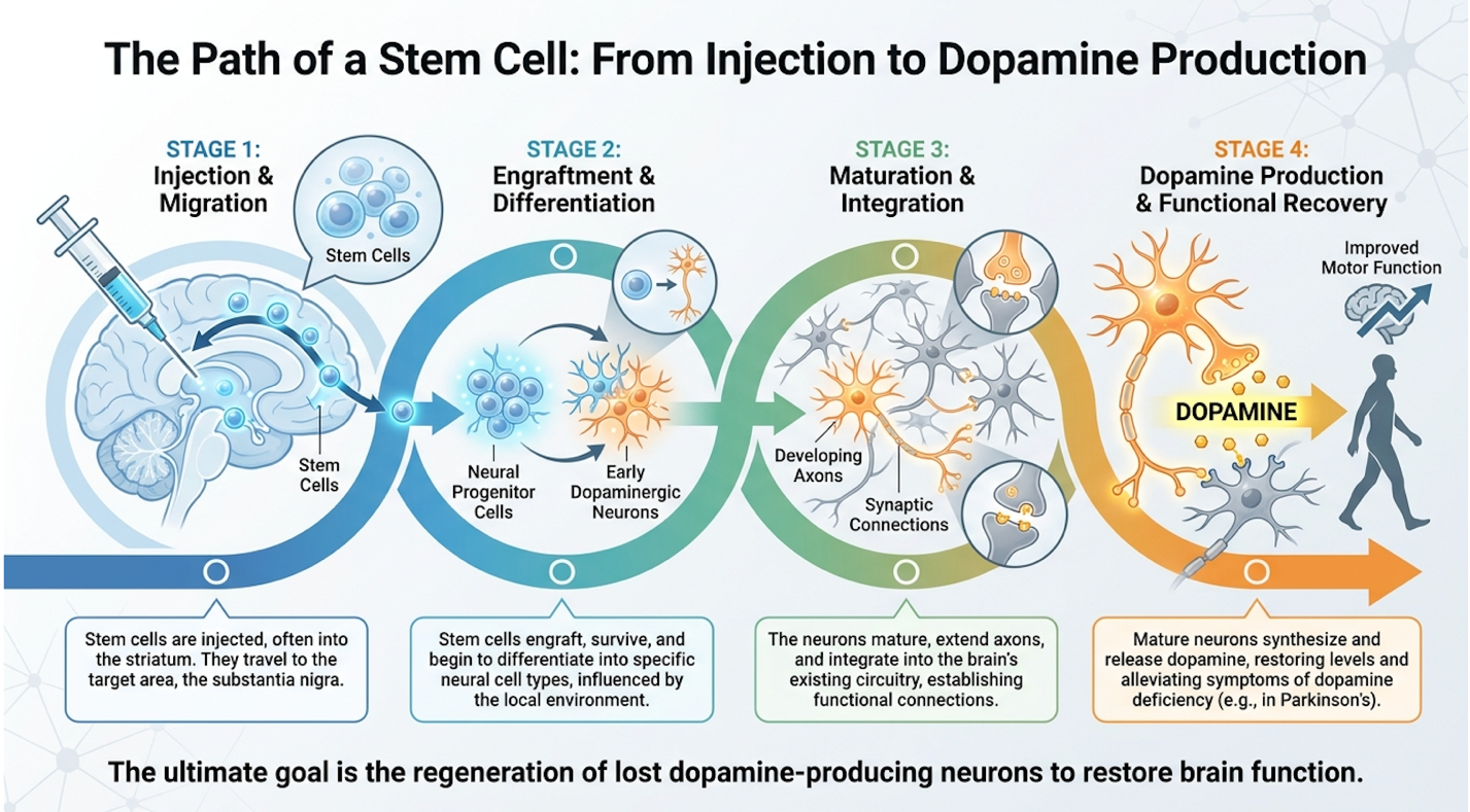
Figure 1: The intricate laboratory-to-brain pipeline required for legitimate dopaminergic progenitor cell engraftment.
While the biological mechanics present a clear theoretical solution, bridging the gap between cellular survival and patient mobility exposes significant clinical hurdles.
This question dominates patient forums constantly: can stem cells reverse Parkinson’s disease? The clinical answer is a definitive, uncompromising no. Current Phase 1 and 2 trials do not aim to reverse the disease entirely. Instead, they focus purely on the motor symptoms. If a patient suffers from debilitating “off” time where medication stops working and they freeze up or experiences violent levodopa-induced dyskinesias, a successful stem cell graft could theoretically smooth out those dopamine fluctuations.
Stem cell therapy for Parkinson’s focuses exclusively on regenerating dopaminergic neurons, not halting the underlying autoimmune degradation (Harvard Stem Cell Institute, 2023). Parkinson’s is a multisystem disorder defined by Braak staging. Lewy body pathology (toxic alpha-synuclein protein clumps) frequently starts in the enteric nervous system or lower brainstem and spreads upward over decades. Injecting cells into the putamen does absolutely nothing for non-
Furthermore, patients often assume they will wake up from surgery ready to walk effortlessly. The biological timeline of cellular maturation shatters this expectation. When dopaminergic progenitors are injected, they are biologically immature. Once deposited, they must survive the initial surgical hypoxia, settle into the host tissue, extend new neurites, and form functional synaptic connections with the host’s existing medium spiny neurons.
This complex biological integration takes a massive amount of time. In the first one to three months, the transplanted progenitors are simply fighting for metabolic survival inside a hostile, post-surgical environment. They do not produce any meaningful dopamine yet. By months four through six, if they survive, the cells finally begin axonal sprouting extending delicate nerve fibers out into the host tissue. It isn’t until the 12-to-18-month mark that these new neurites form mature, functional synapses capable of releasing dopamine into the striatum. The brain reorganizes at a glacial pace, meaning no new endogenous dopamine is manufactured for at least a year and a half.
During this extensive waiting period, patients remain entirely dependent on their standard medication regimen. Early post-operative motor changes observed in the first few weeks are almost universally placebo effects or the result of minor brain lesions caused by the surgical cannula itself a well-documented phenomenon in functional neurosurgery known as the microlesion effect.
This stark discrepancy between cellular integration and visible recovery necessitates a formalized, objective method for evaluating trial success.
When clinical researchers evaluate new restorative neurology trials, they immediately apply The Biological-Functional Distinction (BFD) Framework. Patients reviewing trial data must do the same. This is a vital clinical evaluation model that strictly separates cellular survival metrics
The “Biological” tier looks exclusively at medical imaging. Clinicians use specialized F-DOPA PET scans to measure whether the transplanted cells survived and are successfully taking up fluorodopa. If the scan shows bright uptake in the putamen, the trial is declared a biological success. The cells lived, and they are metabolizing precursors.
However, biological success means absolutely nothing if the patient cannot easily walk to the mailbox. The “Functional” tier measures the Unified Parkinson’s Disease Rating Scale (UPDRS) Part III motor scores during the practically defined “off” state. It tracks unassisted walking time, reduction in daily levodopa equivalent dose (LEDD), and overall quality of life metrics.
To practically define this “off” state, clinical trial protocols require patients to undergo a grueling 12-hour medication washout period overnight. The following morning, neurologists conduct a meticulous UPDRS Part III motor assessment, measuring rigidity, tremor amplitude, and bradykinesia without any pharmacological assistance. If the stem cell graft is truly functioning, it must provide enough endogenous dopamine to carry the patient through this severe washout phase. Furthermore, because the brain’s neuroplastic response to surgical trauma can trigger temporary improvements a well-known placebo effect in neurosurgery—evaluators refuse to declare functional success until the 18-to-24-month mark. This extended timeline guarantees that any observed motor improvements stem from mature synaptic connections rather than transient surgical inflammation.
Historical trial data perfectly illustrates this cruel disconnect. In older double-blind studies, some patients showed a spectacular 40% increase in dopamine uptake on their post-operative PET scans. Biologically, the graft thrived. Functionally, however, those same patients showed absolutely zero improvement in their UPDRS scores compared to the control group. The new axons grew, but they failed to form meaningful synaptic connections with the existing motor network. The cells were alive, but useless. Applying this rigorous framework begins by surviving the initial implantation, which presents immediate and severe physical challenges.
The stem cell injection recovery trajectory is dictated by the invasiveness of intracranial stereotactic surgery and the requisite systemic immune suppression. Unlike outpatient procedures marketed by commercial clinics, legitimate clinical trials require precise burr-hole cranial access under anesthesia. Consequently, the initial recovery is focused entirely on managing acute surgical trauma, cerebral edema, and the immediate neurological aftereffects of brain tissue disruption.
When exploring a stem cell transplant for Parkinson’s pain, patients often picture the systemic agony of a bone marrow transplant used for leukemia. Brain surgery differs entirely. The brain parenchyma itself has absolutely no nociceptors (pain receptors). A surgeon can physically cut into the neural tissue, and the patient will not feel it. The procedural discomfort entirely stems from penetrating the scalp, drilling the skull, and enduring post-operative cranial pressure.
Legitimate trials use stereotactic surgery, an incredibly precise 3D mapping technique used to guide intracranial injections. Before surgery, a rigid titanium stereotactic frame is physically bolted to the patient’s skull under local anesthesia. This frame is then locked into the MRI or CT scanner table to generate a flawless three-dimensional coordinate map of the deep brain structures. The neurosurgeon relies on this map to calculate the exact trajectory for the injection cannula, ensuring it hits the post-commissural putamen with sub-millimeter accuracy while avoiding major cerebral arteries.
The anesthetic approach frequently involves an awake craniotomy. The patient receives local anesthesia for the scalp incision and light sedation to remain calm, but is kept conscious so the surgical team can monitor neurological responses in real-time. By utilizing microelectrode recording (MER), the surgical team listens to single-neuron action potentials. If they advance the cannula and the patient’s speech suddenly slurs or their hand goes numb, the team knows they are dangerously close to the internal capsule and can adjust the trajectory immediately.
While the acute procedural pain is remarkably localized, the systemic physiological shock peaks several days later.
Clinical teams universally warn patients that the worst days after stem cell transplant almost always fall between Day 3 and Day 7. Driving a hollow metal needle deep into the basal ganglia unavoidably causes microscopic bleeding and tears neural tissue. The brain’s mandatory biological response to this trauma is acute localized neuroinflammation.
Because the central nervous system lacks a conventional lymphatic drainage system, this resultant cerebral edema (swelling) compresses adjacent healthy structures inside the rigid cranial vault. By Day 3, this swelling hits its absolute peak.
During this acute phase, the physical reality is grueling. Patients experience profound, crushing fatigue and transient delirium. More alarmingly, their baseline Parkinsonian motor symptoms typically get noticeably worse. Resting tremors might suddenly spike, and severe rigidity often increases. Edema peaks between days 3 and 7 post-surgery requiring intensive neurological monitoring rather than indicating immediate graft failure (National Cancer Institute, 2023). If aggressive systemic immunosuppressants are initiated during this exact window, extreme nausea and gastrointestinal distress compound the misery.
Recovery during this phase does not happen at home. Patients are meticulously monitored in a specialized neurological ICU or step-down ward. Nursing staff perform rigorous checks on pupil dilation, grip strength, and cognition every hour to ensure the cerebral edema is not escalating into a catastrophic intracranial hemorrhage. Clinicians continually monitor the Glasgow Coma Scale (GCS) and track serum sodium levels closely, as brain trauma can trigger cerebral salt wasting—a dangerous complication that further depresses consciousness.
If intracranial pressure spikes dangerously high, physicians may deploy hyperosmolar therapy, administering intravenous mannitol to forcibly draw fluid out of the swollen brain tissue. Families are often terrified during this acute window, as the patient may temporarily lose the ability to speak clearly or swallow safely. This transient bulbar dysfunction requires temporary nasogastric feeding to prevent aspiration pneumonia.
Arterial lines are frequently placed to monitor continuous blood pressure, ensuring perfusion remains tightly controlled to prevent the fragile new surgical tracks from rupturing. National Cancer Institute’s 100-day recovery timeline notes that during the first 100 days post-transplant,
As the acute cerebral swelling slowly subsides, the clinical focus shifts entirely to physical rehabilitation.
When can a patient actually get out of bed? Usually within 24 to 48 hours post-op. This rapid mobilization is not initiated because the patient feels energetic; it is mandated because lying flat drastically increases the risk of deep vein thrombosis (blood clots) in the legs and post-operative atelectasis (lung collapse). Physical therapy is initiated aggressively early, but managed with extreme caution.
This must be emphasized clearly: immediate post-operative mobility is governed entirely by standard surgical recovery metrics, not by the newly injected stem cells. Those dopaminergic progenitors remain completely biologically inert at this stage. Any ability to walk down the hall on Day 4 relies strictly on the patient’s pre-surgery dopamine capacity and their continued standard Parkinson’s medications.
The early post-injection milestones are about basic survival and establishing gross motor stability. Day 2 typically involves simply standing beside the hospital bed to manage orthostatic hypotension a massive drop in blood pressure caused by standing up after brain surgery. Day 4 involves carefully walking the hospital corridor while wearing a secure gait belt. The fall risk during this first week is astronomical. Combining post-operative delirium, lingering anesthesia effects, and existing Parkinsonian postural instability creates a perfect storm for severe physical injury. Hitting the head after recent intracranial surgery is a life-threatening catastrophic event.
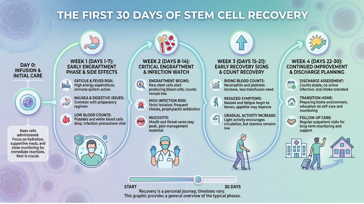
Figure 2: The critical 30-day post-operative window dictates survival; true dopaminergic integration takes months.
When evaluating true surgical success rates, clinicians must look past simply surviving the hospital stay. Reaching the milestone of returning to the pre-operative baseline mobility takes approximately 30 days. Only after that baseline is firmly re-established does the grueling 6-to-18-month wait begin to see if the stem cells actually start manufacturing functional dopamine.
Once safely discharged from acute neurological care, the patient’s home environment must undergo radical modification to protect the newly implanted graft.
To prevent the host immune system from attacking the newly grafted dopaminergic cells, patients undergo intensive immunosuppressive therapy. This pharmacological intervention creates extreme vulnerability to opportunistic pathogens. Consequently, post-transplant care relies on rigid environmental control, transforming the patient’s home into a clinically sterile zone. Failure to adhere to these protocols compromises both the graft’s viability and the patient’s immediate survival.
| 📌 If you’re interested in how mesenchymal stem cells modulate the immune system rather than suppress it, we have an interesting article that discusses mesenchymal stem cell therapy for immune modulation, which you can read via the internal link. |
If a patient receives an allogeneic transplant (stem cells derived from a donor rather than their own body), their immune system will immediately recognize the graft as a foreign biological invader and dispatch T-cells to destroy it. To prevent this violent rejection, patients are placed on heavy immunosuppressant medications typically potent calcineurin inhibitors like cyclosporine or tacrolimus, which forcibly blunt the body’s natural defense mechanisms.
This extreme medication regimen completely wipes out the body’s ability to fight off even a mild common cold, leaving the patient defenseless against latent viruses like cytomegalovirus (CMV). Because of this chemically induced vulnerability, the quarantine after stem cell transplant is incredibly severe. Patients face a minimum of 2 to 4 weeks of strict environmental isolation, a timeline scaled entirely based on specific white blood cell recovery metrics.
This is not a casual lockdown. Visitors are strictly banned. Visits from young children are specifically prohibited due to their statistically high viral loads and poor hygiene habits. Families must physically remove all indoor potted plants and fresh flowers from the home because the damp soil harbors Aspergillus, a common fungal spore that is routinely deadly to immunosuppressed patients. The home’s HVAC system requires specialized high-efficiency HEPA filtration (MERV 14 or higher). strict quarantine periods post-transplant protocols dictate that following a stem cell procedure, patients typically require 2 to 4 weeks of strict hospital quarantine or isolation to protect their heavily suppressed immune systems. (2023).
When a commercial facility heavily markets the “best stem cell clinic for Parkinson’s” but explicitly claims patients can fly home commercially the next day without immunosuppression, they are either injecting dead cells or performing a sham procedure. True allogeneic engraftment requires brutal immune regulation.
| 📌 If you’re wondering how to confirm that injected cells are actually alive and effective, we have an interesting article that discusses the importance of cell viability in UC-MSC stem cell therapy, which you can read via the internal link. |
Environmental control must be flawlessly paired with stringent internal gastrointestinal protection.
Patients are not just fighting airborne pathogens; they are actively fighting the bacteria living naturally in the global food supply. During the heavy immunosuppressive phase, patients are
The stem cell transplant diet restrictions are absolute. Patients face a total ban on unpasteurized dairy, raw or undercooked meats, sushi, and cold deli meats. Listeria monocytogenes a bacteria commonly found on deli turkey that might give a healthy adult a mild stomach ache can trigger fatal sepsis or aggressive graft rejection in an immunosuppressed transplant recipient. Toxoplasma gondii, found in undercooked lamb or pork, presents a similar catastrophic threat.
Produce rules are equally frustrating. Strict neutropenic diets eliminate 100% of raw produce to prevent fatal opportunistic bacterial infections (Loma Linda University Health, 2023). Patients cannot eat raw, unpeeled fruits or vegetables. Fresh berries are entirely forbidden because their porous skins cannot be adequately sanitized. Every single piece of food consumed must be thoroughly cooked to an internal temperature that completely annihilates latent bacteria. strict neutropenic diet guidelines mandate that to prevent foodborne illnesses while severely immunocompromised, patients must adopt a strict neutropenic diet, completely avoiding unpasteurized dairy, raw meats, and unwashed produce. (2023).
Even drinking water must be strictly regulated; well water or unfiltered tap water is forbidden, requiring patients to consume exclusively purified or boiled water to avoid waterborne pathogens like cryptosporidium. Families essentially run a commercial-grade sanitation operation in their own kitchen. Patients have breezed through the physical brain surgery only to end up back in the ICU fighting for their lives simply because they ate an unwashed grape on day 14.
With environmental and dietary vectors strictly controlled, physical exertion becomes the final variable to manage.
A patient’s physical lifestyle comes to a grinding halt during the first three months of recovery. Understanding what not to do after stem cell injection is just as vital as the surgery itself to ensure the graft properly takes root.
First, heavy lifting and high-impact exercises are strictly forbidden. Patients must explicitly avoid the Valsalva maneuver the act of forcefully bearing down or holding the breath while straining,
Patients are also universally banned from taking non-steroidal anti-inflammatory drugs (NSAIDs) like ibuprofen, naproxen, or aspirin. These specific medications thin the blood and strongly inhibit platelet function. In a brain that is still actively healing from a stereotactic needle track, a minor blood-thinning effect can easily trigger a catastrophic intracranial hemorrhage.
Finally, travel restrictions are severe. Boarding a commercial flight is prohibited for at least 3 to 6 months. The recycled cabin air, dense pathogen load, and proximity to hundreds of strangers make airplanes a veritable death trap for the immunosuppressed. If advertisements for stem cell therapy for Parkinson’s disease heavily target American patients for a weekend medical tourism trip abroad, the biological risk is terrifying. Flying home internationally immediately after an experimental transplant is medically reckless.
Understanding these demanding restrictions is crucial for accurately weighing the ultimate question: are the theoretical long-term benefits mathematically worth the severe biological risks?
Evaluating the safety profile of experimental neurorestorative procedures requires setting aside commercial optimism to examine hard clinical data. The reality of surgical stem cell implantation is a delicate balance between generating necessary dopamine and preventing aberrant cellular behavior. Applying the Biological-Functional Distinction (BFD) Framework, patients must critically analyze whether theoretical motor improvements mathematically outweigh the documented risks of brain surgery and immunological rejection.
When clinical teams sit down with patients to review the downsides of stem cell therapy against standard care, they break it down into a rigid, objective comparison. Standard levodopa
The primary “pro” is the potential elimination of “off” time the agonizing hours where standard medication wears off and the patient freezes in place. Successful neuronal grafts have shown the theoretical potential to significantly lower a patient’s daily levodopa equivalent dosages (LEDD), restoring smooth, continuous mobility.
However, the cons are massive and frequently understated by clinics. Patients trade standard medication adjustments for invasive stereotactic brain surgery, carrying a permanent 1-2% risk of severe hemorrhage, deep brain infection, and death. Furthermore, the financial risk is staggering. Since this remains strictly experimental, insurance rarely covers it. The true out-of-pocket trial costs or ancillary fees can easily exceed $100,000, with an exceptionally high failure rate of actual functional graft integration.
Beyond the biological dangers, the psychological toll of a failed graft is devastating. Patients endure months of isolation, brutal medication side effects, and extreme financial drain, only to potentially discover at the 18-month mark that the cells died. This profound disappointment often plunges patients into severe clinical depression, accelerating their overall neurodegenerative decline. Conversely, the potential pros when the graft perfectly survives—are undeniably profound. Successful patients report waking up without the agonizing “morning akinesia” that typically leaves them paralyzed in bed until their first levodopa dose kicks in. The continuous, physiologic trickle of dopamine from the graft creates a stability that oral medications simply cannot replicate.
While levodopa causes systemic fluctuations, stem cells attempt localized, stable delivery, but carry a drastically higher upfront biological risk. The most daunting of these downsides is the extreme biological unpredictability of pluripotent cells once introduced into the brain.
This remains the darkest corner of stem cell research, and one that commercial tourism clinics completely ignore. Patients nervously ask, can stem cells turn cancerous? Yes, they absolutely can.
Biologically, pluripotent stem cells possess an extreme, innate proliferative capacity. They are inherently programmed to rapidly divide and multiply into any tissue type in the human body. In a legitimate Phase 1 clinical trial, scientists spend weeks forcing these cells to differentiate into committed neuronal progenitors before injection. If that laboratory differentiation process is not utterly flawless—if even a tiny fraction of the cells remain undifferentiated—they will continue to multiply wildly once deposited inside the brain.
This aggressive multiplication forms a teratoma, a type of tumor that forms from improperly differentiated pluripotent stem cells. Teratomas are horrifying; because stem cells are intrinsically designed to build a complete human organism, their failure to fully differentiate means they retain their ability to form bone, hair, or cartilage directly against delicate neural tissue. Because teratomas contain completely inappropriate tissues—like microscopic teeth, hair follicles, or bone fragments—growing directly inside the putamen, they trigger massive neurological catastrophes. The only recourse is emergency neurosurgery to excise the mass, a procedure that often causes permanent, severe brain damage. Unregulated therapies carry severe teratoma risks proving that unpurified cell batches can trigger catastrophic tumor formation (FDA, 2023). Legitimate trials use advanced flow cytometry to carefully purify the cell batch, an incredibly expensive step that unregulated commercial clinics routinely skip.
FDA warnings on unapproved stem cell therapies make it clear that unapproved stem cell therapies carry severe risks, including administration site reactions, the multiplication of cells into inappropriate types, and tumor formation. (2023). This is exactly why the regulatory oversight in legitimate trials is so draconian.
| 📌 If you’re wondering how to tell a safe, properly regulated stem cell clinic from a risky one, we have an interesting article that discusses whether stem cell therapy is safe in Thailand, which you can read via the internal link. |
Even if the patient dodges the teratoma bullet, they face the massive risk of Graft-Induced Dyskinesia (GID). Stem cell therapy for Parkinson’s tremors sounds incredible until the new cells misbehave. GID occurs when the surviving stem cells form aberrant synaptic connections and secrete dopamine irregularly. This causes severe, uncontrollable writhing movements that happen even when the patient is completely off their Parkinson’s medication. The patient essentially trades
If the cells successfully integrate without forming tumors or inducing permanent dyskinesia, the clinical focus shifts entirely to their longevity.
If the cell graft survives the initial immune onslaught, how long do stem cells stay in the body? The biological answer is permanent. Successful neural grafts do not “wash out” of the system like a pharmaceutical drug. They physically weave their new axons tightly into the host’s native neural circuitry.
Clinical knowledge from historical fetal midbrain tissue transplant trials conducted in the 1990s demonstrates that dopaminergic grafts can survive and function for 5 to 10 years, and occasionally even longer. clinical data on stem cell graft survival suggests that successfully transplanted stem cell grafts can survive for 5 to 10 years, though patients must be carefully monitored for graft-induced dyskinesias and potential tumorigenesis. (2021).
However, there is a fascinating and grim biological reality to this impressive longevity. Over a decade, the host brain eventually infects the new graft. Autopsy data from old transplant patients proved that Lewy body pathology the toxic alpha-synuclein protein clumps causing the disease eventually spread from the patient’s dying brain directly into the fresh, healthy grafted cells. The disease process inevitably attacks the cure.
Understanding this permanent biological commitment requires a rigorous clinical assessment to determine exactly who should subject themselves to this experimental pathway.
Determining suitability for investigational stem cell therapy demands objective clinical scrutiny. Experimental brain surgery is not a panacea for neurodegeneration, and a vast majority of Parkinson’s patients present with contraindications that render them ineligible for current clinical trials.
Finding the best treatment for Parkinson’s disease means matching the intervention precisely to physiological limits. Use this decision framework to determine trial eligibility. Approximately 80% of patients are disqualified from trials proving physiological resilience remains the ultimate gatekeeper for experimental surgery.
For patients disqualified from experimental cell therapies, Deep Brain Stimulation (DBS) targeting the subthalamic nucleus remains the surgical gold standard for advanced motor fluctuations. Unlike stem cells, DBS does not carry a teratoma risk, and stimulation can be adjusted if side effects occur. Furthermore, continuous levodopa-carbidopa intestinal gels (LCIG) delivered via a pump offer proven, immediate functional benefit without the catastrophic risks of cellular multiplication. Neurologists also increasingly recommend non-invasive focused ultrasound (FUS) for patients with tremor-dominant Parkinson’s who cannot tolerate open brain surgery. These
Stem cell therapy for Parkinson’s disease remains strictly experimental and is not yet a proven, widely available treatment. While early Phase 1 and 2 clinical trials demonstrate that stem cells can survive in the brain and produce dopamine, widespread functional restoration of mobility has not been definitively established. Clinical data shows cellular survival does not guarantee motor improvement. Patients should view this as an investigational approach, not a guaranteed cure.
Stem cells cannot reverse or cure Parkinson’s disease entirely. The therapy specifically targets motor symptoms like tremors and stiffness by replacing lost dopamine-producing neurons in the midbrain. It does not halt the underlying neurodegenerative process, nor does it treat non-motor symptoms such as dementia, depression, or autonomic dysfunction. Because alpha-synuclein pathology continues spreading, disease progression remains inevitable in other areas of the nervous system. Patients must understand this limits the therapy’s scope to mobility alone.
Stem cell therapy carries severe biological and surgical risks that must be managed in highly controlled clinical trial settings. The primary safety concerns include intracranial hemorrhage from the surgical injection, severe infections resulting from necessary immune suppression, and the potential formation of teratomas (tumors). Without rigorous flow cytometry to purify the cell batch, the tumor risk skyrockets. Unregulated treatments offered by commercial clinics lacking strict FDA oversight pose extreme, life-threatening risks to unsuspecting patients seeking experimental interventions.
For patients dealing with advanced motor fluctuations, stem cell therapy for Parkinson’s disease represents the absolute frontier of neurorestorative medicine, prioritizing structural dopamine replacement over systemic medication. However, this investigational surgery requires navigating severe intracranial trauma, extensive immune suppression, and the highly documented biological risk of permanent teratoma formation. The safest approach limits exploration strictly to FDA-monitored Phase 1 or 2 clinical trials, completely avoiding commercial clinics making unverified claims.
Applying the Biological-Functional Distinction (BFD) Framework is essential when evaluating this experimental pathway. Patients and caregivers must aggressively look beyond laboratory metrics of cell survival and demand hard data on tangible, daily mobility improvements before accepting the immense physiological burden of brain surgery.
Consult your treating neurologist to determine if you are a viable candidate for deep brain stimulation before pursuing investigational cellular trials. This article is for educational purposes only and does not replace consultation with a qualified medical professional.