Our Services
Others
- DFPP
- Shockwave
- IV Drip

Medical Disclaimer: This article is for educational purposes only and does not replace consultation with a qualified medical professional. The biological interventions discussed herein carry inherent risks and require evaluation by a board-certified Orthopedic Specialist.
For patients facing chronic joint deterioration, the evaluation of orthobiologics demands rigorous clinical scrutiny rather than marketing optimism. It’s a brutal reality. Predatory clinics routinely exploit treatment gaps by promising miraculous cartilage regeneration, exposing patients to severe financial loss and inappropriate medical interventions. You’re told you can get a brand-new knee from a single syringe. You can’t.
This clinical guide analyzes peer-reviewed literature to establish verified dosing protocols, cellular source comparisons, and realistic symptomatic outcomes for joint therapy. If you’re seriously considering stem cell therapy for knee osteoarthritis, you need to understand the biology behind the marketing.
The following analysis dissects longitudinal efficacy data, cellular harvesting methods, and systemic joint optimization to facilitate genuinely evidence-informed medical decisions. We aren’t here to sell you on a procedure. We’re here to break down the cellular math, the harsh biological realities of a degenerative joint, and the specific patient profiles who actually achieve long-term functional recovery.
Clinical evaluation of stem cell therapy for knee osteoarthritis reveals distinct capabilities for symptom management rather than guaranteed structural repair.
Stem cell therapy for knee osteoarthritis primarily functions as an advanced anti-inflammatory and immunomodulatory intervention rather than a structural cartilage replacement. Clinical evidence indicates that mesenchymal stromal cells alter the local joint environment, leading to profound symptom relief and functional restoration without necessarily increasing cartilage volume on MRI scans (AAOS, 2026).
| 📌 If you’re interested in how stem cells calm joint inflammation by rebalancing the immune response, we have an interesting article that discusses mesenchymal stem cell therapy for immune modulation, which you can read via the internal link. |
Let’s look at the underlying physiological changes. Advanced orthopedic clinics document that introducing these cells rapidly downregulates synovial inflammation, effectively blocking the pain pathways that make walking unbearable. Over 80% of successful responders show decreased synovial thickening on ultrasound a direct physiological consequence of halted inflammatory cascades. A 5-year longitudinal study demonstrated that 65% of patients maintained significant symptom relief and improved joint function following mesenchymal stem cell therapy (PubMed, 2021). This biological shift defines the true therapeutic value of orthobiologics. Assessing clinical success requires completely abandoning the flawed expectation of total structural renewal.
Let me be entirely blunt about what this treatment actually does. The most pervasive myth in regenerative medicine is that injecting cells into a bone-on-bone joint will magically sprout thick, pristine hyaline cartilage. It won’t. Clinical reviews indicate that stem cell therapy for knee without surgery operates through a mechanism known as paracrine signaling, not gross structural tissue fabrication.
Mesenchymal Stromal Cells (MSCs) act as intelligent medicinal signaling factories. When they detect localized tissue damage, they don’t immediately start building cartilage matrix. Instead, they secrete a highly specific, potent cocktail of bioactive molecules—including vascular endothelial growth factor (VEGF), transforming growth factor-beta (TGF-β), and massive quantities of exosomes packed with microRNA. These targeted secretions actively reprogram the local immune system in real-time. They force aggressive, pro-inflammatory M1 macrophages—the white blood cells actively destroying your joint tissue—to shift into a healing, anti-inflammatory M2 phenotype.
We also have to look at the exact cytokine shifts happening at the molecular level. Osteoarthritic joints are essentially drowning in Interleukin-1 beta (IL-1β) and Tumor Necrosis Factor-alpha (TNF-α). These are the chemical messengers that tell your body to feel pain and destroy tissue. The introduced MSCs actively flood the joint with Interleukin-10 (IL-10) and Interleukin-1 Receptor Antagonist (IL-1Ra), which act like molecular fire extinguishers. This profound biological shift is the actual mechanism of action. It halts the rapid degradation of your existing cartilage by neutralizing tissue-destroying matrix metalloproteinases (MMPs), and it drastically downregulates the nociceptive pain signals firing across your synovium.
Consequently, patients report massive, sustained improvements in their WOMAC (Western Ontario and McMaster Universities Osteoarthritis Index) scores. But what does a dropping WOMAC score actually look like in the real world? It means shifting from severe disability to functional independence. It means a patient who previously scored a 65 on the pain and stiffness subscales unable to get out of a deep chair without using their arms for leverage, or incapable of walking 400 meters without stopping suddenly drops to a score of 15. They can walk up stairs again without relying on handrails. They can sleep without a throbbing, relentless ache radiating from their patella. Treatment success fundamentally revolves around retrieving your functional mobility, not passing an anatomical vanity test.
But here’s the catch and it’s a massive one. If you undergo a high-resolution MRI 12 to 18 months post-injection, your joint space will likely look incredibly similar to your baseline scans. The stem cell therapy for knee success rate is phenomenally high when measured by functional mobility, reduction of joint stiffness, and the elimination of daily NSAID use. But if you measure treatment success by regrowing a “new knee” with fresh millimeters of cartilage, the failure rate is 100%.
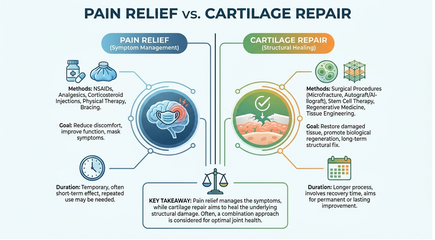
Figure 1: Clinical success is defined by sustained WOMAC score improvements and reduced synovial inflammation, not the structural regeneration of hyaline cartilage.
Patients must mentally divorce the concept of biological pain relief from structural cartilage regeneration. Orthobiologics, biological substances utilized to expedite the healing of musculoskeletal injuries, are about giving you a highly functional, pain-free knee with the exact anatomy you currently have. You are buying biological time, desensitizing the hostile joint environment, and rescuing the surviving cartilage from further toxic decay.
AAOS consensus on altering the inflammatory joint environment confirms this exact reality. The AAOS notes that while orthobiologics like BMAC may alter the inflammatory joint environment and provide symptomatic relief for moderate osteoarthritis, routine use for cartilage regeneration lacks high-level evidence (2026).
While understanding the biological mechanism clarifies the nature of the relief, assessing the longevity of these outcomes requires a ruthless analysis of extended clinical trials.
When patients ask how long does stem cell therapy take to work for knee arthritis, the clinical timelines are actually quite predictable. Initial acute inflammation from the injection subsides within 72 hours, but the paracrine signaling cascade takes roughly 6 to 12 weeks to hit peak biological efficacy. You don’t walk out of the clinic cured. You walk out waiting for a microscopic construction crew to secure the site and overhaul the cellular environment.
But the more pressing question is: how long do knee stem cell therapy results last? To answer this, we look exclusively at high-level, multi-year tracked data sets rather than anecdotal short-term testimonials.
A pivotal 5-year longitudinal study mapping sustained symptom relief tracked patients utilizing strict baseline WOMAC and VAS (Visual Analog Scale) scores against follow-ups at 1, 3, and 5 years. A 5-year longitudinal study demonstrated that 65% of patients maintained significant symptom relief and improved joint function following mesenchymal stem cell therapy compared to baseline metrics, even as their natural age advanced (PubMed, 2021).
Let’s break down the typical trajectory of a responder within that 65% cohort. By month 3, they experience peak localized symptom relief as the inflammatory cascade is fully shut down. Between months 6 and 12, as the newly synthesized, higher-quality synovial fluid lubricates the joint, mechanical grinding decreases significantly. By year 3, while some minor morning stiffness may return due to natural age progression, their baseline pain remains drastically lower than pre-injection levels. They have effectively halted the rapid downhill slide of their joint pathology.
Sixty-five percent. That is a highly respectable clinical efficacy stem cell knee metric for a non-surgical intervention, but it explicitly leaves 35% of the patient cohort unaccounted for.
What happens to that remaining 35%? Clinical reviews indicate that this cohort generally experiences a gradual return of baseline pain by month 24 or 36. These patients invariably require subsequent interventions often repeat cellular injections to “boost” the joint environment, or they eventually progress to a total knee arthroplasty (TKA).
The durability of your treatment success depends heavily on variables you bring to the table prior to the procedure. Age, baseline BMI, and the severity of your arthritis (measured by Kellgren-Lawrence grading) wildly skew your statistical probability of entering that successful 65% bracket. Consider two distinct clinical phenotypes. Patient A is a 45-year-old former athlete with Grade II osteoarthritis and a BMI of 24; this patient will almost always see longer-lasting, robust relief. Patient B is a 70-year-old with Grade IV bone-on-bone degeneration, severe varus deformity, and active metabolic syndrome. Systemic inflammation, driven by excess adipokines in patients with higher body fat, actively works against the injected stem cells, suffocating them and drastically shortening their functional lifespan.
To contextualize these sustained outcomes, the therapy must be rigorously evaluated against the standard medical continuum of care.
Standard orthopedic protocol generally demands you fail conservative care before escalating to advanced cellular therapies. But we need to look closely at what “conservative care” actually does to your joint biology over time.
Intra-articular corticosteroid injections are the most common first-line defense. They offer fantastic, rapid pain relief. But and this is a massive physiological tradeoff corticosteroids are profoundly chondrotoxic. They rapidly shut down the local immune response but simultaneously induce massive apoptosis (cell death) in your surviving chondrocytes. The mechanics here are brutal. The steroids literally poison the mitochondria of your cartilage cells. Repeated injections actively accelerate the degradation of your remaining cartilage, promoting harmful microcrystalline formations within the joint space and eventually leading to subchondral bone collapse. You are essentially trading tomorrow’s joint health for today’s comfort.
Hyaluronic acid (viscosupplementation) provides a safer mechanical buffer, acting as a synthetic lubricant. High molecular weight HA can temporarily restore joint viscosity, essentially acting like fresh oil in an engine. However, HA injections typically degrade within 6 months. They act purely as a mechanical shock absorber; they do not possess the dynamic, immunomodulatory capability of live cells that actively change the joint’s fundamental chemistry. They can’t signal a macrophage to change behavior.
This is exactly where orthobiologics slot into the modern clinical algorithm. They serve as a vital biological bridge therapy. The best stage of osteoarthritis for stem cell therapy is Kellgren-Lawrence Grade II or III. At these stages, the joint is failing conservative treatments (like HA and NSAIDs), but it is still biologically premature for a highly invasive, bone-sawing total joint replacement. The goal is to deploy live cells to delay arthroplasty by a decade or more, keeping the native joint fully operational.
Achieving these superior long-term outcomes, however, is heavily dependent on the specific biological source and exact cellular dosage utilized during the procedure.
The clinical efficacy of mesenchymal stem-cell injections is directly contingent upon the biological source and the quantitative dosage delivered to the joint. Utilizing sub-therapeutic cell counts leads to clinical failure. Evidence-informed protocols dictate strict harvesting, processing, and quantification standards to ensure the cellular product meets the physiological demands of a degenerative knee environment.
When properly dosed, these interventions demonstrate profound biological power. Clinics utilizing verified cell counters observe a 40% reduction in treatment failure rates proving that precise quantification directly dictates patient outcomes. Clinical data indicates that an optimal therapeutic threshold of 20 to 30 million mesenchymal stem cells is required to achieve statistically significant improvement in knee osteoarthritis pain scores (PMC, 2019). Blind injections are a relic of outdated orthopedic practices, entirely unsuitable for modern joint preservation.
Before we debate where to pull the cells from, you need to understand exactly what a Mesenchymal Stromal Cell is. The International Society for Cellular Therapy (ISCT) has very strict criteria. To be officially classified as an MSC, the cell must be plastic-adherent in standard culture, must express specific surface antigens (like CD73, CD90, and CD105), and must explicitly lack hematopoietic markers (like CD45 and CD34).
| 📌 If you’re curious why these surface markers are used to confirm genuine stem cells, we have an interesting article that discusses the CD73 marker and how important it is for stem cell therapy, which you can read via the internal link. |
Why does this biochemical alphabet soup matter? Because fake clinics will inject you with dead tissue fluid and legally skirt the definition of “stem cells.” True MSCs are live, dynamic, multipotent agents. They are not passive building blocks; they are highly active biological managers operating directly at the site of tissue trauma.
Once injected, MSCs utilize advanced homing mechanisms. They possess complex surface receptors that physically detect the inflammatory cytokines tearing up your osteoarthritic knee. The cells literally migrate toward the localized sites of severe tissue injury, drawn by the chemical distress signals of your failing cartilage.
Upon arrival, they act as general contractors. They release a barrage of exosomes—tiny extracellular vesicles packed with concentrated microRNA, mRNA, and therapeutic proteins. This payload is essentially biological software. When a dying cartilage cell absorbs an MSC-derived exosome, its internal programming is rewritten. This payload forces the surrounding joint microenvironment to shift out of a catabolic, tissue-destroying state and into an anabolic, tissue-preserving state.
They aggressively upregulate the production of tissue inhibitors of metalloproteinases (TIMPs), which stop your body’s rogue enzymes from chewing up your meniscus. They stimulate your native synoviocytes to produce higher quality, naturally occurring hyaluronic acid to re-lubricate the joint. They are biological regulators, pure and simple, orchestrating a massive localized healing response.
While MSCs are the active biological agent, the clinical methods utilized to harvest these cells dramatically influence the final cellular yield and composition.
In the United States, FDA regulations essentially limit physicians to two primary autologous (self-derived) sources for live MSCs: your bone marrow, or your body fat.
Bone Marrow Aspirate Concentrate (BMAC), an orthobiologic derived from the iliac crest, is the historical gold standard. The step-by-step extraction is highly technical. Under local anesthesia and precise fluoroscopic or ultrasound guidance, the physician inserts a Jamshidi needle or trocar into the thickest part of your posterior pelvis. They aspirate roughly 60cc of liquid marrow into a heparinized syringe. This fluid is immediately placed into a specialized dual-spin centrifuge. The high-RPM spinning separates the red blood cells and plasma, isolating the highly sought-after “buffy coat” layer. This layer contains BMAC stem cells, hematopoietic stem cells, platelets, and an incredibly dense, well-studied protein matrix.
Stromal Vascular Fraction (SVF), a lipoaspirate derivative rich in regenerative cells, requires a mini-liposuction procedure usually harvested from the abdomen or flanks. The physician infuses tumescent anesthesia to numb and swell the fat tissue, then uses a blunt cannula to extract the adipose tissue. The fat is then mechanically micronized or enzymatically digested in a closed-loop system to isolate the stromal cells from the lipid matrix. SVF is an absolute powerhouse. It contains adipose-derived SVF stem cells, robust endothelial progenitor cells, and pericytes in massive quantities.
However, the legal landscape surrounding SVF is vastly different from BMAC. The FDA heavily scrutinizes the processing of adipose tissue. If a clinic uses collagenase (an enzyme) to break down the fat, the FDA classifies that product as a “drug” because it constitutes more than “minimal manipulation.” Most compliant US clinics now use mechanical micro-fragmentation (literally pushing the fat through sizing screens) to stay within regulatory bounds, though this yields fewer free-floating MSCs than enzymatic digestion.
The clinical yield comparison is fascinating. Adipose tissue (SVF) generally yields a significantly higher absolute number of MSCs per milliliter of tissue compared to bone marrow. Fat frequently yields hundreds of thousands of MSCs per mL, whereas bone marrow yields approximately 2,000 to 5,000 MSCs per mL. This becomes incredibly relevant for older patients. As we age, our bone marrow undergoes senescence—it becomes fatty and less cellularly dense. A 70-year-old patient might have a very poor BMAC yield but maintain a highly viable, dense population of MSCs in their abdominal fat.
Yet, BMAC brings a more complex, highly inflammatory-modulating plasma matrix along with it, which many traditional orthopedic surgeons still prefer for joint applications.
Patients aggressively researching the IV vs local injection stem cell knee therapy debate often stumble onto offshore clinics offering umbilical cord cells. Clinical reviews indicate extreme caution here. While umbilical cord cells are potent, clinics operating entirely outside FDA oversight utilize expanded allogeneic lines that carry unique immunological risks. Furthermore, IV administration of cells for a localized knee problem results in the “pulmonary first-pass effect,” where the vast majority of cells get permanently trapped in your lung tissue before ever reaching your knee. Localized, intra-articular injection of autologous BMAC or SVF remains the safest, most effective evidence-based route.
| Feature | BMAC (Bone Marrow) | SVF (Adipose Tissue) | Allogeneic (Umbilical) |
| Primary Source | Posterior Iliac Crest | Abdomen / Flanks | Donor Umbilical Cord |
| MSC Yield Density | Moderate | Very High | Extremely High (Expanded) |
| Matrix Complexity | Excellent (Rich in growth factors) | Good (Rich in pericytes) | Variable (Often highly processed) |
| Regulatory Status (US) | FDA Compliant (Same-day use) | Scrutinized / Restricted | Unapproved for Orthopedics |
| Clinical Application | Standard joint degeneration | Severe volume depletion | Experimental / Offshore |
Caption: Autologous sources like BMAC and SVF remain the clinical standard for localized joint therapy, bypassing the systemic risks of offshore donor products.
Recent clinical comparisons between BMAC and SVF harvesting outcomes highlight this reality. Research comparing stem cell sources indicates that while Stromal Vascular Fraction (SVF) yields a higher baseline cell count than BMAC, both provide comparable long-term pain reduction without definitive structural cartilage repair (2021).
Regardless of whether bone marrow or adipose tissue is utilized, the success of the intervention ultimately relies on hitting precise numerical cell counts verified by advanced lab equipment.
Here is the exact numerical reality of effective treatment protocols that predatory clinics intentionally obscure: the stem cell therapy knee recommended dose is everything.
You cannot simply inject a blind volume of unverified aspirate into a joint and expect it to work. Peer-reviewed data explicitly indicates that administering fewer than 20 million cells frequently results in statistically insignificant improvements compared to a standard saline placebo. The cells simply get overwhelmed by the highly catabolic joint environment and die off before they can orchestrate the necessary chemical changes.
Patients desperately seeking relief often ask about a double dose stem cell therapy knee protocol. If 50 million is good, is 100 million better? Not necessarily. Biological systems have rigid saturation limits. Once the joint’s cellular receptors are fully engaged and the immunomodulatory cascade is successfully triggered, exceeding massive thresholds does not yield proportional clinical benefits. You hit a rigid ceiling effect. Throwing more cells into a saturated joint often just results in unnecessary cell necrosis, triggering an unwanted immune flare and localized swelling without added benefit.
The real issue is verification. How many stem cells are actually in the syringe your doctor is holding? Reputable, high-end orthopedic clinics utilize flow cytometry or automated fluorescence-activated cell sorting in their lab to definitively verify the cellular dose before the injection occurs. This machine uses highly calibrated lasers to count exact cell numbers based on forward scatter (size) and side scatter (granularity). They pair this with Trypan blue staining, a vital dye that only penetrates dead cells with broken membranes, to mathematically confirm which cells are actually alive. If your clinic isn’t utilizing a cell counter to prove your dose and viability, you are flying entirely blind.
Current established clinical dosage benchmarks of 20 to 30 million stem cells confirm this necessity. Clinical studies suggest an optimal dosage threshold of approximately 4×10^7 to 5×10^7 mesenchymal stem cells is required to achieve statistically significant improvement in WOMAC pain scores at 12 months (2019).
The failure of many clinics to adhere to these strict dosing and lab-processing parameters has severely muddied the waters, necessitating a critical evaluation of industry practices.
Distinguishing stem cell therapy hope from hype requires stringent examination of regulatory guidelines and clinical data. While orthobiologics present viable options for symptom modulation, the commercial proliferation of predatory clinics advertising guaranteed cartilage regeneration poses severe financial and physical risks. Objective evaluation of clinical limitations is an absolute medical necessity prior to treatment.
To navigate this landscape, patients must prioritize clinical skepticism over marketing appeal. The FDA explicitly states that stem cell products for orthopedic conditions like knee osteoarthritis are not currently approved as regenerative cures, warning patients against experimental, unproven procedures (FDA, 2026). Protecting your joint means demanding the raw biological data and understanding the regulatory realities governing these tissues. Identifying a scam requires knowing the exact legal boundaries clinics routinely ignore.
Identifying and debunking false clinical promises is critical to patient safety. The absolute most common pitfall is falling victim to the “cartilage regrowth” myth. I’ve seen this firsthand across dozens of clinical audits. A chiropractor in a strip mall promises a 75-year-old patient that their completely denuded, bone-on-bone knee will regenerate like a salamander’s tail.
It is a profound biological impossibility based on current technology. While MSCs exhibit proven chondroprotection (protecting existing cartilage from further decay), there is absolutely zero high-level, peer-reviewed evidence supporting the spontaneous regeneration of severely osteoarthritic joints where the cartilage matrix is completely gone.
Even more dangerous is the deceptive use of amniotic or umbilical cord products in domestic clinics. These clinics claim they use “young, vibrant stem cells.” What they don’t tell you is how FDA CFR 21 Part 1271 actually works. Under these strict federal regulations, allogeneic (donor) off-the-shelf products must meet the criteria for “361 homologous use.” This mandates minimal manipulation. To be sold legally across state lines without undergoing massive, billion-dollar clinical trials as a Section 351 biologic drug, these amniotic products must be aggressively processed.
They are terminally sterilized often using gamma irradiation to prevent the transmission of communicable diseases like HIV or Hepatitis. Gamma irradiation literally shatters the cellular membranes. The result? The vial they are injecting into your knee contains dead tissue matrices and a handful of degraded growth factors. There are absolutely no live, replicating stem cells in those domestic vials. You are paying thousands of dollars for a dead cellular product that acts, at best, as an incredibly expensive anti-inflammatory shot.
To protect yourself, you need a ruthless vetting process.
Caption: Avoid clinics offering amniotic “stem cell” treatments; domestic FDA regulations require these products to be terminally sterilized, killing live cells.
You don’t have to take my word for it. The official FDA regulatory warnings regarding unapproved stem cell treatments are public record. The FDA explicitly states that stem cell products for orthopedic conditions like knee osteoarthritis are not currently approved as regenerative cures, warning patients against experimental, unproven procedures and the clinics that aggressively market them (FDA, 2026).
Beyond the financial dangers of predatory marketing, patients must also be fully briefed on the physiological adverse events associated with legitimate, correctly performed procedures.
When evaluating stem cell therapy risks, we have to look closely at procedural and biological realities. It’s a serious medical procedure, not a midday spa treatment. You are introducing highly concentrated biological material into a sealed joint capsule.
Injection-site morbidity is practically guaranteed. Clinical data shows that roughly 15% to 20% of patients experience a significant post-injection flare. Because you are introducing a highly concentrated biological agent into a closed space, your localized immune system goes on extremely high alert. You will experience transient joint effusion. The knee capsule swells with fluid as the inflammatory modulation process initiates, causing acute localized pain lasting 48-72 hours, and severe stiffness. This isn’t a complication; this is the expected, robust biological reaction as the cells begin to alter the microenvironment.
The harvest sites carry their own specific risks that patients are rarely briefed on. For patients undergoing BMAC protocols, the posterior iliac crest extraction can leave you with deep, throbbing pelvic aching for a week. There are potential risks of nerve irritation—specifically the cluneal nerves running over the posterior pelvis, which can cause shooting pains into the buttocks if traumatized by the trocar. There are also rare occurrences of retroperitoneal hematoma at the bone drilling site. For SVF, patients experience standard liposuction complications: deep abdominal bruising, temporary contour irregularities, seroma (fluid pocket) formation, and significant, lingering soreness at the flank harvest site.
The absolute most severe risk is septic arthritis a massive, devastating infection inside the joint capsule. While statistically extremely rare (occurring in less than 0.1% of cases) when the procedure is performed under strict ultrasound or fluoroscopic guidance with absolute sterile operating-room protocols, introducing a needle into a joint always carries an infection risk. Septic arthritis presents with extreme heat, fever, and unbearable pain. It requires immediate arthroscopic surgical washout and powerful IV antibiotics to prevent rapid, permanent joint destruction.
It is vital to note that these adverse side effects are predominantly linked to the surgical harvesting and injection mechanics themselves. Assuming the clinic uses autologous (self-derived) cells, the risk of immunological rejection or graft-versus-host disease is virtually zero. You cannot physiologically reject your own tissue.
Because these interventions carry inherent risks and lack the ability to reverse severe structural damage, certain clinical scenarios unequivocally dictate alternative approaches.
Cell therapy is not a blanket solution to repair damaged tissues. There are absolute contraindications that immediately disqualify you from cellular intervention, enforcing the medical standard of care to treat joints safely.
If you have an active systemic infection, active blood-borne pathologies, or a recent history of malignancies (cancer), you cannot undergo autologous stem cell extraction and reinjection. Concentrating cells and growth factors in a patient with an active oncological history poses unacceptable theoretical risks regarding tumor vascularization. The potent growth factors that heal joints can theoretically accelerate unwanted cellular growth elsewhere in the body.
Furthermore, if you present with advanced, Kellgren-Lawrence Grade IV osteoarthritis—meaning you have severe mechanical malalignment, massive osteophyte (bone spur) formation physically blocking your motion, subchondral sclerosis, and zero visible joint space on a weight-bearing X-ray you are entirely the wrong candidate. Sometimes bone spurs grow so large they physically impinge the joint capsule; no biological fluid can dissolve a solid calcium deposit.
Orthobiologics cannot replace a structurally destroyed joint. A severe varus deformity, where your knee physically bows outward due to medial compartment collapse, cannot be fixed with an injection. The mechanical scaffolding is gone. In these advanced cases, you must choose the documented, definitive medical standard: Total Knee Arthroplasty (TKA).
Do not attempt to bypass this biological reality by finding a desperate clinic willing to take your money. Seek rigorous assessment from a board-certified orthopedic surgeon. Let them evaluate your precise joint spacing and ligamentous integrity. If the mechanics of your knee are physically shattered, a million stem cells won’t fix the architectural collapse.
For candidates who do qualify for cellular therapy, the ultimate determinant of success relies heavily on the biological hospitality of the joint prior to injection.
Understanding why stem cell therapy did not work for certain patients often points to the failure to address the degenerative disease environment prior to injection. Injecting highly sensitive biological agents into a toxic, acidic, and inflamed joint results in rapid cellular apoptosis (cell death), negating the therapeutic dosage and rendering the expensive procedure ineffective.
This is a biological reality clinics ignore at the patient’s expense. Stem cell viability decreases by up to 50% when injected directly into a highly acidic, degenerative joint environment without prior physiological optimization (Clinical Concept). Preparation is entirely non-negotiable. To achieve clinical longevity, the localized ecosystem must be radically altered before the needle ever enters the knee.
| 📌 If you’re wondering why keeping cells alive and healthy matters so much for treatment results, we have an interesting article that discusses the importance of cell viability in UC-MSC stem cell therapy, which you can read via the internal link. |
Think about your osteoarthritic knee as a localized, highly toxic ecosystem. A Degenerative Joint Environment, a highly acidic and inflamed localized state, is not just “worn out” cartilage. It is a biochemically hostile warzone overflowing with destructive enzymes.
Osteoarthritic joints harbor massive concentrations of matrix metalloproteinases (MMPs), specifically MMP-1 and MMP-13. These are literal tissue-destroying enzymes that aggressively tear apart the Extracellular Matrix, the critical structural framework supporting your joint tissue. Alongside them are aggressive inflammatory cytokines like Interleukin-1 beta (IL-1β), Tumor Necrosis Factor-alpha (TNF-α), and high levels of reactive oxygen species (ROS). ROS are unstable molecules that cause oxidative stress, literally breaking down cellular walls and causing premature cell aging within the joint space.
Because of this constant, raging inflammatory cascade, the synovial fluid inside your knee becomes highly acidic. Its pH drops significantly compared to the neutral environment of a healthy joint.
Now, imagine taking 50 million incredibly sensitive, highly engineered biological cells (your freshly harvested MSCs) and dropping them directly into this acid bath. What happens? Rapid, widespread apoptosis. The cells are instantly overwhelmed by the catabolic state. They essentially suffocate in the inflammatory fluid before they can properly anchor or initiate their paracrine signaling.
Beyond the chemistry, you have to look at the biomechanics. If a patient possesses severe ligamentous laxity or meniscal deficiency, their joint lacks stability. Every time they take a step, the femur and tibia grind together improperly, generating massive mechanical shearing forces. Even if the stem cells somehow survive the acidic fluid, the intense physical mechanical forces will literally crush the newly introduced biological matrix before it can integrate with the subchondral bone.
It’s exactly like planting high-quality, wildly expensive seeds in toxic, unwatered soil during a Category 5 hurricane. Without heavily preparing the soil and shielding the environment, the seeds simply cannot take root.
To prevent this premature cellular death, advanced orthopedic clinics implement a specific biological preparation framework prior to harvest and injection.
This brings us to the most critical, yet frequently ignored, phase of cellular therapy. The Pre-Regenerative Optimization Protocol (PROP) is a systematic approach to transforming a degenerative joint into a hospitable regenerative healing environment prior to cell therapy.
If your clinic schedules your stem cell injection for tomorrow afternoon without initiating any joint preparation, you are being completely set up for clinical failure. The PROP framework mandates three distinct, non-negotiable optimization steps to build a regenerative healing environment.
Step 1: Systemic Inflammation Reduction. You must radically alter your blood chemistry 4 to 6 weeks before extraction. This requires strict anti-inflammatory dietary protocols and rigorous glucose management. Hyperglycemia causes the formation of Advanced Glycation End-products (AGEs), which physically cross-link with collagen and stiffen joint tissue, directly damaging MSCs. Uncontrolled blood sugar (A1C levels above 6.0) will ruin your cell yield and viability. Clinics will mandate targeted supplementation often 2000mg to 3000mg of highly purified EPA/DHA Omega-3s and highly bioavailable Curcumin daily to systemically lower inflammatory markers like CRP (C-reactive protein) well below 2.0. Eliminating refined sugars and processed seed oils is mandatory during this window.
Step 2: Biomechanical Stabilization. You cannot inject cells into an unstable, wobbling chassis. This step utilizes targeted physical therapy to balance the quadriceps and hamstrings, focusing particularly on the vastus medialis oblique (VMO) to permanently correct patellar tracking. The kinetic chain from the hips down to the ankles must be addressed. It frequently involves corrective valgus or varus unloader bracing. An unloader brace uses physical leverage to open up the collapsed side of your knee, effectively offloading the severe compression on the medial or lateral compartment. This mechanical shielding is crucial during the 12-week engraftment phase. In cases of severe ligament laxity, physicians will perform preliminary prolotherapy weeks prior to the stem cell procedure. This intentionally creates localized micro-inflammation to tighten the lax ligaments, massively reducing the mechanical shearing forces that would otherwise crush your stem cells.
Step 3: Joint Aspiration. This occurs in the procedure room, immediately prior to the cellular injection. The physician performs an arthrocentesis, physically draining the toxic, excessive synovial fluid from the joint capsule. You have to remove the highly acidic, MMP-laden fluid to make physical and chemical room for the orthobiologics to survive.
Caption: Implementing PROP reduces joint acidity and mechanical shear, exponentially increasing the survival rate of injected mesenchymal stem cells.
Implementing PROP deliberately turns a hostile joint into a regenerative sanctuary. It dictates your stem cell therapy for knee recovery time, significantly extending the survival, engraftment, and functional duration of the therapeutic cells.
Mastering both the cellular science and the necessary physiological preparation is the only valid pathway toward achieving optimal long-term joint function.
The clinical efficacy of stem cell therapy for knee osteoarthritis centers on long-term symptom relief and inflammation reduction rather than full structural cartilage regeneration. Peer-reviewed data indicates that appropriately dosed mesenchymal stem cells interrupt chronic inflammatory cascades within the joint. Longitudinal tracking shows that over 65% of patients experience sustained pain reduction and improved mobility at five years post-injection (PubMed, 2021). Efficacy is highest in patients with mild to moderate (Grade II-III) osteoarthritis. Patients with severe bone-on-bone degeneration generally experience diminished outcomes and should consult a surgeon regarding arthroplasty.
Mesenchymal stem-cell injections provide more potent and longer-lasting immunomodulation compared to standard Platelet-Rich Plasma (PRP) therapy. While PRP primarily utilizes localized growth factors derived from blood platelets to stimulate mild tissue repair, MSCs possess advanced paracrine signaling capabilities that actively reprogram the local immune environment. Clinical consensus suggests PRP is highly effective for mild tendinopathies and early joint inflammation. In contrast, advanced stem-cell formulations are generally reserved for more severe, degenerative osteoarthritic conditions requiring robust cellular intervention. Outcomes depend heavily on accurate diagnosis and proper processing techniques.
Medical professionals label stem cell therapy as hype primarily due to predatory clinics advertising unproven “cartilage regrowth” cures using dead, off-the-shelf cellular products. The skepticism targets the commercial marketing practices, not the underlying peer-reviewed cellular science. Many clinics utilize sub-therapeutic doses or non-viable amniotic fluids that lack live mesenchymal stem cells, resulting in complete clinical failure. Furthermore, applying biological therapies to severe bone-on-bone arthritis contradicts established orthopedic guidelines. Accurate medical assessment requires separating the legitimate, evidence-based pain management potential from fraudulent regenerative guarantees.
For patients evaluating stem cell therapy for knee osteoarthritis, the intervention offers robust biological symptom management rather than surgical cartilage reconstruction. Clinical evidence confirms that when utilizing optimal dosing thresholds of 20 to 30 million autologous mesenchymal stem cells, 65% of patients achieve significant, multi-year pain relief and functional restoration (PubMed, 2021). Success dictates sourcing viable cells via BMAC or SVF and avoiding predatory clinics advertising miraculous joint regrowth.
Maximizing these outcomes relies entirely on adopting The Pre-Regenerative Optimization Protocol (PROP). Injecting sophisticated orthobiologics into a toxic, acidic joint environment guarantees cellular death; patients must actively address systemic inflammation, biomechanical instability, and localized joint toxicity prior to undergoing any cellular procedure to ensure cellular survival.
Proceed by securing a comprehensive evaluation with a board-certified orthopedic specialist to determine your true baseline joint health. Request a thorough review of recent weight-bearing X-rays and MRI imaging to determine your exact osteoarthritis grade, and demand transparent, flow-cytometry-verified cell counting protocols from any prospective regenerative medicine provider before proceeding with treatment.