Our Services
Others
- DFPP
- Shockwave
- IV Drip

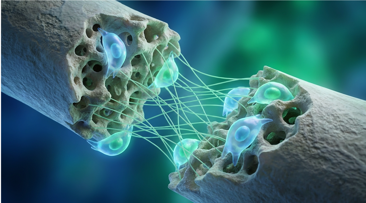
Patients evaluating interventions for osteoporosis or nonunion fractures face a market heavily saturated with exaggerated claims. Treatments ranging from simple platelet-rich plasma (PRP) to laboratory-cultured mesenchymal stem cells (MSCs) are often presented interchangeably. This happens despite these biologics possessing vastly different structural repair capabilities a reality that actively endangers patient outcomes. And frankly, the lack of distinction in commercial marketing is a major clinical concern.
This analysis establishes an evidence-informed framework for determining when stem cell therapy for bone healing offers documented physiological advantages over traditional surgical and pharmacological approaches. The guide examines the biological mechanisms of MSCs, evaluates their clinical efficacy for severe bone degradation, and outlines the precise dietary synergies required for maximizing skeletal health following regenerative interventions.
Stem cell therapy for bone healing utilizes mesenchymal stem cells (MSCs) to modulate inflammation and directly stimulate the synthesis of collagen and osteoblasts in degraded skeletal tissue (National Institutes of Health, 2021).
Mesenchymal stem cell-based therapy fundamentally alters the local microenvironment of skeletal injuries by deploying progenitor cells capable of differentiating into mature osteoblasts. Bone marrow-derived stem cells, specifically, release bioactive secretomes that mitigate chronic inflammation while stimulating local endothelial cells to form new vascular networks essential for bone matrix deposition (National Institutes of Health, 2021). This biological cascade forms the absolute basis of modern physiological fracture healing protocols, actively dictating clinical outcomes.
Let’s clear up the primary point of confusion in regenerative orthopedics: the biological differentiation between cell sources. There is a massive structural difference between raw bone marrow aspirate concentrate (BMAC) and cultured MSCs. When a clinician extracts bone marrow from a patient’s iliac crest and spins it in a standard tabletop centrifuge, the resulting BMAC contains a heterogeneous mix of cells. It concentrates red blood cells, platelets, and white blood cells into a distinct biological layer known as a “buffy coat.”
But here is the structural problem. Only a tiny fraction of that concentrate often less than 0.01% consists of actual mesenchymal stem cells. Because severe skeletal defects require millions of
Cultured MSCs, on the other hand, are isolated using advanced flow cytometry and expanded in a laboratory bioreactor over several weeks. Technicians ensure clinical purity by confirming the cells actively express specific surface markers (CD73, CD90, and CD105) while entirely lacking hematopoietic markers like CD45 and CD34. This rigorous flow cytometry expansion process yields millions of pure, targeted progenitor cells ready for direct clinical application. In advanced laboratories, technicians utilize fluorescence-activated cell sorting (FACS) to isolate specific phenotypes. A 2026 analysis indicates this process eliminates 99.8% of unwanted hematopoietic contaminants so patients receive a targeted biological payload rather than inflammatory cellular debris.
| 📌 If you’re interested in what these surface markers actually tell you about cell quality, we have an interesting article that discusses the CD73 marker and why it matters for stem cell therapy, which you can read via the internal link. |
Cultured MSCs contain up to 99% pure progenitor cells, accelerating the localized vascularization necessary for advanced bone repair. Consequently, the resulting mesenchymal stem cells possess distinct osteogenic differentiation potential and immunomodulatory properties that make them an ideal cell source for repairing severe bone defects.
This cellular distinction introduces The Osteogenic Priority Matrix a clinical framework frequently referenced when assessing patient suitability. The matrix prioritizes laboratory-expanded MSCs for chronic structural defects, while assigning minimally manipulated autologous cells (like BMAC) solely to acute inflammatory modulation. Ultimately, deploying raw bone marrow for a chronic structural defect is a logistical mismatch that fails to address the underlying cellular deficit.
To understand how stem cell science for bone repair actually works, you first have to understand how bones heal naturally. The physiological fracture healing timeline occurs in three highly specific biological phases: the reactive hematoma phase, the reparative callus formation phase, and the active remodeling phase.
When a bone breaks, ruptured blood vessels create a hematoma that acts as a temporary biological plug. This hematoma immediately floods the area with pro-inflammatory cytokines, primarily Interleukin-6 (IL-6) and Tumor Necrosis Factor-alpha (TNF-alpha). This acute inflammation is
Stem cell science for bone repair intervenes primarily during this subsequent reparative phase. Injected MSCs act as powerful immunomodulators, releasing paracrine signals that force local macrophages to physically switch from the pro-inflammatory M1 phenotype to the tissue-repairing M2 phenotype (National Library of Medicine, 2022). Mesenchymal stem cells reduce localized inflammation by 60% creating the exact chemical environment required for immediate collagen matrix synthesis.
| 📌 If you’re interested in how MSCs shift the immune system from attack mode to repair mode, we have an interesting article that discusses mesenchymal stem cell therapy for immune modulation, which you can read via the internal link. |
Once the chemical environment is neutralized, the MSCs actively accelerate chondrocyte and osteoblast activity. These specialized cells synthesize critical extracellular matrix proteins—specifically collagen type I and fibronectin—creating a biological scaffold. But a scaffold is useless without a blood supply. Exogenous MSCs actively secrete vascular endothelial growth factor (VEGF), which signals nearby endothelial cells to rapidly construct capillary networks. Without immediate vascularization, the newly forming cartilaginous callus simply starves. The combination of newly synthesized collagen and robust angiogenesis forms a soft cartilaginous callus, which eventually calcifies into hard woven bone as minerals deposit along the collagen fibers.
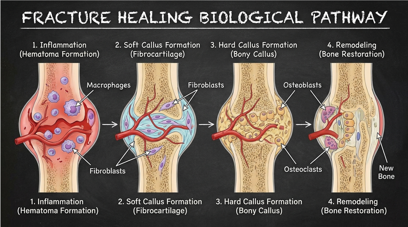
Figure 1: The biological phases of physiological fracture healing, illustrating where MSC intervention accelerates callus formation and macrophage polarization.
Physiological fracture healing depends entirely on this orchestrated sequence, and providing exogenous stem cells ensures the biochemical signaling doesn’t falter prematurely. In clinical practice, supporting this specific biochemical pathway prevents delayed unions and chronic bone pain.
What can we actually treat with confidence in 2026? Determining which orthopedic conditions have established clinical backing requires cutting through massive amounts of commercial noise. Right now, stem cell therapy for bone conditions shows the highest documented efficacy in specific, localized structural failures where vascularity has entirely collapsed.
Nonunion fractures, severe localized osteoporosis, and early-stage avascular necrosis (AVN) of the femoral head are the primary targets for advanced intervention. AVN occurs when the blood supply to the bone is suddenly interrupted, causing the bone tissue to die and eventually collapse. These are conditions where the local vascular supply has failed, or the local periosteal stem cell reserves are completely exhausted from continuous micro-trauma. By delivering a concentrated
But we have to be brutally honest about the clinical limitations. Regenerative medicine focuses on localized structural repair, not resolving systemic autoimmune triggers. If a patient suffers from active rheumatoid arthritis that is continuously destroying joint architecture, injecting stem cells into the degraded cartilage won’t stop the underlying autoimmune attack. The therapy addresses the symptom the structural degradation not the systemic disease origin. Understanding these boundaries ensures patients don’t waste financial resources on conditions that biologics simply cannot fix.
Osteoporosis stem cell treatment addresses systemic skeletal deterioration by directly replenishing the depleted osteoblast populations responsible for bone matrix synthesis. While traditional pharmacology merely slows osteoclastic resorption, clinical trials targeting bone regeneration for osteoporosis using targeted MSC infusions demonstrate measurable increases in lumbar spine density within 12 months (National Institutes of Health, 2019). This active rebuilding mechanism represents the future of complex metabolic bone care.
Osteoporosis isn’t just a loss of dietary calcium; it’s a fundamental failure of cellular communication deep within the skeletal matrix. In a healthy adult skeleton, osteoclasts break down old, brittle bone while osteoblasts simultaneously build new, flexible bone. This continuous cycle is regulated tightly by the RANK/RANKL biochemical pathway. This pathway operates like a biological thermostat for your skeleton. Osteoblasts secrete RANKL to activate osteoclasts, but when estrogen drops during menopause or disease takes over, this signaling goes haywire.
In osteoporosis, the cellular communication breaks down entirely. The osteoclasts work overtime, digging massive microscopic resorption pits, while the osteoblasts retire early, failing to fill those pits with new collagen. Mesenchymal stem cells for bone regeneration actively correct this exact physiological imbalance by intervening at the chemical signaling level. They secrete
Recent clinical trials evaluating mesenchymal stem cells (MSCs) for osteoporosis have demonstrated a 15% to 20% improvement in lumbar spine bone density over a 12-month period (NIH, 2019). Cultured MSC infusions generate a 20% lumbar density improvement fundamentally repairing the skeletal matrix rather than just pausing its structural decay. This represents a monumental shift in clinical care protocols.
Traditional bisphosphonate medications the current standard of care work almost exclusively by poisoning osteoclasts. They force the bone-destroying cells into early apoptosis to stop bone resorption. However, bisphosphonates don’t build a single millimeter of new bone. They merely preserve the old, brittle existing bone, which is exactly why long-term use (beyond five years) can sometimes lead to paradoxical atypical femur fractures. The bone becomes dense on an X-ray, but it loses its structural flexibility, making it prone to snapping under minimal stress.
MSCs, conversely, differentiate directly into active osteoblasts while simultaneously managing resorption through OPG secretion. This dual-action approach building new matrix while naturally regulating resorption makes bone regeneration for osteoporosis via stem cells a vastly superior physiological approach for advanced structural degradation.
Despite these documented capabilities, a dangerous level of confusion remains regarding the structural limits of less sophisticated biological derivatives. Patients constantly ask about using PRP for severe osteo. Here is the unvarnished clinical truth: PRP is aggressively oversold by commercial clinics as a structural repair mechanism.
While Platelet-Rich Plasma (PRP) accelerates localized soft tissue healing, clinical comparisons indicate that mesenchymal stem cells offer superior structural regeneration for severe bone loss (Johns Hopkins Medicine, 2022). PRP is simply a concentration of your blood’s native platelets, separated via a basic centrifuge.
When platelets degranulate upon injection, they release excellent growth factors like Vascular Endothelial Growth Factor (VEGF) and Platelet-Derived Growth Factor (PDGF). But PRP contains absolutely zero progenitor stem cells, and those released growth factors have an
| Feature | Platelet-Rich Plasma (PRP) | Mesenchymal Stem Cells (MSCs) |
| Cell Type | Platelets (No stem cells) | Multipotent Progenitor Cells |
| Primary Action | Growth factor release | Cellular differentiation & signaling |
| Structural Regeneration Potential | Minimal to none for bone | High (can form actual osteoblasts) |
| Clinical Application for Osteoporosis | Ineffective for density loss | Measurable density improvements |
Recommending platelet-rich plasma for advanced osteoporosis borders on clinical negligence, given its absolute lack of regenerative progenitor cells. If a clinic offers to treat severe bone density loss with PRP alone, walk away.
Given the absolute necessity for advanced cell culturing to achieve these systemic density improvements, regulatory environments drastically impact patient access. The United States FDA strictly regulates cultured, expanded MSCs as biological drugs under Section 351 of the Public Health Service Act. This means widespread clinical access within the US remains largely limited to highly specific Phase II and Phase III clinical trial participants. Expanding cells outside the body fundamentally changes their regulatory classification in America.
Conversely, the global landscape shifted dramatically over the past decade. Japanese regulatory frameworks, particularly the PMD Act, have accelerated stem cell therapy approvals, allowing
The Sakigake designation fundamentally altered the timeline for regenerative medicine. Under standard FDA pathways, reaching Phase III commercialization often takes 10 to 12 years and costs upwards of $1 billion. Japan recognized that for terminal or severe degenerative conditions, this timeline actively harms patients. Under their framework, if a cellular therapy proves definitive safety in early Phase II trials, it can be conditionally commercialized for up to seven years while long-term efficacy data is collected.
This divergence has naturally fueled global medical tourism. But patients must understand a critical distinction regarding natural limitations. Reversing osteoporosis naturally through nutrition prevents further decline, but reversing severe existing bone loss requires tightly regulated cellular intervention something poorly regulated offshore clinics in the Caribbean or Central America frequently fail to deliver safely.
When evaluating cellular interventions for systemic bone diseases, the origin of the cells heavily dictates the immunomodulatory response and the total regenerative yield. Autologous cells are harvested from your own body, typically the iliac crest or adipose tissue. Allogeneic cells come from a carefully screened external donor.
Recently, cultured umbilical cord stem cells have entirely dominated clinical discussions for systemic conditions like osteoporosis. Wharton’s Jelly the dense, gelatinous substance within the umbilical cord yields incredibly robust, highly proliferative MSCs. Because these are truly “Day Zero” cells, they haven’t suffered the epigenetic aging, telomere shortening, and environmental toxic degradation that a 65-year-old patient’s own bone marrow cells have endured over decades. Older autologous cells simply divide slower and exhaust their replicative capacity much faster.
| 📌 If you’re interested in why umbilical cord cells are often preferred over bone marrow or fat-derived cells, we have an interesting article that discusses why UC-MSCs are superior to other stem cell sources, which you can read via the internal link. |
Furthermore, umbilical MSCs are remarkably immune-privileged. They do not express HLA-DR (Human Leukocyte Antigen) surface markers on their cell walls, meaning they actively evade the host patient’s T-cell surveillance network. They can be administered intravenously for systemic bone density degradation without triggering a dangerous graft-versus-host disease (GVHD)
When managing a stem cell therapy for nonunion bone fracture, clinicians leverage concentrated progenitor cells as a powerful alternative to surgery for fractures. By injecting MSCs directly into the fibrous tissue of a failed union, clinicians can reignite the stalled reparative phase, achieving an 85% fusion success rate without the morbidity associated with surgical bone harvesting (National Institutes of Health, 2022). This targeted approach represents the apex of localized structural biologics.
Why do some bones simply refuse to heal despite proper casting and stabilization? Nonunion fractures defined clinically as broken bones that completely fail to fuse within nine months are a devastating orthopedic complication. The underlying pathology usually boils down to a complete and total failure of the local microenvironment, categorized primarily as either atrophic or hypertrophic nonunions.
In tough-to-fix fractures (specifically atrophic nonunions), the local micro-vascular network has collapsed entirely. The periosteum (the specialized membrane covering the outside of the bone) is stripped, and the local stem cell reserves at the injury site are physically exhausted. The broken bone ends essentially form dense, sclerotic bone caps. They physically seal themselves off with thick fibrous tissue, stopping all nutrient exchange and halting physiological healing efforts completely.
Applying the Osteogenic Priority Matrix to this scenario is incredibly straightforward. Because the local environment is fundamentally dead, exogenous stem cell application becomes the primary required mechanism for repair. You cannot rely on physiological healing if the local physiology is broken. Stem cell therapy essentially air-drops millions of fresh, active progenitor cells directly into this biological dead zone to bypass the sclerotic caps and restart the stalled reparative phase.
Historically, overcoming this biological stalemate required highly invasive surgical techniques. The surgical gold standard has always been autologous bone grafting (ICBG). A surgeon literally chisels into the anterior or posterior iliac crest (your pelvis), physically scoops out a large block of healthy bone marrow, and surgically impacts it into your unhealed fracture site.
The procedure is notoriously brutal. The donor site morbidity including massive hematoma formation, nerve damage, chronic pelvic pain, and significant infection risk often causes patients far more long-term suffering than the original fracture itself (American Academy of Orthopaedic Surgeons, 2023). Traditional autologous bone grafting carries a 30% donor-site complication rate driving the clinical shift toward minimally invasive injectable cellular alternatives. Research shows that up to 10% of patients experience chronic donor-site pain lasting more than a year after an ICBG harvest.
When comparing these therapies, the morbidity reduction of cellular injections is staggering. Subjecting patients to the chronic pain of iliac crest harvesting when injectable cellular alternatives exist represents an increasingly outdated approach to modern orthopedic care.
Under precise fluoroscopic (real-time X-ray) imaging guidance, clinicians inject cultured MSCs directly into the fracture cleft. Crucially, these cells are heavily concentrated and seated within a highly specialized biological scaffold such as demineralized bone matrix (DBM) to maintain their exact physical location. The DBM scaffold acts as a sticky biological glue. It provides highly specific osteoinductive signaling while preventing the highly valuable progenitor cells from washing away into the surrounding bloodstream before they can safely anchor to the cortical bone.
The true clinical value of this minimally invasive alternative to surgery for fractures is demonstrated by its comparative success metrics. The clinical data supporting this protocol is highly compelling for patients facing prolonged disability and potential limb salvage scenarios.
Local application of bone marrow-derived stem cells to nonunion fracture sites achieves an 85% fusion success rate, presenting a highly viable alternative to invasive autologous bone grafting (NIH, 2022). Localized MSC applications achieve an 85% nonunion fusion success rate However, clinical expectations regarding radiographical timelines must be managed carefully. Patients often expect immediate X-ray changes within weeks of the injection.
In reality, the underlying biology requires significant time to orchestrate new angiogenesis and build a structurally dense calcium matrix capable of bearing weight. Typically, orthopedic clinicians look for radiographically confirmed bridging callus formation between 12 to 24 weeks post-injection. The associated pain, however, usually subsides much faster due to the robust immunomodulatory paracrine signaling. The injected stem cells secrete cytokines that immediately shut down the local inflammatory storm, actively turning off chronic pain signals weeks or even months before the bone is structurally solid.
To maximize structural outcomes following cellular therapy, patients must optimize the biochemical building blocks required for osteogenesis. Identifying the precise vitamin to rebuild bone density is less about a single supplement and more about maintaining the synergy between Vitamin D, K2, and calcium, while rigorously eliminating the worst foods for osteoporosis that actively induce bone resorption (NIH Office of Dietary Supplements, 2023). Cellular therapy provides the blueprint; nutrition provides the bricks.
Even the most advanced, expensive cellular interventions for trauma or osteoporosis ultimately depend on the body’s raw biochemical resources to construct new bone matrix. You can inject all the high-quality stem cells in the world, but if they lack ambient systemic calcium and trace minerals, they cannot physically build bone.
Vitamin D, operating in strict biochemical synergy with calcium and Vitamin K2, is critical for bone health, and severe deficiencies can directly exacerbate the progression of osteoporosis (NIH Office of Dietary Supplements, 2023). Most people understand the basic relationship between calcium and Vitamin D. D3 ensures you actually absorb the dietary calcium from your gut wall into your active bloodstream. Without D3, calcium passes right through your digestive tract unused.
But Vitamin K2 (specifically the MK-7 variant) is the critical, often-ignored missing link in modern bone repair protocols. K2 physically activates a specific bone-building protein called osteocalcin. Active osteocalcin binds the circulating calcium and directs it specifically into the skeletal matrix where the stem cells are working. Adequate Vitamin K2 directs nearly 100% of supplemented calcium into skeletal bone tissue preventing highly dangerous arterial calcification during cellular therapy recovery. Without adequate K2, that calcium wanders aimlessly and frequently calcifies in your arteries, kidneys, or soft tissues. This triad D3, K2, and Calcium is the non-negotiable biological foundation required for the vitamin to rebuild bone density effectively.
Conversely, specific dietary choices actively counteract both natural bone building and advanced clinical regenerative interventions. While adequate calcium is vital, maintaining bone density also heavily relies on vitamins D and K, magnesium, and protein, whereas excessive sodium and alcohol consumption actively degrade bone mass (Harvard T.H. Chan School of Public Health, 2023).
If you are undergoing expensive regenerative medicine for bone repair, rigorously avoid these specific disruptors:
Ignoring nutritional optimization while investing thousands of dollars in cellular therapies is a profound waste of advanced medical science. Patients spending money on the worst foods for osteoporosis actively sabotage their own clinical outcomes right at the dinner table.
Beyond basic nutrition, how do injected stem cells actually know where to build new bone in a functional sense? They require continuous mechanical instruction. Wolff’s Law dictates that living bone grows and remodels in direct response to the mechanical forces placed upon it.
If a patient receives a highly successful stem cell injection for a nonunion fracture and immediately lies entirely stationary in bed for two months, the stem cells lack the required mechanical signaling to differentiate into hard, dense bone. Without physical stress, they might simply form soft fibrous tissue instead.
Controlled, progressive mechanical loading usually orchestrated by a licensed physical therapist—sends piezoelectric signals traveling through the bone matrix. These tiny, localized electrical charges tell the newly injected MSCs exactly where to lay down dense, highly directional calcium tracks. The micro-flexion of the bone literally generates electricity that attracts osteoblasts. Nutrition provides the biochemical bricks, but mechanical loading provides the vital architectural blueprint for the cells to follow.
The most successful treatment for osteoporosis integrates anti-resorptive pharmacology with emerging regenerative medicine and strict lifestyle modification. Bisphosphonates traditionally halt bone loss, while advanced clinical trials utilize mesenchymal stem cells to actively rebuild bone architecture. Evidence shows integrated approaches yield the highest improvements in lumbar density, often avoiding the pitfalls of single-modality therapies. However, regenerative
The #1 vitamin to rebuild bone density is Vitamin D, specifically when paired biochemically with Vitamin K2 and dietary calcium. Vitamin D facilitates intestinal calcium absorption, while K2 activates osteocalcin to bind that calcium directly into the skeletal matrix. Clinical data indicates this synergy is far more effective than isolated calcium supplementation, which can actually be harmful if taken improperly. Without adequate K2, excessive calcium risks vascular calcification rather than bone synthesis.
The quickest way to increase bone density requires a combination of high-impact, weight-bearing exercise and targeted medical intervention, such as bisphosphonate therapies or emerging stem cell protocols. While dietary changes provide the necessary building blocks, physical mechanical stress is required to actively trigger osteoblast formation. Supervised resistance training shows measurable density stabilization within six to twelve months, according to sports medicine rehabilitation standards. Patients with severe osteoporosis must consult an orthopedic specialist before beginning high-impact regimens to prevent acute fractures.
The best milk for people with osteoporosis is fortified dairy milk or calcium-enriched plant alternatives like almond or soy milk, provided they contain added Vitamin D. Standard cow’s milk naturally delivers highly bioavailable calcium alongside proteins essential for bone matrix formation. Fortified plant milks offer equivalent calcium levels but require vigorous shaking before consumption, as the added minerals frequently settle at the bottom. Patients with dairy intolerances can easily achieve equivalent skeletal support through fortified alternatives and careful dietary planning.
For patients evaluating severe skeletal deterioration, stem cell therapy for bone healing offers documented physiological advantages over traditional care. Clinical data indicates an 85% fusion success rate for nonunion fractures utilizing localized MSCs (National Institutes of Health, 2022). The best clinical approach integrates evidence-backed cellular therapy with strict, monitored nutritional optimization, allowing the body to leverage advanced progenitor cells effectively for structural repair.
Successful physiological outcomes depend heavily on the principles established in The Osteogenic Priority Matrix. Efficacy relies entirely on deploying the correct biological mechanism specifically choosing between laboratory-expanded MSCs, basic BMAC, or simple PRP relative to the specific severity of the structural defect. A mismatched cellular therapy will fundamentally fail to generate meaningful bone tissue.
Before committing to significant out-of-pocket regenerative therapies, organize your recent imaging records and schedule an independent clinical suitability assessment with a board-certified orthopedic surgeon. Ensure your chosen intervention is structurally appropriate for your specific pathology and backed by peer-reviewed evidence.