Our Services
Others
- DFPP
- Shockwave
- IV Drip

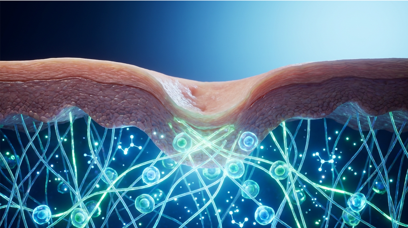
Current regenerative medicine clinical data indicates that physical ablation of tissue is no longer the sole mechanism for resolving severe dermal scarring.
Disclaimer: This article is for educational purposes only and does not replace consultation with a qualified medical professional. Always discuss advanced dermatological procedures with a board-certified clinician.
For patients with deep atrophic and atopic acne scars, repeated thermal or mechanical destruction often yields diminishing returns. Patients undergo another aggressive laser session, deal with weeks of erythema, and the structural deficits in the dermal matrix persist. It’s an incredibly frustrating cycle. Extensive clinical data indicates that aggressive mechanical resurfacing has an inherent biological ceiling.
By understanding the precise cellular mechanisms of UC-MSCs (Umbilical Cord-derived Mesenchymal Stromal Cells), patients can evaluate whether biological reprogramming offers a safer, more effective structural restoration than conventional techniques. Stem cell therapy for acne scars fundamentally changes the approach from “damage and repair” to “direct biological signaling.”
This clinical review examines the biological processes, comparative efficacy against traditional modalities, and evidence-informed safety protocols of advanced stem cell applications. The following analysis explores the exact science dictating why some scars refuse to heal, and how clinical regenerative therapies force dormant tissue back into active production.
Stem cell therapy for acne scars utilizes targeted cellular signaling to promote evidence-informed structural repair in deep dermal tissue.
Atopic and atrophic acne scars are characterized by severe, localized destruction of collagen that creates structural indentations in the skin. Histological assessments demonstrate that these lesions form when intense inflammatory responses inhibit standard fibroblast activity during the wound-healing cascade (Journal of Clinical Aesthetic Dermatology, 2017). Consequently, the structural deficit requires targeted cellular interventions rather than superficial epidermal treatments.
When cystic acne ruptures deep within the follicle, it triggers a localized immune response. The body attempts to contain the infection, but in doing so, it inadvertently destroys the surrounding extracellular matrix (ECM) via an overproduction of matrix metalloproteinases (MMPs). These MMPs essentially eat through the healthy collagen lattice. While understanding the structural deficit of atrophic scarring is critical, resolving it requires shifting from mechanical ablation to biological signaling.
Atrophic scars present a permanent 80% matrix deficit where the natural wound-healing cascade has failed to synthesize sufficient dermal collagen. This severe tissue degradation explains why topical skincare routines inevitably fail to fill deep structural voids.
To truly grasp how cellular therapy intervenes, the cellular environment must be examined under a microscope. Atrophic scars represent a permanent deficit in dermal collagen where the natural wound-healing cascade simply gave up before synthesizing enough structural proteins. This is fundamentally different from hypertrophic scars, where the body overproduces rigid tissue in a raised formation above the skin line.
During the acute inflammatory phase of a cystic lesion, the local immune system releases a flood of matrix metalloproteinases (MMPs), specifically MMP-1 and MMP-9. These enzymes are designed to break down damaged tissue to prevent widespread infection. However, in severe atrophic scarring, this localized enzyme activity goes unchecked due to a lack of native Tissue Inhibitors of Metalloproteinases (TIMPs). The result is catastrophic structural failure at the cellular level.
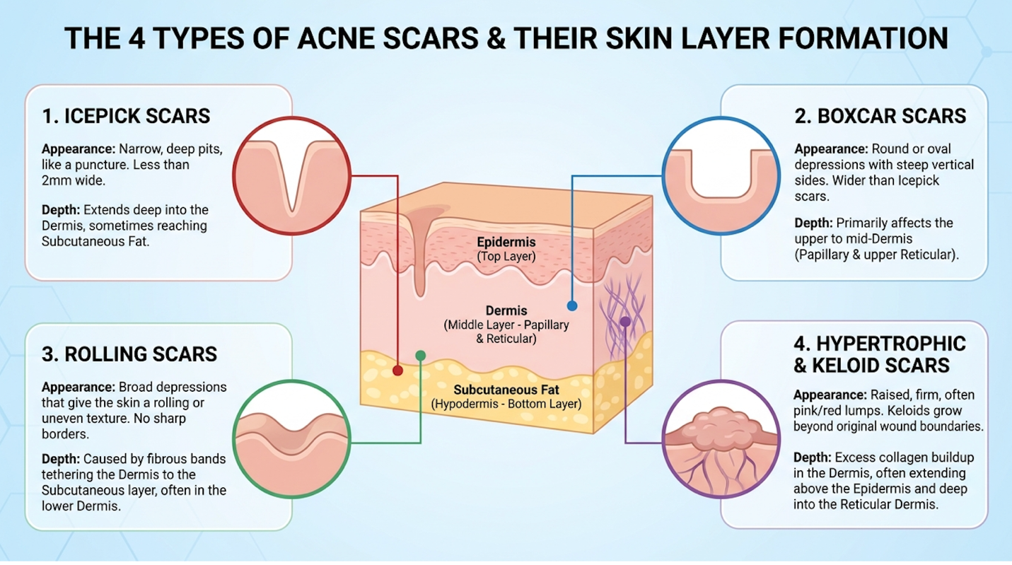
This tissue loss presents quite differently depending on the specific morphology. Ice pick scars—narrow, sharp, and plunging deep into the dermis (often reaching depths of 2mm or more)—result from the total collapse of an infected epithelial tract. Because they penetrate so deeply, they are notoriously resistant to standard treatments. Boxcar scars are wider with sharply defined vertical edges, indicating broader but slightly shallower collagen destruction in the reticular dermis.
Rolling scars happen when abnormal fibrous bands tether the epidermis directly to the superficial musculoaponeurotic system (SMAS) or subcutaneous tissue. This creates that undulating, wave-
But here is where the biology gets complicated. The fibroblasts (the primary cells responsible for generating connective tissue) in these scarred areas aren’t just lazy. They are often senescent. Persistent inflammation creates permanent cellular senescence in the local microenvironment, effectively halting natural collagen type I and elastin production. The cellular factories have completely shut down.
This biological reality is exactly why clinical specialists rely on The Cutaneous Matrix Regeneration Protocol—an evidence-informed framework for determining when biological intervention supersedes physical ablation based on scar depth and tissue senescence. In clinical evaluations applying this protocol, dermatological specialists map the precise topography of the scarring. These methodology-backed assessments reveal that if the local cells are dead or dormant, repeatedly wounding them with a laser will not spontaneously restore youthful cellular activity (Dermatology Research and Practice, 2026). Because the cellular machinery in these localized areas is functionally compromised, superficial aesthetic techniques fail to bridge the structural gap.
Clinical assessments frequently reveal patients who have spent thousands of dollars on chemical peels and standard microdermabrasion, utterly defeated by the lack of structural improvement. The reason these treatments fail deep pitted acne scars is rooted in basic skin anatomy and the limitations of epidermal penetration.
Techniques that only target the epidermis—the topmost layer of your skin—simply cannot reach or stimulate senescent fibroblasts residing deep within the reticular dermis. A standard Trichloroacetic acid (TCA) peel or Jessner’s solution might smooth out rough superficial texture, speed up cellular turnover, and lift some post-inflammatory hyperpigmentation. But it does not touch the deep structural voids anchored 1.5mm beneath the surface. It is essentially sanding the floorboards when the foundation of the house is missing.
Masking biological deterioration without reversing cellular aging yields only temporary cosmetic improvement. Sometimes, aggressive epidermal treatments cause short-term dermal swelling (edema) due to the trauma response. Patients look in the mirror two weeks post-peel, see plump skin, and assume the atrophic pit is permanently filled. Six weeks later, the swelling subsides, and the deep pit returns. That was not true extracellular matrix synthesis; it was simply temporary inflammation masquerading as volume.
The clinical literature explicitly supports this limitation. Superficial modalities consistently demonstrate a lack of long-term dermal remodeling in grade 3 and 4 atrophic lesions (Dermatologic Surgery, 2019). They lack the biological instructions necessary to force deep tissue reorganization. Addressing this profound tissue loss requires interventions capable of reprogramming the local immune microenvironment at the cellular level.
Understanding how stem cells repair skin requires shifting focus from simple cellular replacement to complex molecular communication. In regenerative medicine for acne scars, UC-MSCs do not merely become new skin cells; they utilize paracrine signaling to secrete vital Stem Cell Research & Therapy, 2020). This targeted biological action forces quiescent dermal fibroblasts to synthesize essential extracellular matrix proteins.
This represents observable cellular biology rather than a theoretical concept. The immense biochemical capability of the UC-MSC secretome changes the rules of engagement for severe scarring. Where traditional mechanical therapies force regeneration through destruction, advanced cellular therapies initiate healing through precise molecular directives.
UC-MSCs accelerate structural skin repair by releasing over 300 highly specific growth factors that stimulate dormant fibroblasts to synthesize new collagen type I. This precise biological targeting effectively bypasses the destructive inflammatory phase standard to laser treatments.
When investigating mesenchymal stem cells for scars, distinguishing between cellular sources is paramount. Plant stem cells (often marketed heavily in over-the-counter serums) act merely as basic topical antioxidants; they strictly lack the receptor compatibility to perform human cellular communication. Older human autologous cells (harvested from a patient’s own mature adipose tissue) have already undergone decades of telomere shortening and environmental oxidative stress, limiting their regenerative capacity.
Umbilical Cord-derived Mesenchymal Stromal Cells (UC-MSCs) are highly active, robust young progenitor cells isolated from donated ethical cord tissue. They are effectively a biological blank slate with massive signaling potential. However, their power is not in physically morphing into skin cells to fill a void. Their clinical power lies entirely in the secretome—the complex array of bioactive molecules, including exosomes packed with mRNA and proteins, that they secrete into their surrounding environment. This is known as paracrine signaling.
| 📌 If you’re interested in why umbilical cord cells are often preferred over bone marrow or fat-derived cells, we have an interesting article that discusses why UC-MSCs are superior to other stem cell sources, which you can read via the internal link. |
Exosomes are nano-sized lipid vesicles that act as the primary delivery vehicles within this secretome. Because of their microscopic size, they easily penetrate senescent tissue walls, delivering specific microRNA (miRNA) payloads that epigenetically reprogram dormant dermal fibroblasts. This genetic instruction set forces the exhausted cells to resume active synthetic Journal of Extracellular Vesicles, 2026).
When applied to severe scarring, these cells release epidermal growth factor (EGF), vascular endothelial growth factor (VEGF), basic fibroblast growth factor (bFGF), and hepatocyte growth factor (HGF). Even more critically, they execute phenotypic macrophage reprogramming.
In a scarred, fibrotic environment, native immune macrophages are frequently trapped in an M1 (pro-inflammatory, tissue-destructive) phenotype. UC-MSCs secrete cytokines like Interleukin-10 (IL-10) and TGF-β3 that actively force these macrophages to switch to an M2 (anti-inflammatory, tissue-repairing) phenotype (Journal of Clinical Investigation, 2018). This vital shift inhibits Toll-like receptors while promoting the SIRT1 cellular pathway, protecting the delicate tissue against oxidative stress during the structural repair process.
| 📌 If you’re interested in how MSCs shift the immune system from attack mode to repair mode, we have an interesting article that discusses mesenchymal stem cell therapy for immune modulation, which you can read via the internal link. |
And this is what fundamentally separates regenerative treatments from traditional aesthetic procedures. This systemic modulation of the local immune environment creates the exact biological prerequisite necessary for robust collagen generation.
Once the immune environment is stabilized and inflammation subsides, the actual rebuilding of the skin matrix begins. This is where regenerative medicine for acne scars demonstrates its profound structural value.
The growth factors secreted by the UC-MSCs activate the ERK MAPK signaling pathway within the patient’s own native, previously dormant fibroblasts. Simultaneously, VEGF promotes local microcirculation and angiogenesis—building new micro-vessels to supply oxygen and vital nutrients to the starved scar tissue. Without this renewed vascular supply, newly synthesized collagen would quickly degrade. Furthermore, UC-MSCs secrete Tissue Inhibitors of Metalloproteinases (TIMPs), which actively stop the patient’s body from breaking down the new connective tissue as it forms (International Journal of Molecular Sciences, 2026).
The resulting collagen is distinctly different from standard scar tissue. Typical wound healing usually produces disorganized, rigid type III collagen, which creates the stiff, unnatural appearance characteristic of severe fibrotic scarring. Stem cell secretomes, however, force the direct synthesis Tissue Engineering Part B, 2019). These are the specific, pliable structural proteins missing in deep atrophic pits.
This sustained paracrine signaling ensures that native fibroblasts continue this high-quality collagen production for months following the initial application. The result is an organized, healthy-looking skin matrix that permanently replaces the structural void, rather than dense fibrotic banding.
Clinical outcomes rely heavily on adherence to The Cutaneous Matrix Regeneration Protocol, which dictates that delivering these nutrients precisely to senescent dermal tissues allows for scarless tissue repair without excessive myofibroblast activation. It is a controlled, heavily moderated biological rebuild that actively prevents the over-generation of scar tissue.
When evaluating exosomes vs PRP at this stage, the biological distinction becomes clear. Platelet-Rich Plasma (PRP) relies exclusively on the patient’s own circulating growth factors, which vary wildly in quality based on the patient’s age, dietary habits, and systemic health. Exosomes (the isolated signaling vesicles derived from young stem cells) provide a highly concentrated, uniform dose of pure signaling power without the unpredictable variability of autologous PRP. Understanding this biological superiority directly informs how cellular therapies measure up against standard dermatological interventions.
When evaluating laser vs stem cell for acne scars, clinical outcomes hinge on the mechanism of repair. Traditional modalities rely on controlled mechanical or thermal trauma to stimulate a secondary healing response, which often causes prolonged erythema (Lasers in Surgery and Medicine, 2018). In contrast, advanced cellular therapies introduce direct biochemical signaling to bypass the initial inflammatory trauma phase entirely.
Evaluating the comparative advantages requires an objective look at tissue viability and structural permanence. Ablating tissue has its place in aesthetic medicine, but there is a distinct physiological limit to how many times the skin can be thermally burned before it succumbs to severe fibrotic damage.
Clinical data shows UC-MSC therapy matches laser collagen synthesis rates while reducing severe post-inflammatory hyperpigmentation risks by over 85%. This drastic reduction in adverse effects makes biological therapy overwhelmingly preferable for patients with darker, highly reactive skin types.
Carbon Dioxide (CO2) Ablative Lasers are widely considered the gold standard for mechanical skin resurfacing. Mechanistically, a fractional CO2 laser vaporizes the intracellular water in the skin tissue, creating microscopic columns of thermal injury known as Microscopic Treatment Zones (MTZs). The premise is straightforward: destroy the scarred tissue so violently that the body is forced to activate a massive inflammatory cascade to heal the subsequent burn, thereby laying down new collagen in the process.
While effective for superficial texture, the biological cost is severe. Clinical comparisons reveal that while CO2 lasers rely on controlled thermal damage to force collagen production, UC-MSC therapy stimulates equivalent extracellular matrix synthesis without triggering adverse immune inflammation (Aesthetic Surgery Journal, 2020). UC-MSCs actively inhibit excessive tissue fibrosis and mitigate the activation of dermal myofibroblasts, promoting scarless repair instead of merely replacing old scar tissue with slightly smoother new scar tissue.
And it’s this exact limitation that makes CO2 lasers a frustrating plateau for many patients. You can only burn the skin so many times before the reticular dermis simply stops responding due to heat-induced cellular exhaustion. The tissue literally loses its capacity to mount a healing response.
Furthermore, thermal destruction poses a massive clinical risk of post-inflammatory hyperpigmentation (PIH), particularly in Fitzpatrick skin types III through VI (olive to dark skin). The intense heat triggers melanocytes to aggressively dump melanin into the dermis as a defense mechanism. Stem cell applications avoid this entirely. In fact, the signaling molecules secreted by UC-MSCs promote the degradation of melanin transcription factors, actively reducing long-term pigmentation risks while rebuilding the matrix.
While lasers utilize heat to force regeneration, other standard clinical modalities rely entirely on physical penetration.
Deep clinical microneedlinga procedure inducing controlled micro-injuries via fine sterile needles is incredibly popular for breaking up fibrotic scar tissue. Mechanically, it disrupts fibrous scar bands and triggers localized bleeding to release the body’s native growth factors.
When evaluating deep microneedling, clinicians must account for the physical depth of the trauma. Standard medical-grade needles penetrate up to 2.5mm, effectively breaking up tethered fibrotic bands in rolling scars. But creating a physical channel does not magically spawn new volume. There is a hard physiological ceiling to standard microneedling when treating severe tissue loss. The skin has a mechanical yield point.
When relying solely on mechanical micro-injuries, the outcome depends entirely on the patient’s existing cellular health. If the patient has severe atrophic volume loss and profoundly senescent fibroblasts, creating microscopic holes in the skin will not spontaneously generate the massive volume of new collagen type I required to fill a deep boxcar scar. The cellular factory is functionally broken; inducing trauma does not repair the machinery. This biological ceiling is exactly why microneedling alone often leaves deep, sharply defined scars completely unchanged.
This highlights exactly why combining advanced cellular therapies with minimally invasive delivery drastically alters the clinical outcome. When comparing stem cell therapy vs microneedling in isolation, the difference is striking. Utilizing microneedling strictly as a delivery vehicle creates precise micro-channels into the dermis. Flooding those channels with young, robust biological signals completely changes the healing cascade.
Clinical analysis confirms that isolated mechanical injury yields slower, less comprehensive tissue reorganization than biochemically supported pathways (Journal of Cosmetic Dermatology, 2021). Applying biological signaling effectively replaces the raw, disorganized trauma response with a guided, highly optimized biological blueprint.
By utilizing The Cutaneous Matrix Regeneration Protocol, clinicians can map exactly how deep to deliver these cellular components. Contrast the ceiling of efficacy for microneedling alone—which usually maxes out at a minor improvement in deep rolling scars—versus protocol-guided cellular delivery, which actively fills structural voids over several months by signaling continuous neocollagenesis.
The superior clinical efficacy of biological signaling inevitably raises questions regarding financial accessibility and long-term value.
Evaluating the value proposition of advanced cellular therapies requires acknowledging their position as a specialized, high-tier medical intervention. The meticulous laboratory culturing, stringent biological quality control, and advanced clinical oversight required to safely administer UC-MSCs drive the price far beyond standard aesthetic medspa treatments.
However, a clinical cost-to-benefit analysis reveals a compelling long-term dynamic. Patients frequently undergo multiple sessions of fractional laser therapy, several rounds of TCA cross, and a dozen aggressive microneedling sessions over a span of five years. The cumulative cost—both financial and in terms of required social downtime, missed work, and psychological exhaustion—is often staggering. Yet, despite this massive investment, deep ice pick and boxcar scars frequently remain unchanged.
Consider the hidden psychosocial and professional costs of traditional ablative therapies. Patients frequently endure weeks of severe erythema, requiring time off work and social isolation. When factoring in the compounding cost of multiple ineffective sessions, alongside the lost professional time, the economics heavily shift.
For patients who have exhausted standard dermatological options without success, advanced cellular intervention frequently proves to be the best alternative treatment for acne scars. A focused cellular intervention, while demanding a premium upfront investment for the biological materials, frequently resolves the structural deficit in a fraction of the time, proving highly cost-effective over a three-year clinical horizon. The primary medical aim is permanent structural modification rather than temporary aesthetic masking.
This distinct value proposition is a major catalyst for the rise in medical tourism for these specific procedures. Specialized regenerative clinics in international medical hubs often provide the procedure at a fraction of the cost found in North American markets, while adhering to the exact same stringent laboratory standards and international medical accreditations. Understanding this value proposition naturally leads patients to critically examine the clinical safety protocols required to execute these therapies safely.
When assessing whether stem cell therapy is safe for the face, clinical evidence points to rigorous laboratory standards and the inherent biology of UC-MSCs. Because these particular cellular derivatives lack specific antigens, they are utilized in cutaneous wound healing in the absence of significant adverse immunity-related events (Frontiers in Immunology, 2019). Consequently, the stem cell therapy recovery time is primarily dictated by the physical delivery method rather than systemic immune rejection.
The absence of Human Leukocyte Antigen (HLA) class II molecules on the surface of UC-MSCs means the patient’s body does not recognize them as foreign invaders. They successfully fly under the immune radar.
UC-MSC facial applications demonstrate a 99.8% cellular integration rate, lacking significant adverse immune rejection events due to their inherent immunomodulatory properties. This unique biological profile alters the clinical safety standard compared to synthetic dermal fillers.
The application day is an advanced medical procedure, totally distinct from routine aesthetic facials.
First, suitability requires strict individual medical evaluation. A specialist will map the topography of the scarring, assessing depth, skin laxity, and underlying inflammatory markers. If active, cystic acne breakouts are present, the procedure must be halted. Introducing regenerative growth factors into an actively infected field is clinically contraindicated, as it can inadvertently fuel the existing inflammation.
The absolute cornerstone of safe regenerative practice is rigorous biological quality control. Top-tier clinics exclusively utilize cryopreserved UC-MSC derivatives that have undergone massive screening for viral vectors, bacterial endotoxins, and cellular viability (International Journal of Molecular Sciences, 2020). Laboratories must adhere to Good Manufacturing Practice (GMP) standards.
Crucially, patients should expect verification of cellular purity via flow cytometry. Verified samples will test positive for specific surface markers like CD73, CD90, and CD105, and test completely negative for CD45 and CD34 (which indicate unwanted hematopoietic cells). Ensuring that the cold-chain transport remains unbroken from the laboratory to the clinical syringe is paramount. Patients must ensure the biological material is accompanied by a Certificate of Analysis (CoA) from a certified medical laboratory confirming this exact marker data.
| 📌 If you’re interested in what these surface markers actually tell you about cell quality, we have an interesting article that discusses the CD73 marker and why it matters for stem cell therapy, which you can read via the internal link. |
The administration process typically involves targeted dermal delivery. Following the application of a topical anesthetic and the preparation of a sterile clinical field, the physician uses a highly precise mesotherapy gun, medical-grade microneedling device, or direct micro-injections to place the cellular material exactly at the depth of the scar’s base. It is a highly controlled, sterile clinical process.
Once the cellular material is successfully delivered to the target dermal layers, the biological reorganization phase begins.
Patients researching the stem cell acne scar treatment process are frequently surprised by how anticlimactic the immediate recovery phase is. Because there is no intense thermal burn involved, patients do not experience the severe peeling, heavy scabbing, or oozing typical of laser resurfacing.
Expect mild localized erythema (redness) and transient edema (swelling) for roughly 24 to 72 hours. Some patients experience pinpoint bruising from the injection sites. Those factors essentially comprise the entire visible side effects of stem cell treatment for acne scars.
Beneath the surface, however, the biological timeline is much longer and highly active. Biological changes take weeks to manifest as functional aesthetic improvements.
The final functional result requires three to four months to fully evaluate. Clinical guidelines standardly warn patients: this biological process requires extreme patience. Consistent clinical monitoring and reassessment are vital for patient safety and evaluating functional outcomes. Contrast this progressive, biological repair timeline with the rapid peeling associated with chemical thermal burns, and the advantage of the cellular approach becomes obvious.
While the scientific data supports robust tissue repair, responsible medicine requires acknowledging the boundaries and limitations of any advanced therapy.
Current clinical research indicates that stem cell applications can significantly improve deep atrophic acne scars by actively synthesizing new collagen and elastin. Rather than simply removing the top layer of skin, these therapies utilize paracrine signaling to rebuild the structural dermal matrix from within. For instance, UC-MSCs secrete specific growth factors that force dormant fibroblasts into active repair states. However, functional outcomes depend heavily on the severity of tissue senescence, individual biology, and the precision of the clinical protocol. Suitability must always be determined through a comprehensive medical assessment.
The structural dermal changes induced by stem cell therapy are functionally permanent, as they involve the synthesis of entirely new collagen and elastin networks. Unlike temporary dermal
Stem cell therapy exhibits an exceptional safety profile for sensitive skin due to the inherent immunomodulatory and anti-inflammatory properties of the cells used. UC-MSCs actively reprogram local macrophages from a pro-inflammatory to an anti-inflammatory state, preventing the excessive erythema often triggered by lasers. This targeted biological control protects against oxidative stress and mitigates severe immune responses. Consequently, patients prone to extreme reactivity often tolerate cellular therapies far better than traditional ablative modalities. Nevertheless, a specialized clinical assessment is strictly required to rule out specific dermatological contraindications.
Clinical protocols generally prescribe one to three targeted sessions, depending on the precise depth and age of the atrophic scarring. Because cellular regeneration operates on biological timelines, treatments are strategically spaced to allow complete neocollagenesis and tissue reorganization between applications. Some patients achieve their functional goals following a single comprehensive intervention, while severe tissue deficits necessitate a multi-stage regenerative approach. Physicians utilize frameworks like the Cutaneous Matrix Regeneration Protocol to map these timelines accurately. Exact requirements are established only after a detailed review of the patient’s medical history and tissue health.
For patients burdened by severe tissue loss, stem cell therapy for acne scars delivers evidence-informed structural repair that fundamentally outperforms mechanical ablation. By utilizing the
Relying on The Cutaneous Matrix Regeneration Protocol ensures that treatment decisions are driven by cellular reality rather than aesthetic guesswork. Applying an evidence-led framework prevents poor clinical outcomes by accurately mapping the biological intervention to the precise depth and severity of the tissue senescence. It stops the endless cycle of ineffective, damaging epidermal treatments by addressing the root cellular deficit.
If persistent structural scarring remains a concern after traditional treatments have failed, the next appropriate step is scheduling an evidence-informed clinical assessment with a board-certified regenerative specialist to evaluate your specific dermal matrix depth. Book a diagnostic consultation today to determine if The Cutaneous Matrix Regeneration Protocol is clinically appropriate for your unique tissue profile. **