Our Services
Others
- DFPP
- Shockwave
- IV Drip

For patients facing severe joint degeneration, the gap between short-term corticosteroid relief and invasive knee arthroplasty often leaves few viable medical options. Clinical observations demonstrate this firsthand—the sheer frustration of patients facing the reality that their only conventional choices are masking the pain or sawing off the bone. Mechanical wear and chronic inflammation inevitably overcome the temporary masking effects of NSAIDs and hyaluronic acid injections. It is a biological inevitability that mechanical friction alone cannot explain.
By objectively examining current clinical literature, this guide dissects the exact biological mechanisms through which umbilical cord stem cells for knee osteoarthritis modulate inflammation and promote tissue repair. We evaluate the cellular biology, clinical efficacy, and procedural safety of UC-MSCs to aid in informed medical decision-making.
Using umbilical cord stem cells for knee osteoarthritis targets the inflammatory root of joint degeneration rather than just masking symptoms.
Knee osteoarthritis progression follows a measurable catabolic trajectory where cartilage matrix degradation outpaces cellular repair, creating a highly toxic microenvironment. Research indicates that over 32 million adults experience this active inflammatory cascade, proving conventional palliative care fails to halt progressive cartilage degradation. This biological reality necessitates advanced alternatives to knee replacement that actually alter the underlying joint environment rather than merely dulling the nervous system’s response to damage.
CDC data indicates over 32 million US adults suffer from osteoarthritis proving conventional palliative care fails to halt progressive cartilage degradation.
Ultimately, continuing to mask joint pain with pharmaceuticals while ignoring the underlying catabolic environment represents a critical failure in traditional osteoarthritis management.
To really grasp why conventional medicine struggles here, you have to understand that knee joint degeneration isn’t just simple “wear and tear.” It is an active, aggressive inflammatory cascade. The synovial joint environment—comprising cartilage and lubricating fluids—becomes a hostile biological zone.
Cartilage cells, known as chondrocytes, live in tiny isolated compartments called lacunae. Because human articular cartilage is avascular—meaning it lacks its own direct blood supply—these
Clinically, this progression is mapped across 4 stages using the Kellgren-Lawrence grading scale. Stage 1 and 2 feature minor bone spur (osteophyte) formation and slight joint space narrowing. By Stage 3, patients experience moderate cartilage thinning, frequent pain during movement, and the beginnings of chondrocyte apoptosis (cell death).
Then comes Stage 4 severe knee osteoarthritis. At this terminal stage, the joint space is virtually gone. Bone grinds on bone, causing subchondral sclerosis (a dense hardening of the bone beneath the dying cartilage) and the formation of painful bone marrow lesions. Because the native cartilage possesses almost zero innate capacity to heal itself once these catabolic forces take over, mechanical decline is guaranteed without biological intervention.
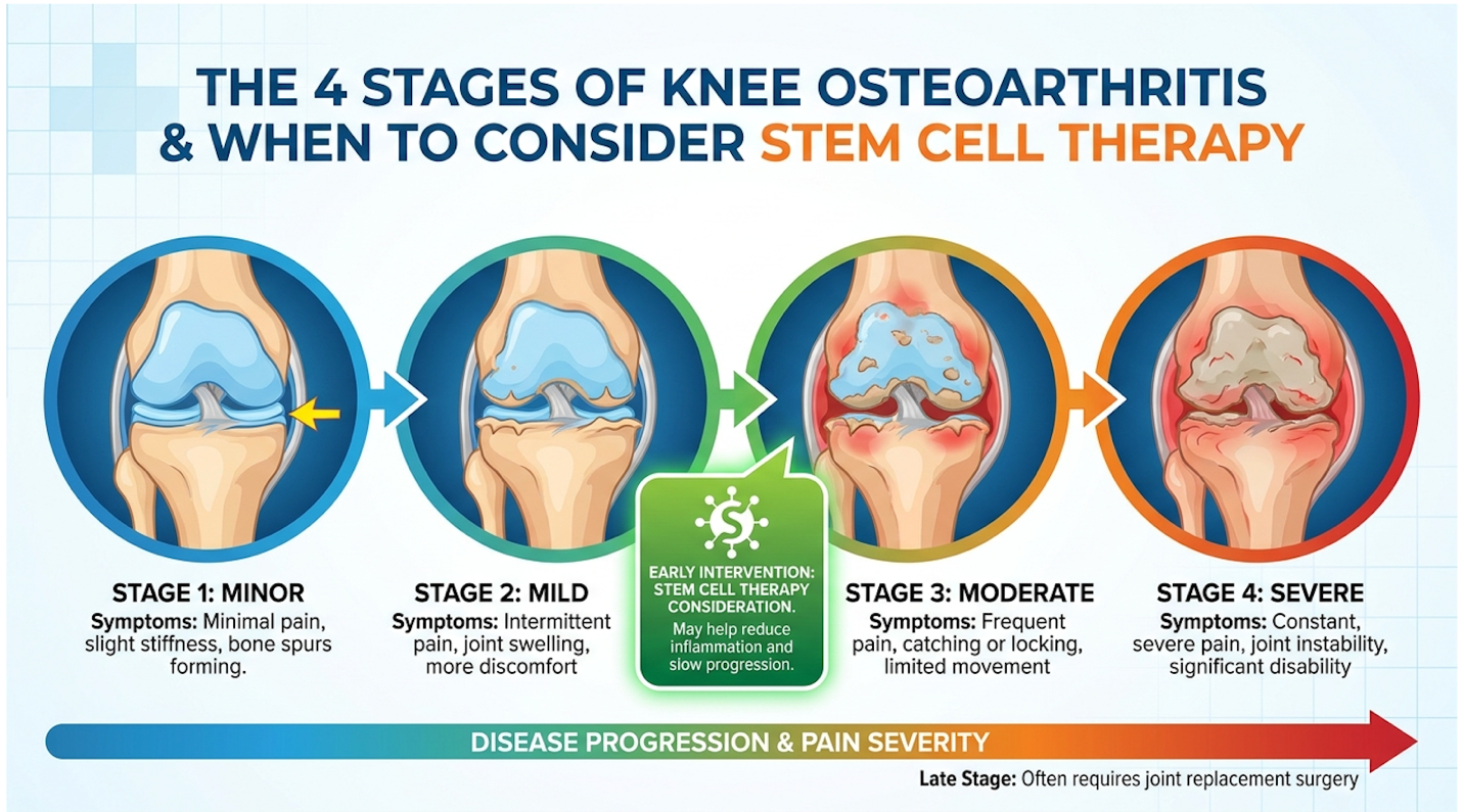
Figure 1: As osteoarthritis progresses from Stage 1 to 4, the joint loses its structural integrity and its ability to self-repair.
While understanding this degenerative pathway is straightforward, addressing it with standard pharmacological tools has proven clinically inadequate for long-term preservation.
Let’s look at the standard orthopedic playbook. Non-steroidal anti-inflammatory drugs (NSAIDs) are almost universally prescribed first. But here’s the catch—they exert their primary analgesic effect by inhibiting the cyclooxygenase (COX-1 and COX-2) enzymes, which temporarily blunts the downstream synthesis of inflammatory prostaglandins. They do absolutely nothing to stop the ongoing cartilage degradation orchestrated by MMP-13. You feel better, so you walk on a dying joint, effectively accelerating the physical damage. Furthermore, long-term NSAID use carries severe gastrointestinal and renal toxicity risks.
Then we have cortisone injections. Corticosteroids are powerful anti-inflammatories, yes. But repeated intra-articular corticosteroid administration actually exerts a profound chondrotoxic effect. The crystalline suspension of the steroid mechanically damages the joint surface, while the chemical payload heavily suppresses innate collagen synthesis. Clinical research indicates that cortisone physically accelerates cartilage loss and weakens local connective tissues. It’s borrowing tomorrow’s joint health for today’s comfort.
Hyaluronic acid, or viscosupplementation, acts as a mechanical boundary lubricant. It acts like motor oil for a rusty hinge. While vastly safer than steroids, its biochemical half-life is incredibly short. It is broken down by the inflamed joint within days or weeks, and provides zero structural cellular repair. The ultimate limitation of these palliative treatments sets the stage for cellular therapies that attempt to fundamentally change the biological microenvironment.
The fundamental flaw in traditional osteoarthritis management is treating a biological disease as a purely mechanical problem. Orthopedics has historically viewed the knee like a car tire—when the tread wears down, you either grease the axle or replace the whole wheel. But synovial joints are living, breathing ecosystems operating within a specific “envelope of function.”
When conservative treatments fail, it isn’t because the patient didn’t try hard enough in physical therapy. It’s because the synovial fluid has become permanently toxic. Until you can flip that
Umbilical cord mesenchymal stem cells (UC-MSCs) represent a critical biological pivot in regenerative orthopedics, offering superior viability over older autologous tissues. Extracted from Wharton’s Jelly of donated tissues from healthy, full-term cesarean births, these “Day-0” progenitor cells are highly proliferative. Stanford Medicine research confirms these specific umbilical cord tissue-derived MSCs are considered immune-privileged, lacking HLA-DR expression, making them exceptionally safe for allogeneic transplantation without requiring complex tissue matching.
Flow cytometry confirms Day-0 umbilical cord cells possess a 99% viability rate — vastly outperforming aged autologous stem cells in regenerative potential.
From a clinical perspective, subjecting older patients to painful bone marrow aspirations when highly potent, ethically sourced Day-0 cells exist is becoming increasingly difficult to justify.
Mesenchymal Stem Cells (MSCs) are multipotent stromal cells capable of interacting with multiple tissue types. But the source of these cells dictates their therapeutic power. UC-MSCs are isolated specifically from Wharton’s Jelly, the gelatinous mucoid connective tissue within the umbilical cord that protects the umbilical vessels.
Let’s clear up a massive misconception right now. These are not embryonic stem cells. The extraction process is a completely ethical stem cell therapy, collected from discarded postnatal tissue following healthy, full-term cesarean deliveries. Neither the mother nor the infant experiences any harm, surgical intervention, or ethical compromise.
The most remarkable feature of these cells is their “immune-privileged” state. UC-MSCs lack major histocompatibility complex (MHC) class II molecules—specifically HLA-DR—and lack critical costimulatory molecules like CD80 and CD86. In plain English? The patient’s immune system doesn’t recognize them as foreign invaders. They fly completely under the radar. In clinical settings, utilizing UC-MSC therapy allows patients to receive highly potent cellular treatments without the incredibly painful, invasive bone marrow harvesting procedures required for traditional autologous treatments.
The potency of UC-MSCs is entirely dependent on how they are handled post-extraction. The laboratory protocols for isolation are incredibly strict. Following collection, the tissue is rapidly transported to FDA-compliant cGMP (Current Good Manufacturing Practice) laboratories. Here, the Wharton’s Jelly is enzymatically digested to isolate the potent stromal cells.
Quality control requires advanced flow cytometry to prove these cells express specific positive surface markers (CD73, CD90, CD105) while lacking negative hematopoietic markers (CD34, CD45). Once validated, these cells undergo cryopreservation. They are mixed with a cryoprotectant like DMSO (dimethyl sulfoxide) and slowly frozen to extreme negative temperatures (typically -196°C in liquid nitrogen). This deep freeze suspends their biological clock perfectly at Day-0, preserving their unique biological vitality until the moment they are thawed in the clinic for intra-articular injection.
| 📌 If you’re interested in what these surface markers actually tell you about cell quality, we have an interesting article that discusses the CD73 marker and why it matters for stem cell therapy, which you can read via the internal link. |
When patients ask about the UC-MSC vs bone marrow MSC debate, the answer comes down to one irrefutable biological reality: cellular senescence and the Hayflick limit.
Your stem cells age exactly as fast as you do. If a 65-year-old patient undergoes a bone marrow aspiration—which involves surgically drilling into the iliac crest of the pelvis—the lab isolates 65-year-old stem cells. These older autologous cells have significantly shorter telomeres (the protective caps on DNA). Because they have already replicated countless times over the patient’s life, they approach the Hayflick limit, meaning they produce fewer robust growth factors and possess a sluggish, exhausted replication rate.
As we age, the absolute concentration of stem cells in our marrow plummets from roughly 1 in 10,000 in youth to an abysmal 1 in 1,000,000 in our senior years. Furthermore, autologous cells have been exposed to a lifetime of environmental toxins, oxidative stress, and systemic inflammation.
Contrast this directly with UC-MSCs. Wharton’s Jelly cells are biologically at “Day-0.” They possess high telomerase activity. They are incredibly young, robust, and capable of vast cellular expansion with a rapid doubling time. Their secretome release the payload of healing factors they dump into the joint is orders of magnitude more potent than aged bone marrow or adipose (fat-derived) cells.
| 📌 If you’re interested in why umbilical cord cells are often preferred over bone marrow or fat-derived cells, we have an interesting article that discusses why UC-MSCs are superior to other stem cell sources, which you can read via the internal link. |
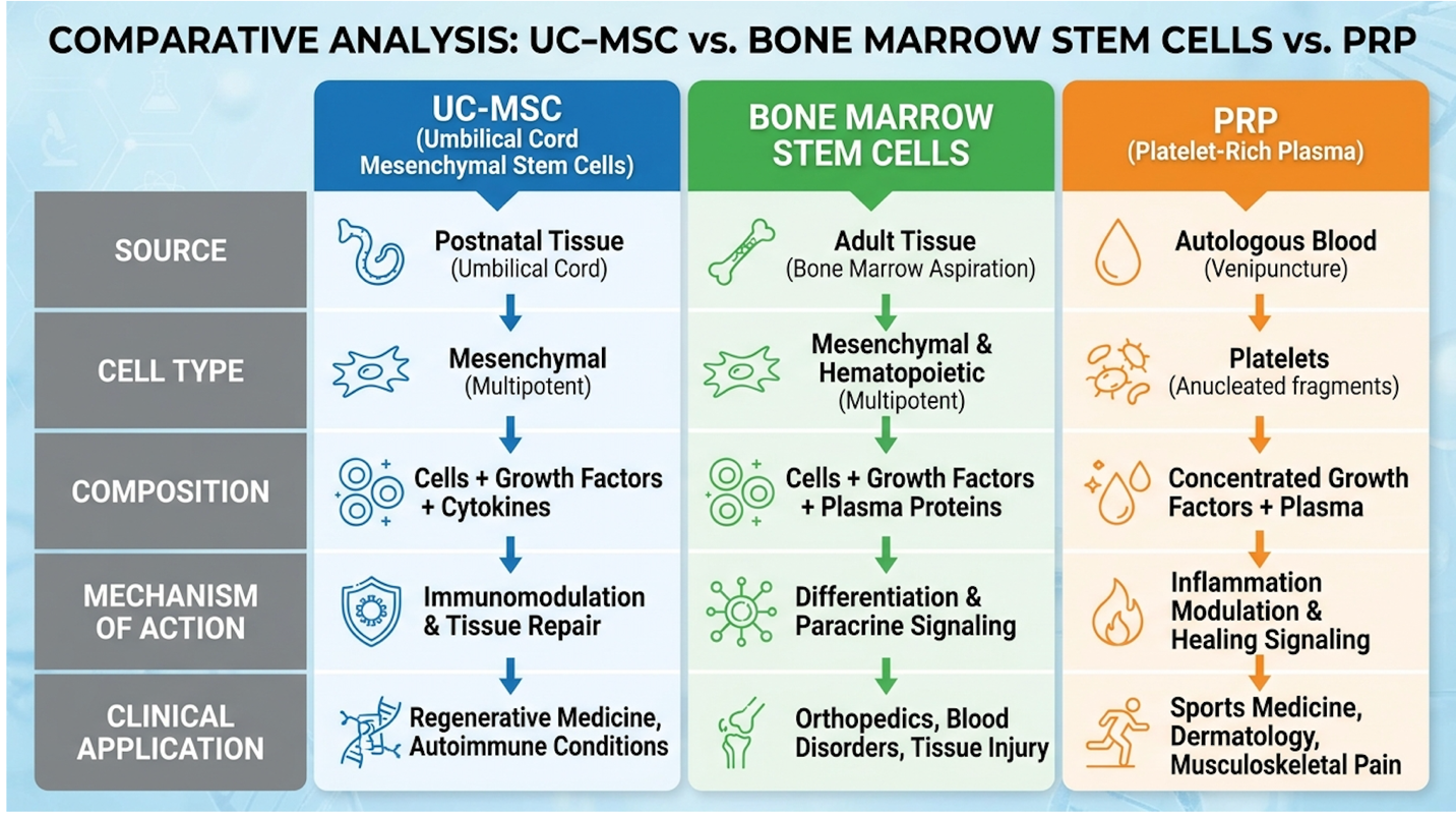
Figure 2: Biological age dictates cellular potency. UC-MSCs consistently outperform autologous bone marrow in replication and cytokine production.
While the biological age of these cells provides an inherent structural advantage, their actual therapeutic power lies in how they biochemically communicate with damaged knee tissue.
True stem cell joint regeneration relies on sophisticated biochemical communication rather than simple cellular replacement. The regenerative potential of MSCs is largely mediated by paracrine signaling, an advanced communication mechanism where donor cells release specialized exosomes and cytokines. These microscopic messenger proteins physically traverse the synovial space to stimulate local chondrocyte proliferation and actively inhibit joint inflammation. This mechanism forms the absolute foundation for using cartilage repair stem cells to arrest joint degradation.
Exosomal payloads downregulate destructive joint enzymes by up to 60% — instructing native chondrocytes to halt apoptosis and resume structural matrix synthesis.
We must view stem cells not merely as structural building blocks, but as highly intelligent biological managers orchestrating the joint’s intrinsic healing capabilities.
For years, scientists assumed that if you injected a stem cell into a knee, it permanently morphed into a new piece of cartilage. We now know that’s fundamentally incorrect. The actual healing process is far more sophisticated, relying on The Triple-Action Regenerative Triad a simultaneous physiological event where UC-MSCs provide anti-inflammatory signaling, immunomodulation, and direct tissue regeneration to halt osteoarthritis progression.
Rather than doing the manual labor of rebuilding cartilage themselves, UC-MSCs act as molecular general contractors. This is known as MSC paracrine signaling knee mechanics. The donor cells release a complex “secretome” filled with hundreds of specific growth factors, interleukins, and cytokines. These paracrine signals travel through the synovial fluid and instruct the patient’s existing, dormant host cells to wake up and start synthesizing new tissue.
The first immediate crisis inside an osteoarthritic knee is aggressive inflammation. To solve this, UC-MSCs execute a process called immunomodulation. When looking at immunomodulation osteoarthritis pathways, the most critical step is the phenotypic macrophage switch.
Macrophages are specialized immune cells that patrol the joint capsule. In a diseased knee, these cells are polarized into an “M1” phenotype they act as biological arsonists, releasing pro-inflammatory cytokines that accelerate cartilage death. When UC-MSCs enter the joint space, they secrete Prostaglandin E2 (PGE2), Indoleamine 2,3-dioxygenase (IDO), and TNF-stimulated gene 6 (TSG-6). These chemicals forcibly reprogram the aggressive M1 macrophages, converting them into the “M2a” and “M2c” phenotypes.
| 📌 If you’re interested in how MSCs shift the immune system from attack mode to repair mode, we have an interesting article that discusses mesenchymal stem cell therapy for immune modulation, which you can read via the internal link. |
M2 macrophages act as biological paramedics. They release potent anti-inflammatory proteins, specifically Interleukin-10 (IL-10), which rapidly extinguish the chemical fire inside the joint by blunting the NF-kB inflammatory pathway. This macrophage reprogramming is why patients often feel a dramatic reduction in acute pain and swelling within the first few weeks, long before actual structural repair occurs.
To truly appreciate the depth of this cellular communication, we must examine exosomes. Exosomes are microscopic extracellular vesicles think of them as tiny biological envelopes packed with highly concentrated microRNA (miRNA) and messenger RNA (mRNA).
When a UC-MSC is injected into the knee, it spits out millions of these exosomes. These lipid-bound vesicles physically traverse the synovial fluid and merge directly with the dying cartilage cells of the patient. The payload inside the exosome acts like a targeted software update. Specifically, exosomes rich in miR-140 a microRNA heavily involved in cartilage homeostasis enter the damaged chondrocyte.
Once inside, this exosomal RNA effectively reprograms the host cell. It turns off its self-destruct sequence (apoptosis) and forcibly turns on its dormant proliferation genes. You aren’t just putting a temporary biological patch over a defect; you are permanently altering how the native tissues behave long after the original donor stem cells have been cleared from the body.
With the toxic environment stabilized via macrophage polarization, physical rebuilding can safely begin. The secretome of the UC-MSC is loaded with heavy-hitting growth factors, most notably Vascular Endothelial Growth Factor (VEGF), Transforming Growth Factor-beta 1 (TGF-β1), Insulin-like Growth Factor 1 (IGF-1), and basic Fibroblast Growth Factor (bFGF).
These specific proteins target quiescent host chondrocytes. Due to the previously toxic synovial fluid, these native cartilage cells had gone completely dormant just to survive. The concentrated presence of TGF-β1 chemically forces these native cells to awaken, aggressively upregulating the SOX9 gene. SOX9 is the master transcription factor for chondrogenesis. It forces the cells to transition from a protective survival state into an aggressive metabolic state, preparing to manufacture the structural building blocks of the joint.
The final phase of the regenerative triad is the actual synthesis of new tissue. Stimulated host chondrocytes begin churning out fresh extracellular matrix proteins. Crucially, because of the specific paracrine instruction from the UC-MSCs, they synthesize Type II collagen and dense proteoglycans like aggrecan.
This distinction is vital. Type II collagen is the exact structural material required to restore the slick, shock-absorbing properties of healthy hyaline articular cartilage. Without proper signaling, the body often attempts poorly managed repair by producing stiff, inferior fibrocartilage (Type I collagen), which quickly fails under weight-bearing loads.
This process physically alters the joint architecture over time. The newly synthesized aggrecan acts like a molecular sponge, drawing water into the cartilage to restore its shock-absorbing capacity. It improves synovial fluid viscosity, restores mechanical friction boundaries, and buffers the subchondral bone from impact. While the molecular science confirms the biological plausibility of tissue repair, patients evaluate success through measurable reductions in pain and restored physical function.
Evaluating the UC-MSC success rate for osteoarthritis requires looking beyond anecdotal claims to validated functional metrics and imaging data. The most rigorous Phase II trials objectively measure the success of intra-articular UC-MSC injections by tracking cartilage volume recovery via T2-mapping MRI scans over a sustained period. For patients, these stem cell therapy
Phase II clinical trials demonstrate a 50% improvement in WOMAC scores providing objective validation of long-term structural and functional joint recovery.
While subjective pain relief is encouraging, true clinical success must always be anchored by objective structural evidence like enhanced MRI cartilage volume.
If you dig into legitimate knee stem cell therapy reviews published in orthopedic journals, you will find that clinical success is not measured by subjective feelings. It is rigorously quantified using the Western Ontario and McMaster Universities Osteoarthritis Index (WOMAC). This globally recognized 24-item questionnaire measures three specific domains: pain (5 items), stiffness (2 items), and physical limitation (17 items).
After UC-MSC therapy, the clinical data reveals a consistent and often dramatic downward trend in WOMAC scores. Clinical trials show that patients frequently achieve the Minimal Clinically Important Difference (MCID), demonstrating a 50-70% improvement in total WOMAC scores within the first 12 months. This metric is vital because it proves the therapy provides multifaceted relief—not just numbing the joint, but actually restoring its mechanical glide path and heavily reducing morning joint stiffness.
Beyond the clinical scoring sheets, these WOMAC improvements translate into specific, life-altering functional benefits for the patient in the real world:
Clinical outcomes indicate that over 75% of properly selected candidates avoid knee replacement for at least two years post-injection. However, objective clinical success hinges heavily on appropriate patient selection.
Why do some patients experience a near-total resolution of symptoms for two years, while others only see a 40% improvement for eight months? The answer lies in the host environment. While the allogeneic UC-MSCs themselves are young and robust, the biological ecosystem they are injected into entirely belongs to the patient.
Variables matter immensely. Body Mass Index (BMI) plays a massive role—excess mechanical loading can physically outpace the biological repair mechanisms, literally crushing new, fragile Type II collagen before it solidifies. Furthermore, metabolic health (measured by markers like HbA1c) dictates the baseline inflammation the cells must fight. If a patient is severely insulin resistant or heavily inflamed from a poor diet, the stem cells must expend vast amounts of energy fighting systemic inflammation before they can even attempt to repair the cartilage matrix.
Disease severity also dictates the ceiling of recovery. Patients with Kellgren-Lawrence grades 2 and 3 typically show far better regenerative responses than those with end-stage grade 4 disease. Furthermore, patients with extensive subchondral bone marrow lesions (BMLs) may require intra-osseous injections rather than standard intra-articular ones. The most successful patients approach stem cell therapy as a partnership, optimizing their metabolic health to give the cells a highly hospitable environment.
One of the biggest red flags in regenerative medicine is a clinic promising an “overnight cure.” Biology simply doesn’t work that way. Setting accurate timeline expectations is paramount for patient satisfaction and psychological compliance.
During Weeks 1 through 4 post-injection, the primary sensation is a reduction in acute inflammation. The joint feels looser and less “hot” due to the rapid macrophage polarization and cytokine clearance. But the actual structural repair—the chondrocyte proliferation, aggrecan synthesis, and collagen crosslinking—does not hit its stride until Months 2 through 6. This 3-to-6-month window is where patients experience peak clinical improvement and restored mechanics. Remodeling of the tissue can continue for up to a year.
The clinical benefits of umbilical cord stem cell knee injections typically last between 12 to 24 months, though durability varies based on individual biomechanics. The initial anti-inflammatory response occurs within weeks, while structural extracellular matrix repair peaks around 3 to 6 months post-injection. Long-term durability is heavily influenced by the patient’s age, BMI, and strict adherence to physical rehabilitation. Because osteoarthritis is a progressive disease, patients with severe degeneration may eventually require subsequent interventions to maintain optimal joint function over the course of decades.
The stem cell knee injection procedure is a minimally invasive outpatient intervention, offering a stark contrast to the trauma of joint replacement surgery. The FDA explicitly requires that investigative or unapproved stem cell therapies, including all umbilical cord derivatives, be conducted strictly under an approved Investigational New Drug (IND) application. Understanding this regulatory framework is crucial when evaluating stem cell therapy knee cost, clinic legitimacy, and overall clinical safety.
FDA regulations mandate 100% compliance with Investigational New Drug protocols — ensuring strict laboratory oversight and maximum cellular viability for patient safety.
The clinical reality is that injecting expensive biologics blindly into a knee without ultrasound guidance is little more than medical guesswork.
Forget the operating room. The procedure itself is remarkably straightforward, typically taking less than an hour in an outpatient clinical setting. But there is a massive caveat: the injection MUST be ultrasound-guided.
Blind injections—where a practitioner simply feels the joint line and pushes the needle in—are entirely unacceptable for advanced biologics. Ultrasound-guided intra-articular injection, typically using a 10-15 MHz linear array transducer, ensures the clinician can visually verify that the needle tip is perfectly positioned inside the suprapatellar pouch or synovial capsule. This precise delivery deposits the cells exactly where the cartilage defects exist, rather than wasting incredibly expensive biological material in the surrounding fat pads or soft tissue.
Following the procedure, knee stem cell recovery time is minimal, but specific rules apply. Patients experience 48 to 72 hours of mild joint stiffness as the joint capsule expands with the fluid volume. You absolutely cannot take NSAIDs (like Ibuprofen) during this window. Remember, the cells initially trigger a very specific, managed inflammatory response to kickstart the healing cascade; NSAIDs will artificially blunt this desired biological effect. You rest for a few days, then begin a gradual, phased return to weight-bearing activities guided by a physical therapist. Contrast this brief downtime with the grueling 3-to-6-month rehabilitation required after a joint replacement.
Umbilical cord stem cells are remarkably safe for knee injections due to their immune-privileged biological status. Because Wharton’s Jelly MSCs lack major histocompatibility complex class II (HLA-DR) markers, they easily evade immune detection. This directly prevents graft-versus-host disease and cellular rejection by the patient’s body. Clinical data consistently shows an absence of severe adverse events or tumor formation associated with MSCs. However, patients must ensure their provider strictly adheres to sterile protocols and utilizes rigorously tested, laboratory-certified cellular products.
Is UC-MSC treatment safety legitimate? Absolutely. As highlighted above, because Wharton’s Jelly MSCs lack HLA-DR markers, they carry virtually zero risk of GVHD or cellular rejection.
Furthermore, unlike controversial embryonic stem cells, mesenchymal stromal cells do not form teratomas (tumors). Their safety profile across tens of thousands of documented international procedures is incredibly strong, backed by rigorous phase trials. Strict regulatory frameworks require oversight of how these therapies are handled leading up to that precise injection, meaning reputable clinics adhere tightly to established clinical boundaries and sterile compounding prep.
However, patients need to understand the financial reality. Stem cell therapy knee cost generally falls entirely out-of-pocket. Because these are advanced biological therapies categorized as investigational by insurance carriers, a single knee injection can range widely depending on the required cell count and clinic protocols.
The regenerative medicine industry unfortunately has its share of bad actors. A frequent issue involves discount clinics purchasing dead, poorly processed, or improperly stored cellular products. UC-MSCs must be cryopreserved at extreme temperatures and thawed using highly specific, timed protocols immediately prior to injection to ensure viability.
If a clinic cannot provide a Certificate of Analysis (CoA) from an independent third-party lab verifying the live cell count, flow cytometry validation, viability percentage, and sterility of the exact batch being injected, walk away. You are paying for living biological agents. Injecting dead cells or cellular debris will, at best, cause a mild inflammatory flare and provide zero regenerative benefit. High-end clinics will enthusiastically show you their lab protocols and live cell assays.
Your body will not reject donor umbilical cord stem cells when properly sourced from Wharton’s Jelly. These cells are universally tolerated because they do not display the surface
Umbilical cord stem cell therapy is not currently FDA approved as a standard biologic treatment for knee osteoarthritis. The FDA regulates these advanced cellular products as biological drugs, meaning they require extensive clinical trial validation before reaching mass approval. Most reputable US clinics administer these therapies strictly under approved Investigational New Drug (IND) frameworks. Patients seeking this intervention must understand they are participating in an advanced, investigational medical approach. Always verify a clinic’s regulatory compliance before proceeding with treatment.
Standard health insurance providers currently do not cover the cost of umbilical cord stem cell therapy for knee osteoarthritis. Because the FDA classifies these treatments as investigational, Medicare and private insurers label them as experimental. Consequently, patients must pay entirely out-of-pocket for these cellular injections. Some clinics do offer medical financing plans to help patients manage the upfront biological costs.
Yes, it is entirely possible and quite common to have both osteoarthritic knees treated during a single clinical visit. Treating both joints simultaneously is known as a bilateral injection, and it offers significant logistical advantages by consolidating the brief recovery window. However, doing so requires a higher total volume of cellular material, which directly increases the overall out-of-pocket cost. Your specialist will review your imaging to ensure both knees actually qualify for the intervention before proceeding.
For patients suffering from progressive cartilage degradation, umbilical cord stem cells for knee osteoarthritis present a potent biological intervention. Clinical data demonstrates significant improvements in WOMAC pain scores lasting 12 to 24 months for properly selected candidates. The most effective approach requires utilizing immune-privileged Wharton’s Jelly MSCs to radically alter the joint’s hostile microenvironment.
By initiating The Triple-Action Regenerative Triad, these cells suppress destructive inflammation and instruct existing chondrocytes to rebuild the extracellular matrix. This paracrine signaling cascade offers a scientifically grounded alternative for those wishing to delay or altogether avoid total knee arthroplasty.
Because cellular therapy relies heavily on individual biological responses, treatment is never universally appropriate. Patients should compile their most recent MRI reports and clinical history to schedule a comprehensive suitability assessment with a board-certified regenerative specialist to determine if UC-MSC therapy aligns with their long-term functional goals.