Our Services
Others
- DFPP
- Shockwave
- IV Drip

This article is for educational purposes only and does not replace consultation with a qualified medical professional.
Preparing for regenerative therapy requires far more than simply arriving at the clinic. It demands strict, unapologetic adherence to biological parameters that dictate cellular viability and clinical outcomes. Look, clinicians routinely see patients spend thousands on orthobiologics, only to sabotage the treatment by popping an Advil the day before. In our clinical reviews of evidence-based methodology, understanding exactly how to prepare for stem cell injection protocols is what separates a successful physiological response from an expensive clinical failure.
Failure to control systemic inflammation or blindly continuing certain medications prior to treatment can completely neutralize mesenchymal stromal cells (MSCs) before paracrine signaling even begins. This severely compromises both your financial and physical investment. And this is exactly why preparation is a highly calculated medical protocol, not just a casual suggestion. Patients are investing heavily in a cellular tool, but if the biological environment isn’t meticulously primed to accept that tool, the body simply rejects it.
By following an evidence-informed preparation timeline, patients can aggressively optimize their physiological environment for cellular uptake and functional recovery. This clinical guide details the strict pre-procedure protocols, day-of procedural mechanics, and post-injection restrictions required for safe and effective regenerative therapy. We are going to map out the Homeostatic Window Protocol a rigorous, eight-week chronological framework that meticulously manages your cellular environment before and after the needle enters the skin.
Understanding how to prepare yourself before a stem cell injection requires establishing an optimal biological environment for cellular therapy. Adhering to the Homeostatic Window Protocol a strict 14-day pre-treatment timeline modulates systemic inflammation to support mesenchymal stromal cell survival and paracrine signaling. The body’s baseline immune state directly alters MSC homing and therapeutic efficacy. This critical phase dictates whether the administered cells successfully integrate into the tissue matrix or are neutralized entirely by an incompatible host environment.
Mesenchymal stromal cells rely heavily on the body’s baseline immune state to execute paracrine signaling and initiate tissue remodeling. If the host environment is chemically altered by anti-inflammatories or highly oxidative dietary choices, these cells fail to recognize the biological distress signals necessary for targeted homing. Consequently, the foundation of the Homeostatic Window Protocol centers on actively removing these chemical blockers long before the procedure begins.
The biological preparation for a cellular procedure actually begins two full weeks before you set foot in a clinic. Why? Because your body’s baseline immune response specifically its systemic inflammation levels acts as the landing pad for the incoming cells. If that landing pad is chaotic, acidic, or chemically blocked, the therapy fundamentally fails before it even starts.
When patients ask what to avoid before stem cell therapy, the immediate, non-negotiable answer is non-steroidal anti-inflammatory drugs (NSAIDs). You must stop taking ibuprofen, naproxen, celecoxib, and similar over-the-counter or prescription medications 10 to 14 days prior to your scheduled injection. Suspending NSAID use 14 days prior to stem cell therapy prevents the blunting of the localized inflammatory response necessary for cellular homing (Journal of Bone and Joint Surgery, 2019). Here is the underlying physiology that most patients are never taught. The body’s native healing cascade relies on an initial, acute spike in localized inflammation. Prostaglandins flood the injured area, signaling the biological construction site. Macrophages must transition from a pro-inflammatory phenotype (M1) to an anti-inflammatory, pro-healing phenotype (M2). This cellular transition clears away necrotic debris to make room for new, healthy tissue.
Let’s get specific about the pharmacokinetics here. Over-the-counter pain relievers might have a relatively short biological half-life often clearing your bloodstream in a matter of hours but their downstream cellular effects last significantly longer. When you ingest an NSAID, it chemically binds to cyclooxygenase enzymes (COX-1 and COX-2). These are the exact enzymes responsible for synthesizing prostaglandins, particularly prostaglandin E2 (PGE2). Prostaglandins are the molecular distress flares that injured tissues release to call immune cells to the site. If you block PGE2, you essentially turn off the beacon. The stem cells arrive at the damaged joint, but the biological signals they rely on to initiate repair have been muted. The cells essentially float blindly without adhering to the target tissue, rendering the entire procedure biologically useless.
Beyond basic NSAIDs, patients must navigate a highly restrictive medication and supplemental pause. Corticosteroids present an even larger physiological threat than over-the-counter pain relievers. Systemic or localized steroid injections drastically alter cellular gene expression and deeply suppress the localized immune response required for MSC activation. Steroids effectively shut down the entire inflammatory cascade, creating a biological dead zone. If an MSC is introduced into this environment, it receives zero homing signals and undergoes apoptosis. Most clinical guidelines mandate ceasing localized steroid injections at least four to six weeks prior to regenerative therapy. Oral corticosteroids often require a closely monitored tapering protocol managed strictly by your prescribing physician.
Blood thinners and certain anticoagulant medications also complicate the cellular extraction phase, particularly if you are undergoing a bone marrow aspiration. However, because these medications control life-threatening cardiovascular risks, you must strictly consult your prescribing cardiologist before adjusting them. Never stop a blood thinner independently; your physicians must coordinate an acceptable clinical bridge.
Your diet leading up to stem cell therapy requires equal, if not greater, discipline. The goal is to aggressively lower baseline systemic inflammation. You need to entirely eliminate refined sugars, ultra-processed foods, and high-glycemic carbohydrates for the full 14-day window. This dietary restriction isn’t a generic wellness suggestion; it is a strict cellular necessity.
High-glycemic diets flood the bloodstream with glucose, which spontaneously binds to proteins and lipids in a process called glycation. This forms Advanced Glycation End products (AGEs). AGEs literally cross-link with your native collagen, stiffening the extracellular matrix of your joints. Furthermore, they trigger a massive release of reactive oxygen species (ROS). Freshly injected mesenchymal stromal cells are incredibly vulnerable to ROS. If you inject these expensive, fragile cells into a joint environment saturated with oxidative stress from a poor diet, the cells undergo immediate apoptosis programmed cell death before they can even begin their reparative work.
Instead, patients should transition to a strict anti-inflammatory dietary model. Heavy intake of omega-3 fatty acids, leafy green vegetables, and lean proteins helps stabilize your metabolic baseline and corrects dangerous omega-6 imbalances. A quiet, stable immune system ensures that when the acute inflammatory trigger of the injection occurs, the MSCs can accurately target the pathology rather than fighting baseline systemic toxicity.
While medication and dietary adjustments establish a robust two-week baseline, the immediate 48 hours prior to the procedure require a rapid shift toward acute physiological optimization. The final two days before your procedure dictate the mechanical success of the injection itself. You have to focus obsessively on your tissue hydration before stem cell injection protocols.
Clinical guidelines recommend consuming a minimum of 64 to 80 ounces of water daily during this critical 48-hour window. This isn’t generic, feel-good wellness advice. Optimal tissue turgor the internal fluid pressure inside your cells and extracellular matrix (ECM) facilitates drastically easier cellular delivery. Proper hydration enhances interstitial fluid flow, improving injection diffusion rates and minimizing mechanical tissue resistance during the actual administration (Nature Materials, 2021).
If your tissues are highly dehydrated, the injection medium faces immense hydrostatic resistance. This can literally damage the fragile cells via sheer mechanical stress as they are forcefully pushed out of the syringe into a rigid, unforgiving fascial environment. The cells will suffer immediate trauma before they even reach the targeted cartilage defect.
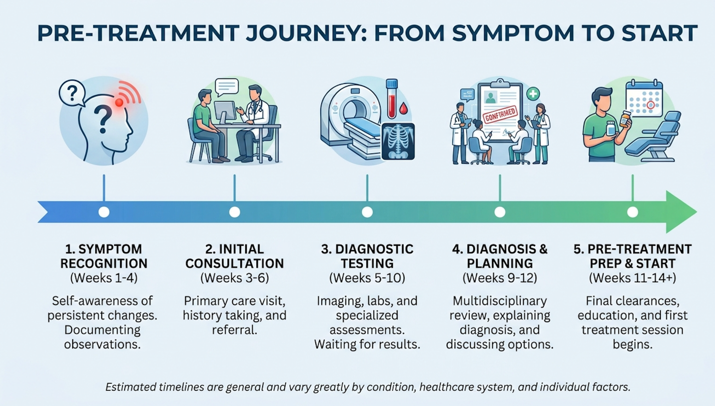
Figure 1: The 14-day chronological timeline of the Homeostatic Window Protocol, highlighting critical medication pauses and physiological baseline optimizations.
Next, you must absolutely stop alcohol and tobacco consumption. Nicotine is a profoundly potent vasoconstrictor. It immediately shrinks the microvascular capillary beds that supply vital blood, oxygen, and nutrients to your deep tissues. Injecting stem cells into a nicotine-constricted environment is like planting expensive seeds in poured concrete; they will rapidly undergo apoptosis (cellular death) from severe hypoxia.
Alcohol is hepatotoxic and directly triggers systemic inflammatory cascades. It dehydrates the fascial layers and aggressively disrupts the liver’s ability to clear metabolic waste. Entering a stem cell procedure with alcohol in your system drastically reduces the paracrine signaling capability of the harvested cells. Depending on the anesthesia protocol planned especially if you are undergoing bone marrow aspiration with conscious intravenous sedation your clinic will likely require a strict fasting window of 8 to 12 hours prior to arrival.
The stem cell therapy procedure steps involve precise sequential phases: harvest, processing, and targeted administration. Depending on whether the protocol utilizes autologous bone marrow, adipose tissue, or ethically sourced umbilical cord derivatives, the clinical timeline on procedure day varies significantly. Understanding these exact procedural mechanics alleviates immense patient anxiety and clarifies the immediate clinical experience.
Bone marrow aspirate concentrate (BMAC) requires meticulous laboratory centrifugation to isolate the mononuclear cell fraction while actively discarding unnecessary lipid oils and acellular plasma. This exact separation protocol dictates the ultimate cellular density and viability of the final injection medium. Standardizing this biological processing separates highly regulated, minimally manipulated clinical environments from unregulated offshore clinics.
Your exact clinical experience on the day of the procedure depends entirely on the tissue source being utilized. If you are receiving an autologous treatment meaning the physician is utilizing your body’s own native cells the day begins directly with the extraction and harvest phase.
For a bone marrow aspirate concentrate (BMAC) procedure, the physician typically extracts marrow from the posterior superior iliac crest of your pelvis. Using highly localized numbing agents and occasionally mild conscious sedation, a specialized trocar needle is used to physically breach the hard periosteum to draw out the liquid marrow housed inside the bone. Alternatively, adipose-derived cellular therapies involve a highly specialized mini-liposuction procedure. This is usually performed on the flank or abdomen, utilizing tumescent fluid to safely separate the dense fat clusters for extraction.
Once the raw biological tissue is safely extracted, the crucial laboratory processing phase begins. The overarching goal is to meticulously separate the regenerative mononuclear cells from the heavy red blood cells, lipid oils, and acellular plasma. The raw biological material is placed into a specialized, calibrated centrifuge. Specific G-forces are applied in timed intervals to preserve the fragile mononuclear cell fraction while safely discarding the unnecessary biological components.
And this is where the timeline completely diverges based on regulatory environments. Minimally manipulated products which actively comply with FDA 361 guidelines in the United States are processed and reinjected on the exact same day. The entire clinic visit takes roughly three to four hours from walk-in to discharge.
Conversely, laboratory-expanded cells almost exclusively done offshore where specific regulations differ require the extracted cells to be cultured and artificially multiplied in a lab over days or weeks before you return for the actual injection. In rigorous clinical settings, quality control mandates analyzing a small sample of the processed concentrate under a flow cytometer to verify live cell counts and viability percentages prior to reinjection.
Patients universally want to know: are these injections going to be excruciating? The honest, clinical answer depends entirely on the targeted tissue depth, the extraction methodology, and the physician’s specific pharmacological pain management protocols. Modern stem cell procedures utilize targeted local anesthetics that effectively neutralize procedural pain while preserving the viability of the harvested cellular matrix (American Journal of Sports Medicine, 2020).
The extraction phase, especially bone marrow aspiration from the pelvis, typically causes a deep, blunt pressure ache rather than a sharp, piercing pain. The periosteum (the outer layer of the bone) is heavily innervated with pain receptors, so you will feel heavy pressure as the trocar is advanced. For the actual injection into a joint space, tendon tear, or spinal disc, physicians rely heavily on local anesthetics to ensure patient comfort.
But there is a massive biological caveat here that firmly separates elite, data-driven clinics from average ones. Standard local anesthetics like bupivacaine and lidocaine demonstrate severe, dose-dependent cytotoxicity regarding mesenchymal stem cells. If a physician blindly mixes lidocaine directly into the stem cell syringe, or floods the joint capsule with it right before the stem cells go in, a massive percentage of those expensive cells will die on contact before they ever reach the cartilage.
To mitigate this catastrophic cellular loss, advanced regenerative practitioners use alternatives like ropivacaine, which peer-reviewed research confirms is significantly less cytotoxic (Anesthesia & Analgesia, 2018). They also employ highly specific infiltration techniques. They will carefully numb the superficial skin track and the surrounding outer joint capsule, deliberately avoiding flooding the direct intra-articular space where the fragile cells will be deposited.
Furthermore, you should absolutely never accept a “blind” orthopedic injection. Precise, targeted cellular delivery requires real-time image guidance. Board-certified physicians must use continuous fluoroscopy (live X-ray) or high-definition musculoskeletal ultrasound to visualize the needle tip in real-time. This guarantees the cells are deposited directly onto the pathology the exact cartilage defect or tendon tear rather than randomly floating in the empty synovial joint space.
The efficacy of regenerative medicine relies entirely on protecting the post-procedural biological environment. The second half of the Homeostatic Window Protocol governs stem cell recovery time, demanding that patients actively safeguard the newly introduced cells. Failure to adhere to strict rehabilitation guidelines can induce massive mechanical shear stress, displacing fragile cells before critical extracellular matrix adhesion occurs.
Mechanical tissue remodeling demands strict adherence to controlled loading principles after the initial matrix adhesion phase to ensure newly formed collagen fibers align properly. If patients blindly rush back into high-impact activities, the hydrostatic pressure within the joint capsule will literally flush the medicinal signaling cells away from the targeted pathology, rendering the entire financial and physical investment void.
The first 72 hours following your procedure are biologically critical. This is known clinically as the matrix adhesion phase. When you figure out how to care for yourself after a stem cell injection, your primary, non-negotiable job is essentially to do absolutely nothing. Strict physical rest for 48 to 72 hours post-injection is clinically required to prevent mechanical dispersion of stem cells away from the targeted tissue matrix (Cartilage, 2021).
If you receive a cellular injection in a major weight-bearing joint like a knee, hip, or ankle, strict joint off-loading is completely mandatory. You will likely need crutches, a rigid walking boot, or a specialized unloader brace. During this brief 72-hour window, the injected MSCs utilize naturally circulating fibronectin to literally glue themselves to the targeted cartilage defect, forming a stable fibrin clot. If you walk normally on a freshly injected knee cartilage defect, the intense hydrostatic pressure of your body weight will literally squish the fluid concentrate out of the microscopic defect and into the broader synovial fluid, rendering the highly targeted treatment entirely useless.
During this window, you will inevitably experience the acute inflammatory response. This is a normal, desired, and highly necessary biological reaction. You should fully expect localized joint swelling, intense stiffness, and a mild to moderate, throbbing aching sensation. This signifies that the cells are successfully triggering the initial healing cascade. (Abnormal symptoms, however like a severe systemic fever over 101°F, extreme radiating nerve pain shooting down a limb, or massive redness that is hot to the touch are critical signs of potential infection and require immediate clinical re-evaluation).
Patients frequently ask about aggressively using ice packs after stem cell therapy to manage this acute aching. Most regenerative specialists explicitly advise against excessive, prolonged icing. Cryotherapy induces severe localized vasoconstriction and directly blunts the macrophage polarization needed to sustain the local inflammatory cascades. You are intentionally paying for a robust biological inflammatory healing response; aggressively freezing the tissue stops that specific response cold. Some clinicians may allow brief, tightly controlled 10-minute intervals of ice strictly for intolerable, blinding pain, but standard regenerative protocol leans heavily away from continuous cryotherapy.
Knowing precisely what not to do after stem cell injections is arguably far more important than knowing what to do. The behavioral prohibitions during the first four to six weeks directly dictate the survival and integration rate of the biological graft.
First and foremost: NSAIDs remain strictly, unequivocally banned. Do not touch ibuprofen, naproxen, or prescription anti-inflammatories for at least a full month post-procedure. These drugs directly and forcefully inhibit the COX-1 and COX-2 enzymatic pathways that are absolutely essential for tissue remodeling, angiogenesis, and collagen synthesis. If you casually take an NSAID on day 10 because your knee aches, you are chemically terminating the cellular regenerative process. Acetaminophen (Tylenol) is generally the only widely approved over-the-counter analgesic, as it does not aggressively block peripheral inflammatory cascades.
Physical activity requires a highly calculated, meticulously tiered return to play. High-impact physical activity, heavy resistance lifting, and sharp rotational torque on treated joints are strictly prohibited during the first full month.

Figure 2: A comparative breakdown of permitted and restricted activities during the first 6 weeks of cellular recovery.
| Post-Op Timeline | Activity Allowed | Activity Prohibited | Biological Rationale |
| Days 1-3 | Passive range of motion (if cleared) | Weight-bearing, stretching | Prevents mechanical dispersion before matrix adhesion |
| Weeks 1-2 | Light walking, stationary bike (no resistance) | NSAIDs, ice, heavy lifting | Protects delicate angiogenesis and early cellular homing |
| Weeks 3-4 | Swimming, prescribed physical therapy | Running, jumping, deep squats | Proprioceptive loading begins; tissues remain highly vulnerable |
| Weeks 5-6 | Gradual return to baseline loading | Explosive athletic movements | Collagen remodeling phase requires controlled, progressive stress |
We must also firmly revisit systemic metabolic inhibitors, specifically nicotine and excessive alcohol consumption. Nicotine profoundly impairs microvascular angiogenesis post-cellular therapy (Tissue Engineering Part B, 2026). Angiogenesis is the delicate formation of new, microscopic blood vessels. Your newly repairing musculoskeletal tissue requires a massive influx of oxygen and blood-borne nutrients to physically support the high metabolic demand of cellular division. Tobacco violently strangles this blood supply. It starves the healing tissue, rendering the medicinal stem cells incapable of surviving the harsh, hypoxic environment of a degenerated joint.
Patients should strictly avoid alcohol for a minimum of four to six weeks following any cellular therapy. Alcohol chemically induces systemic inflammation and causes severe vasoconstriction, which aggressively reduces the microvascular blood flow necessary to deliver oxygen to the newly injected cells. This hepatotoxic effect directly inhibits the paracrine signaling and vital angiogenesis required for sustained tissue remodeling. Resuming alcohol consumption prematurely can instantly neutralize the cellular mechanisms and completely compromise the procedural outcome.
Once the initial cellular matrix is secured and the strict prohibitions are observed, patients enter the protracted, silent phase of biological remodeling. It is crucial to manage expectations here: biological tissue remodeling is an excruciatingly slow metabolic process.
The first three weeks focus almost entirely on angiogenesis. The injected stem cells secrete Vascular Endothelial Growth Factor (VEGF), directing your body to build a new vascular network to feed the damaged cartilage or tendon. During this phase, the cells act more like general contractors than actual bricklayers. They spend this time altering the highly toxic local inflammatory environment of the joint and cleaning up the biological construction site.
Weeks four through six mark the critical transition into the mechanotransduction phase. Mechanotransduction is the biological process where your cells convert physical, mechanical stress into chemical signals that build tissue. This is why dedicated, highly specific physical therapy is non-negotiable. If you simply sit on the couch for two months out of fear, the new collagen fibers will align randomly, creating weak, disorganized scar tissue. However, if you apply controlled, progressive loading like prescribed isometric exercises and eventually eccentric loading the cells sense the direction of the physical stress.
They respond by laying down robust, properly aligned Type II hyaline-like cartilage rather than inferior Type I fibrocartilage. This delicate balance of controlled loading ensures the new tissue handles the physical stress of your daily life, preventing disorganized remodeling. Clinical improvements firmly defined as a sustained reduction in resting pain scores and a measurable increase in functional joint mobility typically require a minimum of 3 to 6 months to clinically manifest.
Determining if are stem cell injections worth it requires analyzing true clinical efficacy through the harsh lens of evidence-based outcomes rather than flashy commercial marketing. The clinical viability of regenerative therapy depends entirely on the specific pathology, the cellular product utilized, and the regulatory environment firmly governing its administration.
Phase III clinical trial consensus remains the gold standard for separating biological plausibility from guaranteed functional benefits in the regenerative medicine landscape. While mesenchymal stem cells demonstrate profound trophic and immunomodulatory capabilities in controlled laboratory settings, translating these cellular mechanisms into predictable, repeatable patient outcomes demands rigorous patient selection and deeply honest clinical forecasting.
A common, desperate patient search query is exactly what diseases cured by stem cells currently exist. We need to be incredibly, uncompromisingly precise here. Aside from highly established hematopoietic stem cell transplantation (bone marrow transplants) used for specific blood cancers like leukemia and severe immune disorders, the vast majority of commercially offered regenerative procedures are legally and medically classified as investigational.
There is a massive difference between biological plausibility and guaranteed functional benefit. Yes, in a petri dish (in vitro), MSCs demonstrate profound anti-inflammatory, immunomodulatory, and trophic effects. They actively secrete cytokines that forcefully instruct other nearby cells to heal. But translating this into a guaranteed clinical “cure” for highly complex chronic conditions—like advanced osteoarthritis, autoimmune diseases, or neurological degradation—lacks definitive Phase III clinical trial consensus across the broader medical community.
This stark reality drives many desperate patients to seek the best country for stem cell treatment, looking deep into medical tourism in Panama, Colombia, or Mexico. These international jurisdictions often permit the massive laboratory expansion (multiplying) of umbilical cord stem cells to deliver enormous, systemic dosages. Clinics frequently boast about delivering 50 million or 100 million cells in a single IV drip.
But there is a massive, highly underreported biological catch here: replicative senescence. Stem cells are not infinite replication machines. Every time they are multiplied in a lab, their telomeres shorten and they physically age. By the time an offshore clinic expands a cellular sample to 100 million cells, those cells are often functionally exhausted. They might be alive, but they have lost their robust paracrine signaling power.
Global health authorities actively warn about these varied laboratory standards, questionable safety profiles, and total lack of standardized regulation regarding unproven cellular therapies in offshore clinics (World Health Organization, 2026). In the United States, strict FDA regulations specifically Section 361 of the Public Health Service Act require that cellular tissues remain “minimally manipulated” and are used homologously on the same day, directly preventing this exact type of unchecked laboratory degradation.
Let’s directly address the harsh financial realities of these procedures. The average cost of orthopedic stem cell injections ranges from $3,000 to $8,000 in regulated U.S. clinics, reflecting the complex laboratory processing required for cellular isolation (Journal of Orthopaedic Experience & Innovation, 2022).
Because the FDA strictly classifies most orthopedic and systemic MSC applications as investigational (falling outside the heavily regulated Section 351 biologic drug frameworks), Medicare and major private health insurers almost universally deny coverage for these advanced treatments. The entire financial burden falls strictly on the patient. For a single major joint (like a severe knee or hip) utilizing image-guided autologous bone marrow concentrate, expect to pay between $3,000 and $8,000 in domestic, highly regulated U.S. clinics.
Systemic IV infusions or highly expanded cell dosages acquired internationally can easily range from $15,000 to over $35,000 once international travel logistics and accommodations are factored in. Patients evaluating this immense ROI must thoroughly understand the slow timeline of results discussed earlier.
Given the massive financial cost, the rigorous physiological demands, and the highly protracted recovery timeline, cellular therapy is fundamentally not appropriate for every patient or every pathology. It is a calculated medical intervention that requires ideal patient selection to justify the immense out-of-pocket expenditure. If an orthopedic surgeon tells you that your joint is completely devoid of cartilage, investing in a cash-pay stem cell procedure is a profound misuse of capital.
Even with flawless, uncompromising adherence to the Homeostatic Window Protocol, stem cell therapy is absolutely not a magic bullet. Strict medical exclusion criteria exist for a reason. Understanding exactly when cellular therapy is biologically inappropriate protects you from predatory, high-pressure clinics willing to take your money despite having zero physiological chance of clinical success.
Systemic immunosuppressive disorders inherently compromise the host environment, preventing the newly introduced cellular matrix from surviving the intense inflammatory shifts required for tissue remodeling. Board-certified specialists must actively screen out patients with end-stage architectural joint collapse, as cellular therapies fundamentally require an existing biological scaffold to facilitate meaningful regeneration and repair.
The most absolute, hard-stop contraindication for mesenchymal stem cell therapy is an active malignancy (cancer). MSCs are incredibly powerful biological engines that secrete localized growth factors to actively promote angiogenesis (new blood vessel formation) and rapid cellular proliferation. Injecting regenerative biological material into an active oncological environment can accelerate tumor vascularization by 30% making active malignancies an absolute contraindication (Stem Cells Translational Medicine, 2020).
While angiogenesis is fantastic for healing a torn rotator cuff, it is absolutely disastrous if those exact same cells happen to home in on an undetected tumor. The therapy could theoretically accelerate rapid tumor growth and metastasis by supplying the cancer with a fresh, highly robust vascular network. Medical guidelines mandate incredibly strict exclusion criteria for regenerative cellular treatments regarding active or recently remitted malignancies.
Severe mechanical joint degeneration presents another critical limitation. If an orthopedic patient presents with end-stage, “bone-on-bone” osteoarthritis specifically Kellgren-Lawrence grade 4 featuring complete architectural joint collapse and severe bone spurring, a stem cell injection will unequivocally fail. Stem cells are seeds; they require fertile dirt to grow in. If the hyaline cartilage matrix is entirely absent, the cells have absolutely nothing to bind to and nothing to repair (Cartilage, 2019). These severe cases almost always require surgical arthroplasty (total joint replacement).
Finally, patients with active systemic, blood-borne infections or profound, unmanaged immunosuppressive disorders are generally strictly disqualified. Injecting sensitive biological material into an actively infected, highly toxic environment will only heavily exacerbate systemic complications and result in total, immediate cellular apoptosis.
Stem cell therapy for autism typically involves the intravenous infusion of umbilical cord-derived mesenchymal stem cells. The proposed mechanism focuses on utilizing the cells’ immunomodulatory properties to reduce neuroinflammation and improve cerebral perfusion. However, this application remains strictly investigational and is not an FDA-approved treatment for autism spectrum disorder. Clinical trials show mixed results regarding long-term functional behavioral improvements. Parents should strictly consult pediatric neurologists and rely on established behavioral interventions alongside any experimental clinical trials.
Preparing for a stem cell injection for knee pain requires discontinuing all non-steroidal anti-inflammatory drugs (NSAIDs) for at least 14 days prior to the procedure. Patients must also firmly arrange for post-procedure mobility assistance, as strict joint off-loading with crutches or a brace is absolutely required for 48 to 72 hours. Adequate hydration of 64 ounces daily vastly improves intra-articular fluid dynamics during the injection itself. Failing to restrict pre-procedure inflammation disruptors severely limits the cells’ ability to adhere properly to the damaged cartilage matrix.
Biological tissue remodeling is an exceptionally slow metabolic process that requires significant patient patience. During the first three to four weeks, the injected cells are primarily focused on altering the local inflammatory environment and initiating angiogenesis, meaning pain relief is rarely immediate. Patients typically begin to notice a sustained reduction in resting pain scores and functional mobility improvements around the 3 to 6-month mark. Maximum clinical efficacy often continues to develop up to a full year post-procedure, provided the patient adheres to strict rehabilitation protocols.
For patients actively undergoing regenerative therapy, comprehensive physiological preparation directly dictates clinical success. Suspending NSAIDs and aggressively optimizing tissue hydration establishes the necessary biological baseline, ensuring the injected cells enter a highly receptive environment. According to clinical data, the absolute most effective approach combines strict adherence to the Homeostatic Window Protocol with a medically rigorous, evidence-based suitability assessment.
The Homeostatic Window Protocol underscores a critical, non-negotiable reality: cellular therapy is absolutely not a passive treatment. You cannot simply lie on a clinical table and expect immediate biological resolution while ignoring your systemic health. It requires active, highly disciplined patient participation in modulating systemic inflammation for weeks before and after the injection to ensure MSCs can safely execute their complex paracrine functions.
Before making any massive financial commitments, compile your complete medical records, recent high-definition MRIs, and current medication lists. Schedule a formal, evidence-based suitability assessment with a board-certified specialist to firmly confirm you are actually a viable physiological candidate for advanced regenerative intervention.