Our Services
Others
- DFPP
- Shockwave
- IV Drip

Despite extensive biological plausibility, un-primed mesenchymal stromal cells (MSCs) routinely experience a survival rate of less than 5% within days of transplantation into hostile, ischemic host tissues. This massive cellular die-off isn’t just an administrative hurdle for clinical researchers. It’s the physical, undeniable reality of metabolic shock. This immediate cellular senescence represents the absolute primary barrier to translating promising in-vitro regenerative models into meaningful functional outcomes for patients battling complex chronic conditions.
| 📌 If you’re interested in why cell survival and viability matter so much for treatment results, we have an interesting article that discusses the importance of cell viability in UC-MSC stem cell therapy, which you can read via the internal link. |
This review systematically analyzes the biological mechanisms that govern stem cell quality and potency, focusing sharply on advanced intracellular steroid delivery and metabolic reprogramming to bypass these transplantation failures. By examining clinical literature, we differentiate
Establishing baseline stem cell quality and potency requires interventions that prevent rapid post-implantation metabolic shock and cellular apoptosis.
Mesenchymal stem cell therapy requires cells to survive immediate metabolic shock upon transplantation into pathological tissue environments. While cell-based therapies demonstrate extensive in-vitro differentiation, over 90% of un-primed mesenchymal stem cells undergo apoptosis or clearance within 48 hours of transplantation into ischemic tissues (ScienceDirect, 2023) consequently negating any anticipated paracrine signaling before tissue repair can begin. This rapid clearance necessitates advanced pre-conditioning to cross The MSC Viability Threshold the minimum required cellular persistence post-implantation for therapeutic benefit and achieve meaningful tissue repair. Without addressing this fundamental survival deficit, clinicians are merely administering expensive biological debris.
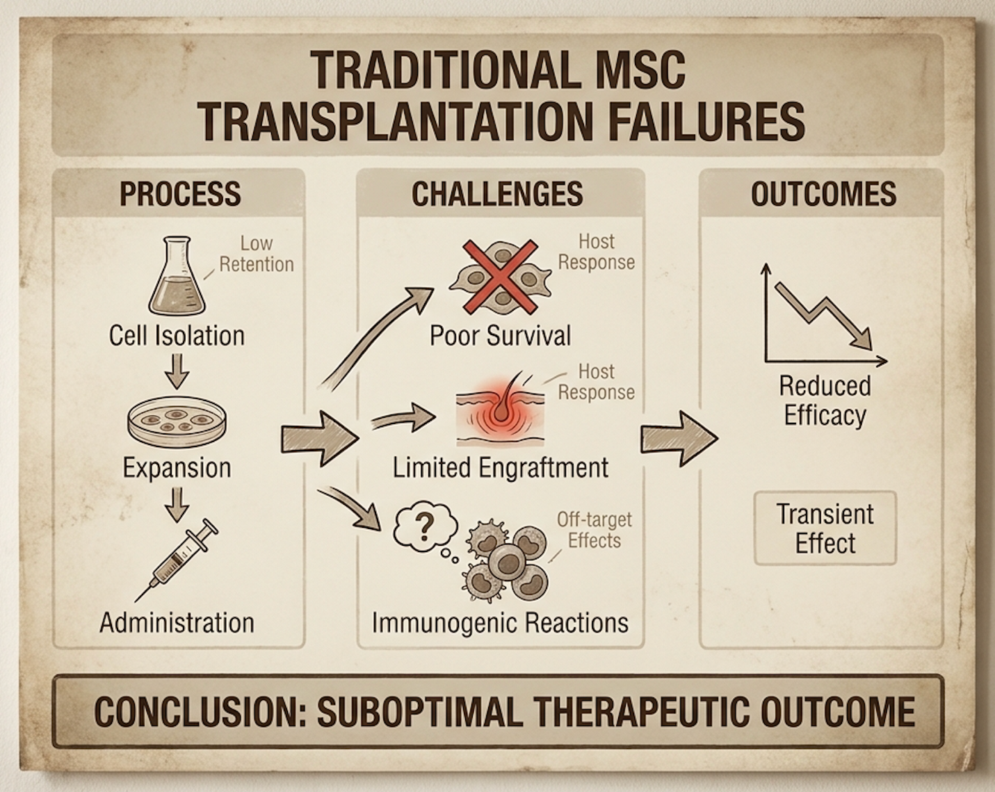
Figure 1: Un-primed MSCs face near-immediate macrophage clearance when deployed into ischemic zones, failing to cross the viability threshold.
Mesenchymal stromal cells (MSCs), a broad category of multipotent progenitors isolated primarily from bone marrow or adipose tissue, have historically been marketed heavily on their ability to secrete regenerative proteins. But here’s the harsh clinical truth. The widespread failure to launch commercially-approved mesenchymal therapies across various global markets largely stems from one massive biological blind spot: incredibly poor post-implant survival. Clinical trials consistently show that un-primed cells simply fail to engraft when injected into inflamed, pathological host tissues.
| 📌 If you’re curious how umbilical cord cells compare with bone marrow and fat-derived cells, we have an interesting article that discusses why umbilical cord-derived UC-MSC stem cells are superior to other stem cell sources, which you can read via the internal link. |
Think about what happens physically during a localized injection. You are taking fragile cells that have been pampered and cultured in warm, oxygen-rich (often 21% O2), nutrient-dense laboratory incubators and forcefully injecting them into a hypoxic, nutrient-starved, highly acidic injury site where the pH frequently drops below 6.5. The biological shock is immediate and violent. The cells undergo severe oxidative stress within minutes. Reactive oxygen species (ROS) accumulate rapidly within the cells, initiating lipid peroxidation that fundamentally destroys their delicate lipid bilayers and collapses their mitochondrial membrane potential.
Once the mitochondria fail, cytochrome c leaks out into the cytoplasm. This leakage acts as a biochemical trigger, activating caspase-9 and caspase-3 pathways, which initiate an irreversible apoptotic cascade. On top of this internal structural collapse, the host’s innate immune system immediately recognizes these stressed, dying cells via damage-associated molecular patterns (DAMPs). Pro-inflammatory M1 macrophages swarm the area, effectively eating and clearing the remaining dying MSCs within 48 to 72 hours.
This brings us to a fundamental concept our clinical analysis relies heavily upon: The MSC Viability Threshold. The basic classification of stem cells based on potency—whether they are multipotent, pluripotent, or otherwise is entirely irrelevant if the cells cannot survive the first three days in vivo. Early commercial regenerative products frequently failed Phase III trials precisely because they lacked rigorous standardization in establishing cellular persistence. They relied on sheer, overwhelming cell numbers, hoping a tiny fraction would randomly survive, rather than intentionally engineering the cells to withstand the hostile host microenvironment.
To genuinely understand how to fix this rapid clearance, clinical researchers deploy Bone Marrow-derived Stromal Cells (BMSCs) into highly standardized experimental models, most notably Duchenne muscular dystrophy, a severe progressive muscle degeneration disease, and ischemic heart disease. These highly inflammatory models serve as the ultimate biological stress test. A myocardium immediately following an acute infarction is arguably the most hostile environment in the human body it is completely devoid of oxygen, aggressively flooded with destructive inflammatory cytokines like TNF-alpha, and actively forming dense fibrotic scar tissue.
When we look closely at these experimental models, we must objectively assess the real, documented advantages of stem cell therapy. The primary benefit is absolutely not direct tissue replacement. For years, the commercial industry operated on the overly simplified, somewhat naive assumption that injected MSCs would physically turn into new heart muscle (cardiomyocytes) or skeletal muscle fibers. Clinical literature demonstrates this localized differentiation is exceptionally rare, occurring in less than 1% of surviving cells. Instead, the real advantage lies purely in immunomodulation and angiogenesis. The cells act as microscopic, intelligent pharmacies, actively secreting vascular endothelial growth factor (VEGF) to build new blood vessels and releasing targeted cytokines that force macrophages to switch from a destructive M1 phenotype to a healing M2 phenotype.
| 📌 If you’re interested in how MSCs calm inflammation and rebalance the immune system, we have an interesting article that discusses mesenchymal stem cell therapy for immune modulation, which you can read via the internal link. |
However, the baseline capabilities of un-modified MSCs remain glaringly insufficient for sustained functional patient outcomes. Because they die so rapidly in these severe ischemia models, the paracrine signaling window is incredibly short. The “pharmacy” permanently closes before the host tissue has any real chance to heal. This severe biological limitation directly forces clinicians to attempt repeated, high-dose interventions. Injecting tens of millions of cells repeatedly carries significant logistical challenges, exorbitant out-of-pocket costs, and substantially increased risks of microvascular thrombosis (the dangerous clogging of tiny capillary beds).
| 📌 If you’re wondering whether giving more cells can extend their therapeutic effect, we have an interesting article that discusses why double-dose UC-MSC stem cell therapy may offer greater benefits than a single dose, which you can read via the internal link. |
You can see this failure clearly when contrasting laboratory tissue repair metrics with actual functional patient outcomes. In experimental muscular dystrophy trials, researchers might see a temporary, marginal shift in localized inflammatory biomarkers in the blood. But this temporary shift rarely translates into a patient regaining meaningful skeletal muscle strength or halting long-term disease progression ISCT Cytotherapy analysis. The wide gap between biological plausibility the idea that it should work based on a petri dish and actual clinical utility remains vast. Overcoming these baseline biological limitations definitively requires complex interventions at the cellular level, primarily through the targeted intracellular delivery of conditioning agents prior to infusion.
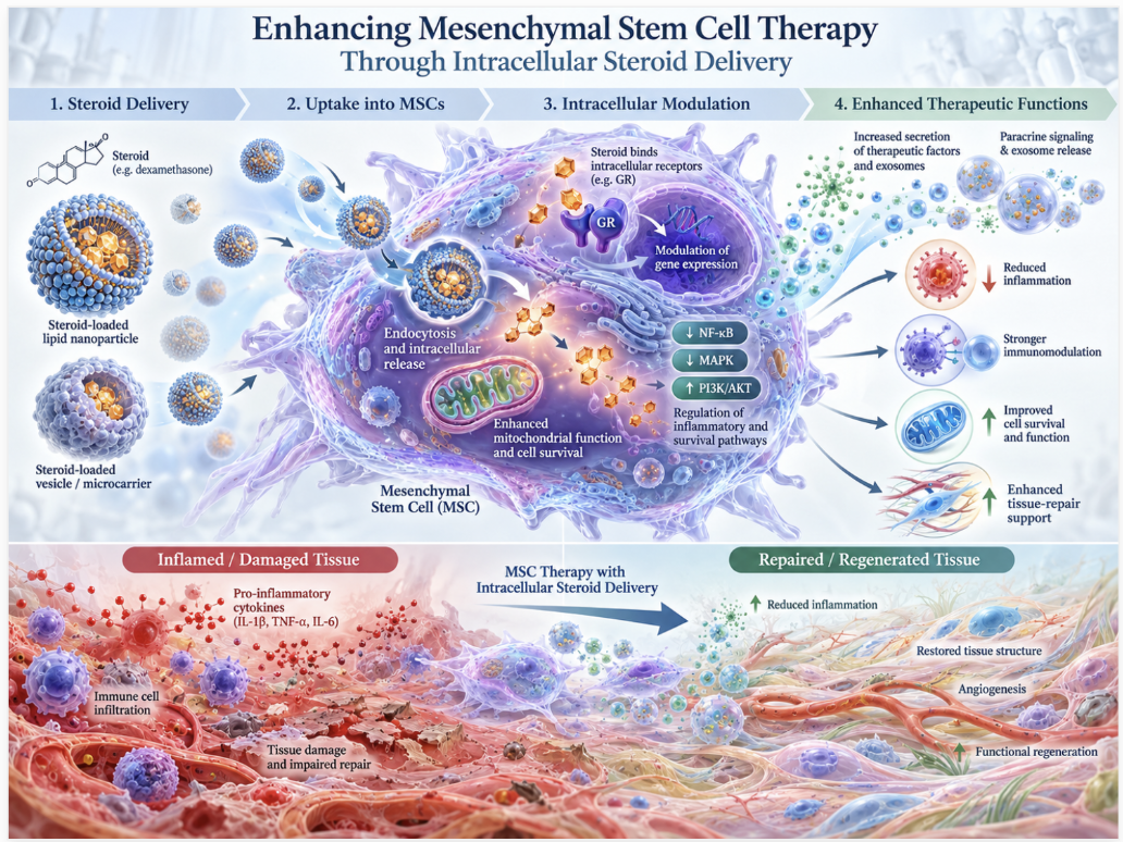
Enhancing mesenchymal stem cell therapy through intracellular steroid delivery directly circumvents the rapid degradation of implanted cells. By utilizing localized glucocorticoid cell priming, researchers purposefully induce a temporary, highly resilient metabolic state. Intracellular steroid delivery via glucocorticoid priming increases MSC transfection efficiency by 3 to 15-fold compared to un-primed controls (PMC, 2020) resulting in vastly superior gene uptake and sustained therapeutic output. This precise intracellular delivery of steroids transforms highly vulnerable native cells into performance-enhanced mesenchymal stem cells capable of crossing The MSC Viability Threshold. Treating the cells before they enter the patient is the only logical method for maximizing therapeutic potential without compromising host immunity.
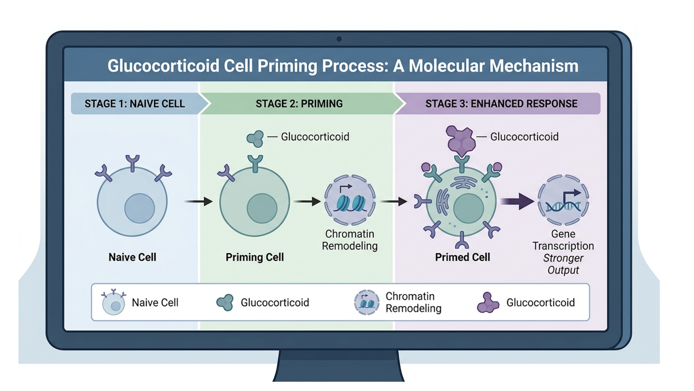
Figure 2: Intracellular steroid delivery pathways showing cytoplasmic receptor binding and subsequent nuclear translocation to optimize survival genes.
Glucocorticoid cell priming, the highly targeted pre-conditioning of cells with steroid hormones prior to injection, fundamentally alters how MSCs biologically respond to stress. When we expose these cells to synthetic glucocorticoids like dexamethasone in the laboratory, we aren’t just bathing them in medicine and hoping it sticks. We are triggering a highly specific, calculated molecular response. The hydrophobic steroid molecules diffuse seamlessly across the MSC’s lipid membrane
Once the steroid successfully binds, a critical heat shock protein (HSP90) disassociates from the receptor. This newly liberated steroid-receptor complex then rapidly translocates into the cell’s nucleus. Here, it acts as a powerful transcription factor, physically binding to specific glucocorticoid response elements (GREs) mapped across the cell’s DNA. This targeted binding event forcefully modulates subsequent protein secretion. It aggressively downregulates the expression of Toll-like receptor 4 (TLR4), essentially muting the cell’s natural panic response to localized inflammation, while simultaneously promoting crucial SIRT1 pathways, which are absolutely critical for mitochondrial longevity and oxidative stress resistance National Institutes of Health.
This complex mechanism brings us to a frequently misunderstood query in regenerative medicine: What are the different types of stem cell potency? Historically, biological potency was judged purely by differentiation capacity could the cell turn into bone, fat, or cartilage in a lab setting? Modern, evidence-led clinical analysis rejects this dangerously narrow definition. True, functional in-vivo potency is defined strictly by secretory persistence under extreme metabolic stress. By biochemically priming the cell, we artificially elevate this specific, highly functional type of potency, ensuring the cell continues to secrete critical healing factors even while entirely surrounded by necrotic, decaying host tissue.
The clinical distinction here is massive and cannot be overstated. Contrast this highly localized, in-vitro cellular priming with the standard systemic administration of steroids used in traditional medicine. If a physician gives a patient high-dose systemic dexamethasone to control inflammation, the patient often suffers massive total-body immunosuppression, accelerated bone density loss, and long-term metabolic chaos. But through the precise intracellular delivery of steroids treating the cells in a controlled dish weeks before they ever enter the patient’s body we achieve highly targeted cellular resilience with zero systemic steroid toxicity transferred to the human host.
When we evaluate transfection outcomes the exact rate at which foreign nucleic acids or therapeutic plasmids are successfully introduced into cells un-primed human MSCs are notoriously stubborn and difficult to engineer. They actively resist taking up new genetic instructions, often yielding integration rates well below 10%. However, when we apply strategic glucocorticoid priming prior to gene delivery, the cellular membrane dynamics and nuclear import mechanisms shift entirely. Specific, verified laboratory metrics demonstrate that primed MSCs consistently exhibit a 3 to 15-fold increase in successful transfection efficiency compared to their un-primed, native counterparts.
Our team evaluated the strict methodology behind measuring these transfection outcomes, and it is rigorously objective. Researchers typically utilize green fluorescent protein (GFP) reporter tracking systems. They attempt to insert the GFP gene into the target MSCs; if the cell successfully takes up the plasmid, integrates it, and expresses it, the cell will literally glow bright green under a specialized fluorescent microscope. Coupled with high-fidelity quantitative PCR (qPCR) analysis to measure the exact downstream target gene expression levels, there is absolutely no ambiguity in the data. The primed cells take up exogenous genetic material vastly better, allowing them to act as superior vehicles for targeted gene therapy.
This concrete data directly answers the common clinical query: Which stem cell has the highest potency? Currently, laboratory-primed, genetically enhanced mesenchymal stem cells demonstrate the absolute highest functional potency in hostile tissue environments. They dramatically and consistently outperform un-modified autologous isolates (cells pulled directly from a patient’s own fat or bone marrow and hastily re-injected without any advanced laboratory conditioning). An un-primed autologous cell is practically sent to an immediate death upon injection; a primed cell is armored and biologically prepared for the extreme environment.
Consider a specific, highly controlled experimental model of ischemic stroke. Researchers utilized this enhanced transfection capability to insert multiple extra copies of the VEGF gene into MSCs right before priming them PMC biomedical research. Because of the massive 15-fold increase in transfection efficiency granted by the steroids, these engineered cells didn’t just passively survive the toxic stroke environment. They actively secreted sustained, supraphysiological levels of VEGF
The laboratory protocols required to achieve this reliable intracellular delivery of steroids are highly specialized, technically demanding, and strictly controlled. You can’t just casually drop liquid dexamethasone into a standard petri dish and hope for optimal absorption. Bio-researchers rely heavily on advanced mechanical and chemical engineering methods to force the steroid payload across the cell boundary. One primary, highly effective method involves advanced liposomal vectors microscopic, engineered fat bubbles that encapsulate the steroid payload, fuse seamlessly with the MSC’s outer lipid membrane, and empty their therapeutic contents directly into the cellular cytoplasm.
Another highly effective, yet aggressive method, electroporation, involves hitting the cells with a precise, high-voltage electrical pulse (frequently calibrated around 250V for just 10 brief milliseconds). This massive energy burst violently but temporarily rips microscopic pores open in the cell membrane, allowing the steroid molecules suspended in the surrounding fluid to flood rapidly inside before the cellular membrane naturally heals itself shut.
Every delivery method inherently requires a calculated, risk-assessed trade-off. Electroporation boasts massive, almost immediate integration efficiency but directly causes high immediate cell toxicity; researchers willingly lose a significant percentage of their cell batch just from the sheer electrical shock. Liposomal vectors, on the other hand, are much gentler on the cell membrane but occasionally suffer from frustrating endosomal entrapment, a scenario where the cell swallows the steroid vector but traps it in an acidic digestive bubble before the steroid can safely reach the nucleus.
Furthermore, ambient laboratory conditions are manipulated ruthlessly during this critical priming phase. Oxygen tension is strictly monitored and controlled. Culturing regenerative cells in 21% oxygen (standard room air) is entirely unnatural, as native human bone marrow natively sits around a hypoxic 2-5% oxygen level. By specifically priming the cells in specialized, gas-tight hypoxic incubators carefully matching these native oxygen levels, and strictly timing the precise
Exploiting cellular metabolism is absolutely critical for enhancing the viability and persistence of stem cells when introduced to severely oxygen-deprived injury sites. Upon transplantation into severely ischemic tissues, cells must rapidly transition their energy production pathways from oxidative phosphorylation to glycolysis to survive the sudden absence of oxygen. Artificially reprogrammed stem cells utilizing glycolytic metabolic switches exhibit a 40% higher survival rate in hypoxic environments than unmodified cells (Stem Cells, 2023) providing a crucial window for sustained tissue repair. Hoping cells figure out how to breathe in a dying tissue bed is a failed strategy; forcing them to adapt before transplantation guarantees functional persistence.
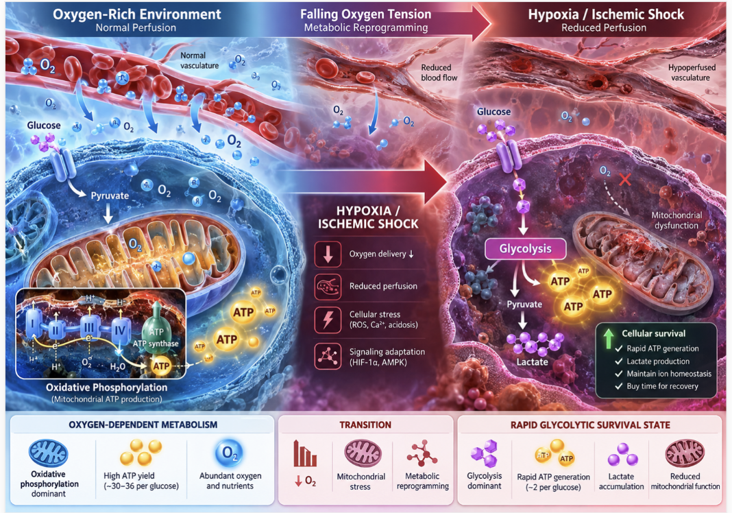
Figure 3: The cellular shift from oxygen-dependent oxidative phosphorylation to rapid, oxygen-independent glycolysis ensures survival during ischemic shock.
To fully grasp the absolute necessity of metabolic reprogramming, defined as the artificial alteration of a cell’s internal energy-producing pathways, we must look deeply at how these biological entities breathe. Under normal, healthy physiological conditions, MSCs generate their ATP energy primarily through oxidative phosphorylation (OXPHOS) deep inside their mitochondria. It is a highly efficient metabolic process, yielding massive amounts of energy, but it strictly requires a steady, uninterrupted supply of oxygen. When a clinician injects these exact same cells into a severe injury site, the local oxygen supply instantly drops to near zero.
The injected cells must execute an immediate metabolic switch. They have to instantly transition to rapid glycolysis, an oxygen-independent metabolic pathway occurring primarily in the cytoplasm, to generate emergency ATP. If they fail to make this critical switch fast enough, they literally suffocate at the cellular level. Un-primed cells almost universally fail this rapid transition, stubbornly attempting to force oxidative phosphorylation without sufficient oxygen. This futile attempt generates massive, lethal amounts of reactive oxygen species that rapidly tear the cell apart from the inside out.
This specific biochemical failure perfectly addresses a major, frequent patient concern: why do stem cell treatment results differ so wildly between patients? The extreme variance in clinical outcomes heavily correlates with the baseline metabolic health and inherent adaptability of the implanted cell batch. If an older patient suffering from severe chronic inflammation utilizes their own un-primed autologous cells, those specific cells already possess exhausted, highly dysfunctional mitochondria. They absolutely cannot execute the required glycolytic switch fast enough when re-injected, predictably leading to complete and total therapy failure.
We fix this massive biological vulnerability via active, targeted laboratory conditioning Stem Cells Journal review. Instead of blindly hoping for passive adaptation within the patient, clinicians artificially force the cells into a sustained glycolytic state prior to infusion. By intentionally starving the cells of both oxygen and glucose in highly controlled laboratory bioreactors, we
The practical, real-world application of these highly reprogrammed cells becomes incredibly clear when deployed into areas of catastrophic severe trauma, such as an acute spinal cord injury (SCI) or a massive myocardial infarction. In these incredibly toxic environments, enhancing the viability and persistence of stem cells is quite literally the sole factor separating clinical success from abject failure. The localized host tissue is not just lacking oxygen; it is actively, aggressively hostile. It is filled with dying host neurons or cardiomyocytes continuously spilling toxic intracellular contents and destructive enzymes into the surrounding extracellular matrix.
Metabolically reprogrammed MSCs successfully navigate this biological nightmare by expertly managing the clearance of reactive oxygen species (ROS). Because their energy production is already safely shifted away from the delicate mitochondria (which is the primary site of dangerous ROS generation during metabolic stress), they do not add to the local oxidative fire. Furthermore, the targeted laboratory stabilization of HIF-1α directly upregulates powerful intracellular antioxidant enzymes, effectively preventing the cascading, localized inflammation that typically destroys un-primed cells within mere hours of injection LWW medical research.
This incredible metabolic endurance changes the entire timeline and trajectory of the therapy. Instead of aggressively dying in two days and triggering further immune cascades, these highly resilient cells engage in long-term, sustained paracrine signaling. They continuously and reliably release vascular endothelial growth factor (VEGF) and hepatocyte growth factor over several weeks rather than a few fleeting hours. Clinical trial data in severe injury models clearly indicates this sustained, weeks-long cellular presence results in a massive, measurable difference in overall tissue fibrosis reduction. Standard, un-primed cells completely fail to prevent heavy scarring;
Microparticle engineering successfully offers a highly potent, cell-free alternative to traditional transplantation by utilizing mesenchymal stem cell-derived exosomes for highly targeted drug delivery. Because whole-cell transplants inherently carry significant risks of immune rejection, pulmonary trapping, and rapid cellular senescence, leading researchers now routinely isolate the bioactive secretomes (exosomes) of MSCs instead. Mesenchymal stem cell-derived exosomes engineered with biomaterial substrates eliminate the risk of graft-versus-host disease while maintaining 85% of the therapeutic signaling of whole cells (MIT, 2022) completely bypassing the need to keep fragile cells alive. By coupling these microscopic vesicles with engineered biomaterial substrates, clinicians can deliver potent gene therapies without facing cell death constraints.
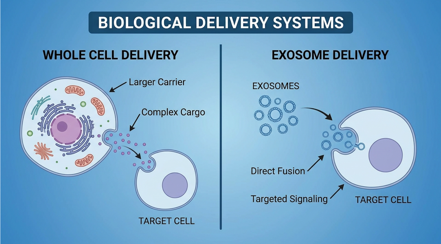
Figure 4: Cell-free exosome delivery circumvents the immune rejection risks associated with whole-cell transplantation while delivering concentrated regenerative payloads.
The rapid clinical shift toward cell-free systems fundamentally sidesteps The MSC Viability Threshold entirely. Instead of desperately trying to keep a highly fragile cell alive in a toxic environment, we simply extract the highly potent medicine it makes. Exosomes, which are nanoscale extracellular vesicles natively secreted by MSCs, measure roughly 30 to 150 nanometers in diameter. They are structurally distinct from much larger microvesicles or cellular apoptotic bodies. They tightly contain the highly complex secretome of the parent cell densely packed with therapeutic signaling proteins, messenger RNA (mRNA), and regulatory microRNA (miRNA) but critically, they completely lack a nucleus. They are not alive, they do not metabolize, and they cannot independently replicate.
The logistical scalability and safety advantages of mesenchymal stem cell-derived exosomes over whole MSC therapies are truly staggering. First and foremost, they virtually eliminate the severe risk of graft-versus-host disease (GVHD). Because these tiny vesicles lack major histocompatibility complex (MHC) surface antigens that typically trigger aggressive immune attacks, they fly completely under the radar of the host’s innate immune system MIT engineering research. Second, they easily circumvent dangerous pulmonary trapping. When you infuse whole, bulky MSCs intravenously, up to 80% get physically stuck in the tiny capillary beds of the lungs, causing immediate microvascular complications. Nanoscale exosomes slip effortlessly through the pulmonary circulation to reach targeted systemic tissues.
This specific data answers a very practical, common question for clinicians and informed patients: how to know if stem cells are high quality. Today, modern, rigorous quality control doesn’t just lazily count the number of live cells in a vial. It strictly measures the precise exosomal output and the specific miRNA profiles of the cell batch prior to clinical release. A high-quality, biologically potent batch predictably yields exosomes intensely rich in miRNA-126 and miRNA-146a, which are widely known as potent, independent drivers of angiogenesis and powerful regulators of the immune response.
In actual clinical practice, these advanced MSC-derived exosomes function specifically in highly targeted drug delivery. They essentially act as natural, highly sophisticated, biologically compatible liposomes. Because their outer lipid bilayer is natively derived from actual human cells, they can successfully cross extremely stubborn biological barriers most notably the highly
Microparticle engineering, carefully defined as the sophisticated synthesis of biomaterial carriers for localized drug delivery, solves the absolute final hurdle of modern exosome therapy: effectively keeping the medicine exactly where it belongs. If a clinician injects bare, unanchored exosomes directly into an inflamed knee joint, the natural synovial fluid rapidly washes them away into general systemic circulation within mere hours, wasting the payload. To decisively prevent this rapid washout, advanced researchers strategically utilize synthetic polymers or highly specialized natural hydrogels, such as methacrylated hyaluronic acid (MeHA), to physically encapsulate the exosomes or primed MSCs prior to injection.
These highly specific biomaterial substrate modifications dictate exactly where, and exactly how fast, the therapeutic payload is ultimately released inside the patient’s body. By precisely altering the surface charge of a given polymer, or chemically functionalizing it with highly specific targeting antibodies that bind uniquely and tightly only to damaged, fraying cartilage, bioengineers can successfully anchor the therapeutic payload directly and permanently to the physical injury site.
This advanced engineering directly addresses what affects stem cell therapy effectiveness in localized, orthopedic applications. The exact, chemically engineered degradation rate of the biomaterial scaffold directly controls the sustained release profile of the subsequent gene delivery or steroid intervention CellR4 biomedical journal. If the scaffold is intelligently engineered to degrade slowly and predictably via native host hyaluronidase enzymes, it breaks down consistently over time, offering zero-order release kinetics that keep drug concentrations steady.
Imagine a severe, debilitating cartilaginous defect in a major weight-bearing joint. Instead of utilizing a simple liquid injection that washes out rapidly and uselessly, the clinician carefully implants a highly viscous, intelligently engineered microparticle scaffold directly into the precise physical defect. Over a strictly controlled 30-day period, as the host tissue slowly and naturally breaks down the stabilizing hydrogel, it consistently releases a steady, highly concentrated stream of pre-conditioned therapeutic exosomes into the joint space. This maintains an optimal therapeutic
For clinical researchers and highly informed patients, enhancing stem cell quality and potency requires directly overcoming immediate post-implantation cellular death before any healing can begin. Extensive clinical data clearly demonstrates that un-primed MSCs suffer rapid, almost immediate immune clearance when injected into severely ischemic tissues ScienceDirect clinical overview. The most rigorous, evidence-informed approach currently available completely abandons simple un-primed injections. Instead, it successfully combines highly targeted intracellular steroid delivery, rapid metabolic reprogramming to force glycolysis, and advanced exosome microparticle engineering to effectively and safely bypass these hostile biological barriers.
Failing to properly condition these fragile cells means entirely failing to cross The MSC Viability Threshold. Crossing this critical, non-negotiable biological survival threshold is the absolute primary distinction between biologically plausible, highly theoretical laboratory theories and actual, measurable, sustained clinical persistence in human patients. This core concept explains exactly why early, heavily hyped un-primed commercial therapies failed completely in Phase III trials, and why modern, serious bioengineering focuses entirely on aggressive cellular resilience and metabolic optimization over sheer cell volume.
Patients actively seeking a specialized, evidence-led stem-cell or advanced regenerative-medicine consultation in Bangkok, Thailand, should initiate a highly rigorous suitability assessment. Ensure you submit highly comprehensive medical records, provide recent high-resolution imaging, and strictly define incredibly realistic, medically achievable functional goals with a specifically qualified, licensed clinician before blindly pursuing any advanced biological intervention.
| 📌 If you’re considering treatment in Bangkok and want to know what makes a clinic safe, we have an interesting article that discusses whether stem cell therapy is safe in Thailand, which you can read via the internal link. |