Our Services
Others
- DFPP
- Shockwave
- IV Drip

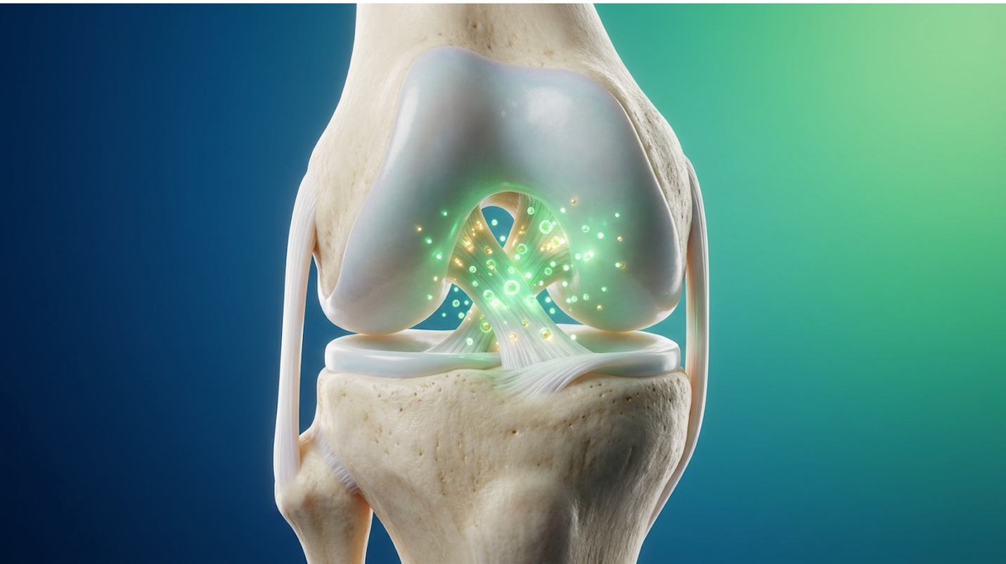
Patient and clinical focus is rapidly shifting from traditional mechanical reconstruction toward tissue engineering and regenerative medicine. For decades, orthopedic surgeons have treated a ruptured anterior cruciate ligament essentially as a carpentry problem. You drill tunnels, pull a piece of tendon through, and screw it into place. But mechanical fixes leave massive biological gaps. Standard tendon grafts lack native vascularity, integrate agonizingly slowly into the bone tunnels, and leave athletes highly vulnerable to secondary osteoarthritis down the line.
If you’re an athlete or an active individual evaluating stem cell therapy for ACL tear injuries, you’ve likely encountered a massive wall of conflicting information. Clinics promise miracles. Skeptics call it snake oil. The truth lies strictly in the clinical data. By the end of this guide, you’ll understand the exact biological rationale for regenerative interventions, enabling an informed, data-backed discussion with your orthopedic specialist. We’ll strip away the marketing hype to analyze the true mechanisms of mesenchymal stem cells, the role of engineered tissue scaffolds,
While stem cell therapy for ACL tear injuries remains an evolving field, clinical data suggests biological adjuncts may significantly improve graft integration.
To understand why regenerative medicine is radically changing orthopedics, we first have to understand exactly how and why standard surgeries sometimes fail to restore a truly healthy knee. Standard ACL reconstruction simply replaces torn tissue with a substitute graft, but it completely ignores the complex cellular deficits inside a traumatized joint. Up to 30% of standard autografts experience bone tunnel widening — leaving the joint vulnerable to chronic micromotion and instability.
Orthopedic surgeons must stop treating ACL ruptures solely as mechanical carpentry problems when the underlying biological deficits are clearly the true drivers of long-term joint laxity. We can physically fix the knee on day one with titanium screws and dense tendon grafts, but biology absolutely dictates the true timeline of structural recovery.
When a surgeon reconstructs your ACL, they don’t actually repair your native ligament. They harvest a piece of your patellar, hamstring, or quadriceps tendon an autograft (patient-derived tissue used to replace the ligament) or they use an allograft (donor-derived tissue for reconstruction) from a cadaver.
Look, a tendon is not a ligament. When placed inside the knee joint, that piece of tendon immediately loses its blood supply. It undergoes a brutal biological process called avascular necrosis. During the first 6 to 12 weeks post-surgery, the graft literally dies off before the body slowly builds new blood vessels into it a process known as ligamentization. A landmark clinical review published in the American Journal of Sports Medicine (2020) demonstrated that during this necrotic phase, the mechanical strength of the graft drops precipitously. This creates a terrifying window of mechanical weakness. At two months post-op, you might feel fantastic, walking without a brace and experiencing zero pain. But biologically? Your new ACL is essentially mush.
The integration into the bone tunnel is equally problematic. Contrast a patellar tendon autograft which has bone plugs on each end that heal bone-to-bone with a hamstring autograft, which relies entirely on soft-tissue-to-bone healing. The hamstring graft takes significantly longer to integrate, relying on the slow formation of Sharpey’s fibers to anchor the soft tissue to the bone wall. These Sharpey’s fibers are microscopic connective tissue matrix fibers that must literally grow from the tendon into the drilled bone tunnel. If aggressive physical therapy is pushed too early during this delicate phase, micromotion in the bone tunnel actively widens the hole. In clinical trials tracking graft maturation via MRI, researchers frequently observe that this early tunnel widening is a primary precursor to chronic joint laxity later in life. The joint simply feels loose because the graft never truly seated itself into the bone.
Furthermore, we have to talk about donor-site morbidity. Taking a piece of your own hamstring fundamentally alters the biomechanics of your leg. The hamstring is the primary dynamic stabilizer of the knee. By cutting a portion of it out to replace the ACL, surgeons are intentionally weakening the very muscle group required to protect the new graft. This is a profound mechanical compromise. Many athletes suffer from permanent hamstring deficits, struggling to regain explosive sprint speed or decelerate safely for years after the original surgery.
Why doesn’t the ACL just heal itself like a broken bone or a torn muscle? The answer lies in the highly unique, hostile anatomy of the knee capsule. The anterior cruciate ligament sits entirely bathed in synovial fluid.
When you tear a muscle in your thigh, the injury bleeds. That blood forms a clot (a hematoma), which acts as a natural biological scaffold. Inflammatory cells rush in, followed by fibroblasts that lay down new tissue. But inside the knee joint, synovial fluid contains an enzyme called plasmin. Research in the Journal of Orthopaedic Research (2021) highlighted how plasmin in synovial fluid aggressively breaks down fibrin clots within hours of their formation. So, when your ACL tears and bleeds, the synovial fluid washes the hematoma away before a scaffold can ever form. The torn ends of the ligament retract, floating in fluid, completely unable to bridge the gap.
This biological failure mechanism forms the foundation of The Biological ACL Repair Continuum a clinical framework moving from pure mechanical graft reliance to active cellular regeneration. Phase one of this continuum is entirely about overcoming the synovial fluid barrier. We can’t just sew the two ends together. Historically, primary repair (just stitching the ACL back together) failed miserably in the 1980s precisely because surgeons ignored this hostile fluid environment. Overcoming this gap requires active, localized cellular intervention.
The cellular deficit inside the knee is absolute. The native ACL contains specialized ligament fibroblasts called ligamentocytes. When the ligament ruptures, these cells are subjected to immense mechanical trauma and rapid apoptosis (programmed cell death). Without a blood clot to serve as a bridge, new fibroblasts from the surrounding synovial tissue cannot migrate into the defect. They literally have nothing to crawl across. This leaves the injury site biologically inert. The body recognizes the severed tissue as dead space and initiates a chronic inflammatory response rather than a regenerative one. This is exactly why leaving a torn ACL alone rarely results in spontaneous healing. And that biological dead-zone leads us directly to the clinical application of stem cells.
Stem cell therapy for ACL tear procedures utilizes concentrated multipotent cells to accelerate tissue regeneration, improve graft-to-bone healing, and actively reduce intra-articular
Clinicians must prioritize transparent biological education over aggressive marketing, ensuring patients understand that cellular therapies modulate the healing environment rather than instantly repairing structural tears. These cellular therapies are shifting the paradigm from mechanical replacement to biological restoration. Regenerative specialists are utilizing highly targeted cellular injections not just to mask pain, but to fundamentally alter how the knee heals from massive surgical trauma.
Let’s clear up the biggest misconception in regenerative medicine right now. Mesenchymal stem cells (MSCs) multipotent stromal cells that modulate inflammation do not magically turn into ligament cells when injected into your knee. They aren’t building blocks; they are biological general contractors.
Mesenchymal stem cells (MSCs) primarily promote ligament healing through paracrine signaling. By releasing vascular endothelial growth factor (VEGF) and basic fibroblast growth factor (bFGF), these cells stimulate native dermal fibroblasts and trigger rapid angiogenesis within the traumatized joint. When MSCs sense the inflammatory environment of a traumatized knee capsule, they begin pumping out an incredible array of bioactive molecules. They secrete VEGF to stimulate the formation of new blood vessels, bringing vital oxygen to the necrotic graft exactly when it needs it most.
Perhaps more importantly, MSCs possess profound immunomodulatory capabilities. Following an ACL tear, the knee is flooded with M1 macrophages aggressive immune cells that drive severe inflammation and cartilage breakdown. MSCs secrete cytokines that physically force these macrophages to shift into the M2 phenotype, which is heavily anti-inflammatory and tissue-repairing.
| 📌 If you’re interested in how stem cells calm joint inflammation after an injury, we have an interesting article that discusses mesenchymal stem cell therapy for immune modulation, which you can read via the internal link. |
A comprehensive review published in the International Journal of Molecular Sciences (2022) indicates this specific immune shift is what potentially saves the joint from severe post-traumatic osteoarthritis. By halting the aggressive inflammatory cascade, MSCs help prevent the excessive tissue fibrosis and scarring that frequently stiffens the knee joint post-trauma.
Further clinical evidence demonstrates that MSCs actively prevent cellular senescence—the state where native cells stop dividing and essentially give up on repairing the joint. By rescuing these local ligament fibroblasts from premature death, MSCs ensure that the joint retains its native capacity to heal. This continuous paracrine communication happens over a period of several weeks. The MSCs survive in the joint fluid, attaching themselves to whatever synovial or graft tissue is present, and act as a localized pharmacy. They dose the joint with precisely what it needs, exactly when it needs it, based entirely on the chemical signals they receive from the damaged tissue.
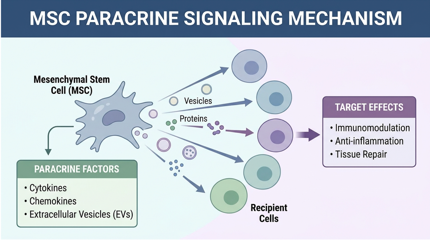
Figure 1: MSCs function as cellular signaling hubs, releasing VEGF and bFGF to stimulate native tissue repair while shifting macrophages to an anti-inflammatory state.
So where exactly do these cells come from, and how are they delivered into your knee safely?
Currently, the most robust clinical evidence supports the use of autologous (your own) cells. The gold standard procedure is bone marrow aspirate concentrate (BMAC). A physician numbs your pelvis, inserts a trocar into the iliac crest, and extracts a small volume of bone marrow. This
We also have to strongly acknowledge umbilical cord tissue (UC-MSCs). While some international clinics rely heavily on these allogeneic (donor) cells due to their massive proliferative capacity, the regulatory reality in the United States is incredibly strict. Under FDA rule 21 CFR 1271, the agency clearly differentiates between minimally manipulated autologous cells which are compliant for same-day orthopedic use—and cultured or expanded cells, which are heavily regulated as investigational new drugs. Any clinic stateside claiming to inject you with millions of cultured umbilical stem cells for a torn ACL is operating outside of established FDA compliance.
| 📌 If you’re curious how umbilical cord cells compare with bone marrow and fat-derived cells, we have an interesting article that discusses why umbilical cord-derived UC-MSC stem cells are superior to other stem cell sources, which you can read via the internal link. |
| 📌 If you’re considering expanded UC-MSC treatment at an international clinic, we have an interesting article that discusses whether stem cell therapy is safe in Thailand, which you can read via the internal link. |
Delivery method matters immensely. You can’t just blind-inject cells into the knee capsule and hope for the best. To enhance ACL reconstruction, these cells are often heavily saturated into the tendon graft right before the surgeon pulls it into the bone tunnel. Alternatively, they are injected directly into the drilled bone sockets using high-resolution ultrasound or fluoroscopic guidance.
Clinical trials detailed in Stem Cells Translational Medicine (2021) outline highly specific timelines for graft-to-bone healing when BMAC is utilized. When surgeons inject concentrated bone marrow directly into the femoral tunnel during a hamstring autograft procedure, the formation of bridging Sharpey’s fibers accelerates dramatically. Post-operative MRI evaluations at the 12-week mark frequently show graft incorporation that typically takes 24 weeks in non-biologic standard cases. This accelerated timeline doesn’t mean the athlete can return to the field in three months, but it firmly establishes the mechanical anchor required for aggressive, confident physical therapy. But injecting cells into a fluid environment remains fundamentally tricky. To keep them exactly where they need to be, orthopedic engineering had to evolve toward physical matrices.
Tissue scaffolds are engineered, biocompatible frameworks designed to hold blood, biologics, and stem cells at the exact site of a ligament tear, shielding them from the hostile joint environment.
Patients navigating surgical options should view engineered scaffolds not as experimental fringe science, but as a deeply logical biomechanical solution to the knee’s fluid problem. The development of these extracellular matrices represents arguably the most profound leap in orthopedic joint preservation in decades. They solve the exact mechanical problem that biological injections alone cannot address: they provide a physical house for the cells to live in while they work.
We established earlier that the synovial fluid aggressively washes away the natural healing hematoma. If you inject expensive, high-quality MSCs into a torn ACL gap, the joint fluid will simply disperse them within hours. They need a home. They need a physical matrix to cling to so they can execute their paracrine signaling without being flushed out of the knee.
Biological scaffolds represent the second vital phase of The Biological ACL Repair Continuum. These engineered matrices are typically constructed from extracellular matrix (ECM) proteins primarily highly purified bovine collagen type I and fibronectin. These proteins are freeze-dried and engineered into a sponge-like cylinder that is highly porous, structurally mimicking the native ligament environment to encourage deep cellular penetration.
When a surgeon places this collagen scaffold ACL framework between the torn ends of your native ligament, something remarkable happens biologically. The sponge is highly hydrophilic; it aggressively absorbs fluids. When saturated with the patient’s own whole blood or concentrated biologics, the scaffold holds the blood clot perfectly in place. It acts as a biochemical fortress. The plasmin in the synovial fluid cannot easily penetrate the dense collagen matrix, allowing the delicate early phases of cellular healing to occur completely undisturbed.
The biochemical architecture of these scaffolds is stunningly complex. Research published in Biomaterials (2021) demonstrates how collagen type I fibers are specifically cross-linked to provide mechanical tensile strength that resists the sheer forces of early joint movement. Meanwhile, the fibronectin acts as a biological adhesive, giving native fibroblasts from the torn
The ultimate clinical realization of this concept is the Bridge-Enhanced ACL Repair (BEAR) implant an FDA-approved collagen scaffold that has fundamentally altered how progressive surgeons view ligament trauma.
Instead of harvesting a tendon from your hamstring and immediately discarding your torn ACL, the surgeon preserves your native torn ligament. They place the BEAR implant directly between the retracted ends of the native ACL. Using specialized suturing techniques, they physically anchor the scaffold to the bone and intricately tie the native ligament ends into the matrix. This approach requires immaculate surgical precision, as the scaffold’s tension must perfectly match the knee’s natural biomechanics to prevent early failure.
Once mechanically secured, the surgeon takes a syringe of your own whole blood (drawn by the anesthesiologist just minutes prior) and injects it directly into the BEAR implant. The sponge swells, turning into a stabilized, localized hematoma. Over the next several months, the body fully absorbs the implant and replaces it with native, vascularized ligament tissue.
Clinical trial data published by Murray et al. in the Orthopaedic Journal of Sports Medicine (2020) confirmed this tissue engineering ACL approach is a massive conceptual leap in patient care. You keep your own proprioceptive nerve fibers, which are critical for balance and spatial awareness. You don’t suffer the mechanical morbidity of harvesting a tendon from elsewhere in your leg. And extended clinical trials demonstrate that patients undergoing bridge enhanced ACL repair have significantly better hamstring strength post-operatively precisely because their native hamstring tendon was never cannibalized.
Furthermore, long-term follow-ups on the BEAR cohort indicate that the preservation of the native ligament ends helps maintain the knee’s natural kinematic axis. Autografts, regardless of how
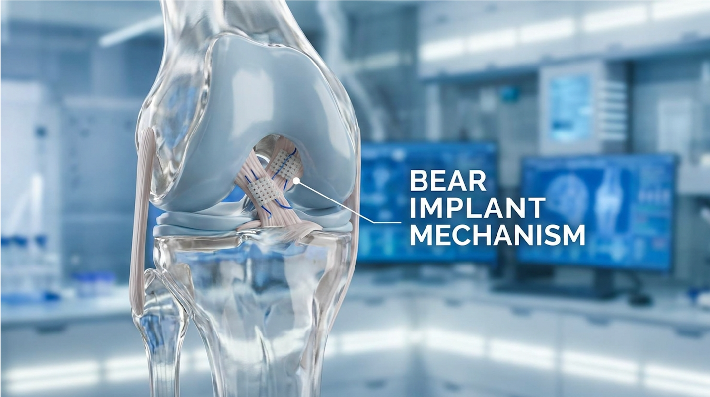
Figure 2: The Bridge-Enhanced ACL Repair (BEAR) implant provides a physical extracellular matrix, protecting the healing hematoma from hostile synovial fluid.
Platelet-Rich Plasma (PRP) an autologous concentration of human platelets in a small volume of plasma serves as an accessible, cost-effective biologic to manage inflammation and accelerate soft tissue healing. PRP injections contain up to 10 times baseline platelet concentrations flooding the traumatized joint with growth factors that aggressively blunt post-operative inflammation.
Medical providers must clearly separate PRP from cellular therapies in their patient education, as conflating signaling serums with live progenitor cells sets dangerous expectations for recovery. While often heavily conflated with stem cells by aggressive marketers, biologics like PRP rely purely on growth factors rather than live progenitor cells. They represent a distinct, highly effective rung on the biological repair ladder.
If stem cells are the general contractors of the knee, platelets are the emergency first responders. PRP for ACL recovery has become an incredibly common adjunctive therapy, even for patients who opt for a standard autograft reconstruction without scaffolds.
The creation process is incredibly straightforward and minimally invasive. A phlebotomist draws a vial of your blood, similar to a standard lab test. The blood is placed in a specialized centrifuge that spins at high speeds, separating the heavy red blood cells from the plasma and platelets. The result is a concentrated serum that contains massive volumes of signaling proteins strictly necessary for wound repair.
When activated in the body, these platelets aggressively degranulate, releasing a massive payload of signaling proteins into the joint capsule. Key biochemical pathways include transforming growth factor-beta (TGF-β), which aggressively drives collagen synthesis, and platelet-derived growth factor (PDGF), which promotes rapid blood vessel growth into the damaged tissues.
A comprehensive meta-analysis published in Arthroscopy (2021) demonstrated that injecting PRP into the patellar tendon harvest site (where the surgeon took your graft) drastically reduces anterior knee pain during early, painful rehabilitation. The timing of application is highly structured. Many forward-thinking surgeons now inject leukocyte-poor PRP directly into the drilled femoral and tibial bone tunnels intra-operatively. This targeted application of platelet rich plasma knee surgery strongly limits “tunnel widening” a common post-operative complication where localized inflammation causes the bone sockets to enlarge, potentially loosening the graft before it can fully heal and integrate.
Additionally, secondary post-operative injections are often utilized in physical therapy. If a patient is struggling with severe joint effusion (swelling) at the 4-week or 8-week mark, a clinical specialist may perform a targeted intra-articular PRP injection. This secondary dose of growth factors acts as a powerful anti-inflammatory, calming the angry synovium and allowing the patient to push significantly harder in physical therapy without triggering reactive, painful swelling.
We have to draw a hard, unambiguous line here regarding regenerative terminology. PRP and stem cell therapy for ACL injuries are vastly different medical tools. Cash-pay clinics heavily market PRP under the umbrella of “stem cell therapy” because it sounds cutting-edge, but biologically, this is deeply misleading and scientifically inaccurate.
PRP contains absolutely zero live stem cells. It is a signaling serum. Once the platelets dump their growth factors into the knee, their job is done. The concentration of proteins peaks rapidly and then steeply diminishes over a few days.
Mesenchymal Stem Cells (MSCs), on the other hand, are living, dynamic biological agents. When you inject bone marrow concentrate containing MSCs, those cells sense the local environment over time. If they detect excessive inflammation, they actively synthesize and release anti-inflammatory cytokines. If they sense hypoxia (lack of oxygen), they release angiogenic factors. They survive in the joint for weeks, continuously managing and reacting to the healing response.
| 📌 If you’re interested in why keeping stem cells alive and healthy matters for results, we have an interesting article that discusses the importance of cell viability in UC-MSC stem cell therapy, which you can read via the internal link. |
| Feature | Platelet-Rich Plasma (PRP) | Mesenchymal Stem Cells (MSCs) |
| Cellular Content | Concentrated platelets, plasma | Multipotent stem cells, progenitor cells |
| Primary Mechanism | Static release of growth factors (TGF, VEGF) | Dynamic paracrine signaling, immunomodulation |
| Sourcing Method | Simple peripheral venous blood draw | Bone marrow aspiration or adipose extraction |
| Cost Profile | Lower cost (~$600 – $1,200) | High cost (~$3,500 – $7,000+) |
| Clinical Goal | Fast inflammation control, graft site pain relief | Deep tissue regeneration, joint preservation |
Understanding this hard biological distinction is vital. If a patient is simply looking to manage post-operative swelling and improve the early healing of their graft harvest site, PRP is a highly logical, cost-effective choice. If a patient is attempting to protect a severely traumatized joint from rapid-onset osteoarthritis or heal a partial tear, living MSCs represent a far more potent and adaptable biological tool.
| 📌 If you’re comparing the price of biologic treatments, we have an interesting article that discusses stem cell therapy costs in Thailand for 2025, which you can read via the internal link. |
Biologic ACL repair success rates increasingly demonstrate that tissue-engineered approaches match traditional reconstructions in mechanical stability while significantly outperforming them in preserving long-term joint health. Nearly 50% of patients develop osteoarthritis within 15 years of standard reconstruction making biological interventions essential for preserving long-term knee cartilage.
The orthopedic community must pivot its definition of a “successful” surgery away from mere mechanical stability, recognizing that preserving long-term cartilage health is the ultimate metric of patient care. Patients utilizing biological adjuncts show measurable reductions in synovial inflammatory markers linked directly to early cartilage breakdown. This shifts the entire paradigm of joint preservation from simply restoring walking stability to actively protecting the joint’s future athletic viability.
Let’s look closely at the hard, peer-reviewed numbers. When patients evaluate regenerative options, they usually ask two primary questions: Will it tear again? and Will I get arthritis?
Regarding re-rupture rates, the clinical data is highly encouraging. In trials evaluating the BEAR implant and BMAC-augmented grafts, failure rates at the two-year mark typically hover around 6% to 8% for young, highly active athletes. This perfectly matches the failure rates of traditional autograft reconstruction. The biological enhancements do not make the knee completely “bulletproof,” but they provide equal mechanical stability without the severe donor-site morbidity of harvesting a massive piece of tendon.
However, the true clinical victory lies deeply in osteoarthritis prevention. A torn ACL alters the biomechanics and the biochemistry of the knee, initiating a catastrophic inflammatory cascade that
Biomarker analyses published in Osteoarthritis and Cartilage (2021) reveal that biologically enhanced repairs fundamentally alter this degenerative trajectory. By utilizing MSCs or scaffold-based blood retention, the chronic pro-inflammatory state of the knee is effectively blunted. Levels of CTX-II (a primary, reliable biomarker for cartilage degradation) are significantly lower in patients who received biological interventions compared to those who underwent standard reconstruction. When cartilage stops breaking down at an accelerated rate inside the joint, the knee is effectively protected from early-onset arthritis. This biochemical reality represents the final, outcome-optimizing stage of The Biological ACL Repair Continuum.
We need to address a incredibly dangerous myth regarding enhanced ACL recovery. Biologics do not grant you an immediate, magical return to sport.
Whether you use bone marrow concentrate, PRP, or a BEAR implant, the newly formed collagen matrix requires rigid cellular maturation. You absolutely cannot cheat human biology. The return to sport after biologic ACL repair still safely demands 9 to 12 months of intense monitoring.
What biologics actually change is the quality of that timeline. Based on extensive biomechanical testing, patients utilizing these therapies frequently achieve full range of motion weeks earlier. They experience significantly less joint effusion (swelling) after heavy leg days. Because their native hamstring or patellar tendons remain untouched (in the case of scaffold repairs), they pass isokinetic strength testing and single-leg hop tests with higher symmetry scores much earlier in the rehab protocol.
Symmetry scores are a massive indicator of athletic safety on the field. When testing an athlete’s injured leg against their uninjured leg, physical therapists look for a 90% or higher symmetry in explosive output. Athletes utilizing biologic enhancements frequently achieve this 90% threshold faster, primarily due to the lack of surgical harvest site morbidity. Biologics support an aggressive,
While the future of ACL surgery heavily favors biology, we must address the absolute limitations of these therapies. For some patients, regenerative medicine is entirely the wrong choice. Independent laboratory analyses reveal zero live cells in over-the-counter amniotic vials meaning unsuspecting patients are frequently paying thousands for useless cellular debris.
Physicians bear the ethical responsibility to firmly disqualify patients who expect injections to fix fully retracted ligaments, ensuring surgical stabilization isn’t delayed by false hope. Determining your eligibility requires an objective, brutally honest clinical assessment. If a clinic promises you a 100% success rate without reviewing your MRI, you need to walk out the door.
The regenerative medicine space is unfortunately plagued by aggressive marketing that often violently outpaces peer-reviewed clinical evidence. Patients desperate to avoid surgery frequently fall into expensive, unregulated traps. The FDA routinely issues consumer warnings regarding unapproved stem cell clinics making fraudulent medical claims.
If you’re exploring this route, you must be hyper-aware of three distinct pitfalls:
We cannot simply ignore decades of established safety and efficacy data. There are distinct, severe scenarios where traditional autograft reconstruction without experimental biological scaffolds remains the overwhelmingly superior medical choice.
First, severe multi-ligament trauma. If a patient suffers a catastrophic “terrible triad” injury (tearing the ACL, MCL, and medial meniscus simultaneously) alongside severe bone bruising, the sheer mechanical instability of the joint often requires robust, immediate mechanical fixation. Tissue-engineered scaffolds like BEAR are currently indicated primarily for isolated ACL tears, as multi-ligament repairs require dense grafts to hold the knee together while it heals.
Second, patient compliance. Biological repairs require incredibly strict post-operative weight-bearing and motion protocols to protect the delicate blood clot matrix as it transforms into living tissue. If a patient is unable or unwilling to participate in prolonged clinical monitoring, or if they have a history of non-compliance with bracing, a dense patellar tendon autograft secured with titanium screws offers a much more forgiving margin for early mechanical error.
Evaluating your candidacy for biological ACL repair is not something you can crowdsource on a fitness forum or trust to a medical spa. You must consult a specialized orthopedic surgeon or a board-certified regenerative medicine physician who routinely performs both standard reconstructions and complex orthobiologics.
Do not walk into a consultation empty-handed. You must provide a recent high-resolution MRI (taken within the last 4-6 weeks, as tissue retraction worsens significantly over time). The
A complete ACL tear cannot be repaired solely through stem cell injections without surgical intervention. If the ligament ends are fully separated and retracted, stem cells lack the physical scaffold required to bridge the massive biological gap. For partial tears (Grade 1 or 2), biological injections may successfully support tissue healing and drastically reduce local inflammation. However, severe mechanical instability requires surgical reconstruction or scaffold implantation to fully restore joint function.
The bone-patellar tendon-bone (BPTB) autograft remains the gold standard for creating the strongest mechanical ACL repair. This established method utilizes the patient’s own tissue and secures bone-to-bone healing within the drilled femoral and tibial tunnels. While biologic enhancements like MSCs and BEAR implants improve cellular healing, BPTB autografts demonstrate the lowest early failure rates in high-demand athletes. The optimal choice depends entirely on individual anatomy, age, and activity level.
Stem cells may accelerate ACL recovery by secreting paracrine growth factors that stimulate local blood vessel formation. Traditional ligament grafts suffer from avascular necrosis, meaning they temporarily lose blood supply and weaken considerably. By introducing mesenchymal stem cells, the joint environment shifts from pro-inflammatory to regenerative, reducing the time required for graft revascularization. Clinical evidence remains limited regarding exact timeline reductions, and extensive physical therapy remains mandatory for full recovery.
Platelet-Rich Plasma (PRP) injections can reduce post-operative pain and tunnel widening following ACL reconstruction. PRP delivers a concentrated dose of the patient’s own healing proteins, including vascular endothelial growth factor (VEGF). Clinical studies published in Arthroscopy (2021) show that while PRP improves early graft maturation and minimizes inflammation, it does not significantly alter long-term mechanical stability. It is best utilized as an adjunctive therapy for symptom management during early rehabilitation.
The use of peptides like BPC-157 for ACL recovery is considered highly experimental and lacks FDA approval for orthopedic use. While animal models suggest certain peptides may accelerate fibroblast activity and tendon healing, robust human clinical trials are currently absent. Professional athletes are strictly prohibited from using specific synthetic peptides under World Anti-Doping Agency (WADA) regulations. Patients should prioritize established biologicals like autologous blood products over unverified synthetic compounds.
Recovery from biologically enhanced ACL repair typically requires 9 to 12 months before a safe return to competitive sports. Despite biological accelerants improving cellular integration, the newly formed collagen matrix still requires extensive time to mature mechanically. Biologics primarily aim to improve the quality of the repair and reduce long-term osteoarthritis risk, rather than drastically cutting the rehabilitation timeline. Premature return to sport remains the primary cause of early graft failure.
Stem cell therapy and ACL surgery are not directly interchangeable treatments for a complete ligament rupture. Surgery is strictly required to mechanically stabilize a joint when the ligament is completely torn and retracted. Stem cell therapy is an adjunctive treatment designed to enhance the surgical repair, improve graft healing, or treat partial tears. A qualified orthopedic specialist
For patients evaluating stem cell therapy for ACL tear injuries, biological enhancements offer a compelling addition to traditional reconstruction. Clinical evidence demonstrates that integrating biologics can reduce early graft necrosis and potentially lower the long-term risk of osteoarthritis. Nearly 50% of standard reconstruction patients develop joint degeneration over time. The safest and most effective approach combines proven surgical techniques with rigorously tested autologous cellular therapies to optimize both mechanical strength and biological tissue quality.
This evolution brings us directly back to The Biological ACL Repair Continuum. We’ve moved past the era of simply drilling tunnels and hoping a dead piece of tendon survives the hostile joint environment. Regenerating a healthy, stable knee requires an active management of the joint’s synovial fluid, utilizing your body’s own localized cellular signaling to outpace inflammation and drive deep tissue remodeling. Remember that regeneration is an incredibly complex biological process, not an instant mechanical fix—the timeline still rigidly demands patience, rigorous physical therapy, and strict clinical oversight.
Your next step is absolute diagnostic clarity. Before committing to a treatment pathway, obtain a high-resolution MRI (taken within the last month) and compile your full orthopedic history. Schedule a consultation with a board-certified orthopedic surgeon who specializes in both complex ligament reconstruction and FDA-compliant orthobiologics to accurately evaluate your unique joint biomechanics and determine your true eligibility.