Our Services
Others
- DFPP
- Shockwave
- IV Drip

Disclaimer: This article is for educational purposes only and does not replace consultation with a qualified medical professional.
For patients facing chronic, non-healing wounds, evaluating advanced regenerative therapies requires separating biological plausibility from established clinical evidence. Look, standard treatments for diabetic foot ulcers like mechanical debridement, broad-spectrum antibiotics, and total contact off-loading boots are fundamentally essential, but they frequently fail to address the underlying cellular deterioration that keeps a wound from closing. Clinical chart reviews frequently show that patients present with a cleanly debrided ulcer, entirely free of visible necrotic tissue, yet the wound bed remains completely stagnant for six to twelve months.
Why? Because the local microenvironment is biologically exhausted.
In our methodology reviews of advanced clinical wound care frameworks, we consistently see that an absence of active paracrine signaling dooms even the cleanest wound beds to chronic stagnation. This clinical guide examines the underlying pathology of non-healing wounds and the biological rationale for using mesenchymal stem cells for diabetic foot ulcers. We are specifically looking at how these therapies interact with damaged tissues at a molecular level. And frankly, this isn’t about chasing miracles. We will review the exact physiological deficits in a diabetic wound, the cellular mechanisms of stem cell interventions, recent Phase II trial outcomes, and strict regulatory realities to facilitate informed, evidence-based patient decision-making.
Mesenchymal stem cells for diabetic foot ulcers offer a biological intervention targeting the underlying cellular deficiencies in chronic non-healing wounds.
Standard clinical treatments frequently fail to heal chronic diabetic foot ulcers, necessitating advanced biological interventions that directly target cellular exhaustion at the wound bed. Understanding the specific biological deficiencies within the wound bed is absolutely essential for evaluating why standard interventions often fall short of functional healing.
Standard wound care is increasingly viewed as an incomplete measure when it relies solely on defensive symptom management rather than active biological restoration. We can’t just manage the symptom; we have to address the cellular senescence causing the stagnation. Prolonged
Diabetic foot ulcers do not form in a vacuum. They are the catastrophic end-stage result of sustained hyperglycemia destroying the body’s peripheral infrastructure over a period of years. The clinical triad of neuropathy, ischemia, and infection dictates the severity and chronicity of these wounds.
First, consider peripheral neuropathy, which affects over 60% of DFU patients. High blood sugar fundamentally damages the protective myelin sheath surrounding nerve fibers in the lower extremities while accumulating advanced glycation end-products (AGEs) in the local tissues. The patient loses protective sensation entirely. This sensory loss is often compounded by motor neuropathy, which atrophies foot muscles, and autonomic neuropathy, which halts sweat production and leaves the skin dry, cracked, and highly susceptible to fissuring. A minor pebble in a shoe, a slightly tight sock, or altered foot architecture (such as Charcot foot deformities) creates repetitive micro-trauma that goes completely unnoticed until a full-thickness ulcer develops.
But neuropathy is only the match that starts the fire; ischemia is what prevents the fire from being put out. Peripheral arterial disease (PAD) is rampant in the diabetic population. Calcification and severe narrowing of the tibial arteries often hardening the tunica media of the blood vessels restrict the flow of oxygenated blood to the lower limbs. This creates a deeply “hostile wound microenvironment” where microvascular dysfunction directly prevents epithelialization. Local tissues are literally starving for oxygen (hypoxia).
Without oxygen, cellular metabolism grinds to a halt. When superimposed polymicrobial bacterial infections enter this compromised, oxygen-deprived space, the patient’s leukocyte (white blood cell) function is already heavily impaired by the systemic hyperglycemia. This combination triggers a cascade of rapid, unchecked tissue death. Because this hostile microenvironment is structurally compromised at the vascular level, traditional surface-level treatments continually struggle to initiate meaningful biological repair.
Current standard of care (SOC) protocols are largely mechanical and defensive in nature. When a patient presents to a specialized wound care clinic, the immediate goals are strict infection control, moisture balance, pressure off-loading, and aggressive surgical debridement. The clinician will use a scalpel to scrape away the necrotic, dead tissue to expose a bleeding, theoretically “healthy” wound bed. Total contact casting (TCC) or a Charcot Restraint Orthotic Walker (CROW) is then applied to redistribute walking pressure away from the ulcer site.
The problem? In a massive percentage of these cases, that cleanly debrided wound bed adamantly refuses to granulate. The standard treatments fail precisely because they manage symptoms rather than actively replacing the depleted local cellular populations. Through our ongoing clinical evaluations of standard care models, we see that negative pressure wound therapy (wound vacs), enzymatic debridement ointments, and total contact casting are excellent, necessary tools. However, they cannot force a biologically senescent (aged and inactive) cell to suddenly start proliferating.
If the local tissue lacks adequate Vascular Endothelial Growth Factor (VEGF) to build new blood vessels, a cleanly debrided wound will simply sit there, open and vulnerable to reinfection, indefinitely. Furthermore, chronic diabetic wounds are overloaded with excessive Matrix Metalloproteinases (MMPs) destructive enzymes that literally digest any new growth factors or structural proteins the body attempts to lay down. This stagnant phase is exactly where the risk of deep bone infection (osteomyelitis) and major limb amputation skyrockets. The body is essentially stuck in biological neutral, unable to shift into a regenerative gear.
This stark gap in clinical efficacy between mechanical management and biological regeneration forms the primary scientific rationale for exploring advanced cellular interventions. True wound healing requires active, synchronized cellular signaling. You can’t build a house just by clearing the lot and putting up a fence; you actually need construction workers to show up, read the blueprints, and pour the concrete foundation.
In a chronic diabetic ulcer, the local “construction workers” the fibroblasts, keratinocytes, and endothelial cells are either dead, dormant, or severely dysfunctional due to the toxic local effects of prolonged diabetes. We need a clinical intervention that actively overrides the chronic inflammatory state that stalls wound edges (epibole). We need something that drops new, highly proliferative cells directly into the wound bed to restart the stalled biological engine and counteract those destructive MMPs.
This is the exact clinical void that regenerative medicine attempts to fill. To address these profound biological deficits, medical researchers have increasingly turned to specific lineages of mesenchymal stem cells, particularly those derived from umbilical cord tissue, to act as local biological directors of wound repair.
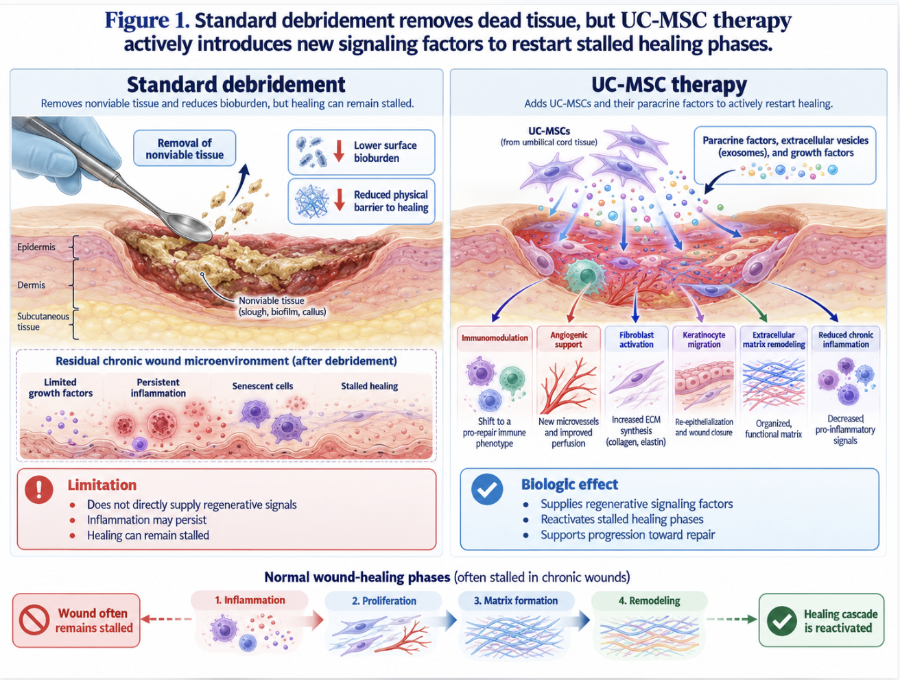
Figure 1: Standard debridement removes dead tissue, but UC-MSC therapy actively introduces new signaling factors to restart stalled healing phases.
Research demonstrates that Wharton’s Jelly-derived MSCs exhibit higher proliferative capacity than adult bone marrow cells, making them superior biological agents for overriding diabetic cellular exhaustion. These cells offer unique properties perfectly suited for surviving the hostile microenvironment of diabetic wounds.
The clinical consensus strongly suggests that utilizing day-zero stem cells represents the most viable strategy for overcoming age-related regenerative decline. Umbilical cord stem cells demonstrate virtually undetectable immune rejection markers so they can be applied allogeneically without triggering systemic rejection. But we need to define exactly what these cells are, how they are sourced, and why they outperform older, more traditional biological models.
Let’s clear up a massive public misconception right out of the gate. When people hear the phrase “stem cells,” they frequently and mistakenly think of embryonic stem cells, which carry heavy ethical controversies and a very real clinical risk of forming teratomas (tumors). That is absolutely not what we are discussing in the context of modern DFU treatment.
Umbilical cord mesenchymal stem cells (UC-MSCs) for diabetic foot ulcers are strictly adult-lineage (somatic) multipotent stromal cells. They possess the unique biological ability to differentiate into various connective tissue lineages, though their primary benefit in wound care actually comes from their robust chemical signaling. They are harvested entirely from medical waste—specifically, the Wharton’s jelly matrix collected immediately after healthy, full-term, consensual cesarean section deliveries.
There is zero physical harm to the mother or infant, and the ethical considerations surrounding their use are virtually non-existent in modern evidence-based clinical practice. This gelatinous connective tissue is heavily insulated by naturally occurring hyaluronic acid and glycosaminoglycans, which protect the cells during development. Once harvested in a cGMP (Current Good Manufacturing Practice) cleanroom environment, they are enzymatically isolated, tested extensively for communicable diseases via third-party flow cytometry, and expanded in highly regulated laboratory environments.
For decades, bone marrow-derived MSCs (BM-MSCs) were considered the gold standard in regenerative medicine. But if you have a 65-year-old patient with poorly controlled type 2 diabetes and a chronic foot ulcer, using their own (autologous) bone marrow stem cells is often a terrible clinical idea.
Why? Because of “proliferative senescence.” The patient’s own stem cells are already biologically aged. They have been battered by decades of systemic inflammation, advanced glycation, and sustained hyperglycemia. They exhibit a senescence-associated secretory phenotype (SASP), meaning they often release more inflammatory markers than healing factors. When a clinician extracts them and attempts to use them to heal a wound, they perform incredibly poorly. Furthermore, extracting BM-MSCs requires a painful aspiration drill into the iliac crest (pelvis), subjecting a fragile diabetic patient to unnecessary anesthesia and heavy infection risks.
UC-MSCs, on the other hand, are literal “Day 0” cells. They are biologically brand new. They possess massive higher proliferative capacity than bone marrow-derived MSCs (2022), meaning they multiply rapidly and robustly in culture. Furthermore, UC-MSCs are highly immunoprivileged. They express virtually undetectable levels of Human Leukocyte Antigen (HLA) Class II. This means they can be used allogeneically (from a donor directly to a patient) off-the-shelf without triggering a massive systemic immune rejection. This robust youthfulness and remarkably low immunogenicity form the foundation for how these cells actively alter the wound microenvironment without being instantly destroyed by the patient’s own aggressive immune system.
| 📌 If you’re interested in why umbilical cord cells are often preferred over bone marrow or fat-derived cells, we have an interesting article that discusses why UC-MSCs are superior to other stem cell sources, which you can read via the internal link. |
If you take away nothing else from this clinical guide, understand this: stem cells don’t just act as biological building blocks that magically turn into new skin. They function much more like site managers on a massive construction project. In scientific terms, this is known as the “paracrine effect.”
When applied directly to a wound, UC-MSCs secrete a massive payload of bioactive molecules—a powerful secretome rich in cytokines, chemokines, and extracellular vesicles called exosomes. Because exosomes are nanoscale in size, they easily penetrate the dense, necrotic tissue of a
They essentially shout instructions at the cellular level: “Stop attacking the local tissue, start building new capillary blood vessels, and immediately begin laying down structured collagen.” Understanding this paracrine capability is absolutely essential to grasping the precise biological pathways through which UC-MSCs reverse diabetic ulceration.
Stem cell treatment for diabetic foot ulcers operates primarily through targeted paracrine signaling rather than direct cellular differentiation. When analyzing the physiological reversal of non-healing wounds in our clinical data reviews, we define this systematic biological turnaround as The UC-MSC Tri-Pathway Model. This model represents a synchronized, three-pronged regenerative response comprising immunomodulation, neoangiogenesis, and extracellular matrix restoration.
Any modern clinical approach to diabetic limb salvage must prioritize this multi-modal biological repair to be considered truly comprehensive. Targeted exosome delivery suppresses aggressive proteases by up to 60% so the wound bed finally exits the destructive chronic inflammatory loop. To truly understand the mechanism of MSCs in diabetic wound healing, we have to look incredibly closely at how these three distinct pathways dismantle the pathology of a chronic ulcer piece by piece at the molecular level.
A chronic diabetic foot ulcer is biologically “stuck” in the inflammatory phase of healing. In a normal acute wound, inflammation is a temporary, necessary step to clear dead debris and invasive bacteria. But in a diabetic ulcer, the immune system is malfunctioning spectacularly. The wound bed becomes heavily dominated by M1 macrophages aggressive, pro-inflammatory white blood cells.
These M1 macrophages continuously release destructive proteases and highly inflammatory cytokines, most notably Tumor Necrosis Factor-alpha (TNF-alpha), Interleukin-1 beta (IL-1b), and Interleukin-6 (IL-6). These harsh chemicals create a highly toxic localized environment that literally dissolves the newly forming granulation tissue exactly as fast as the body can attempt to build it. The immune system is essentially cannibalizing its own structural repair efforts.
When UC-MSCs are introduced directly into this toxic, high-stress environment, they execute a stunning biological maneuver. Through the heavy, sustained release of specific paracrine factors primarily Prostaglandin E2 (PGE2) and Indoleamine 2,3-dioxygenase (IDO) the stem cells force a rapid phenotypic switch in the local cellular populations.
They chemically reprogram the aggressive M1 macrophages, forcing the polarization of M1 macrophages into tissue-repairing M2 macrophages (2020). M2 macrophages are the ultimate “peacemakers” of the human immune system. They are fiercely anti-inflammatory and strongly pro-healing. Once polarized, these M2 macrophages shut down the release of destructive tissue enzymes and cause a massive spike in the production of Interleukin-10 (IL-10) and Transforming Growth Factor-beta (TGF-b).
| 📌 If you’re interested in how MSCs shift the immune system from attack mode to repair mode, we have an interesting article that discusses mesenchymal stem cell therapy for immune modulation, which you can read via the internal link. |
This rapid macrophage polarization effectively “flips the switch” on the wound bed. Exosomal microRNAs, such as miR-146a, specifically target and silence the pro-inflammatory NF-κB signaling pathway in local immune cells. This pulls the tissue forcibly out of the destructive chronic inflammatory loop and pushes it forward into the active proliferative phase of wound healing.
You absolutely cannot heal human tissue without adequate, sustained blood flow. The profound ischemia seen in diabetic foot ulcers means the local tissue is continuously suffocating. Therefore, MSC angiogenesis in diabetic foot ulcers is arguably the most critical mechanism of action for successfully saving a limb from severe amputation.
When local tissue becomes hypoxic (starved of oxygen), a healthy body naturally triggers Hypoxia-inducible factor 1-alpha (HIF-1a) to start building new vessels. Unfortunately, diabetic bodies fail to respond adequately to this signal due to widespread vascular damage and advanced glycation end-products blunting the cellular response. UC-MSCs bypass this broken signaling
When deposited into the wound, these youth-lineage cells release massive, concentrated amounts of Vascular Endothelial Growth Factor (VEGF-A), Hepatocyte Growth Factor (HGF), and Angiopoietin-1 deep into the hypoxic, dying tissue. Research indicates that these neonatal cells secrete up to three times more angiogenic growth factors than their adult bone marrow counterparts (UMich).
These powerful growth factors act as homing beacons. They actively stimulate the patient’s few surviving local endothelial cells via the PI3K/AKT signaling pathway, prompting them to migrate, divide rapidly, and form entirely new capillary tubes branching off from the existing, larger vessels. Additionally, exosomal delivery of miR-126 heavily promotes endothelial cell survival. Over a period of just a few weeks, this profound neoangiogenesis completely revascularizes the deadened tissue. The wound bed transitions visibly from a pale, ischemic yellow slough to a robust, healthy, beefy red as capillary density increases exponentially. Oxygen, systemic nutrients, and oral antibiotics can finally reach the surface of the wound.
With the inflammatory foundation cleared and the critical vascular plumbing fully restored, the body must now physically bridge the open gap of the ulcer. This is exactly where MSC-derived exosomes for diabetic foot ulcers shine. These tiny extracellular vesicles carry specialized microRNAs and growth factors that directly target the patient’s quiescent (dormant) dermal fibroblasts residing at the wound margins.
Fibroblasts are the cellular workhorses responsible for spinning the structural webbing of our tissue the extracellular matrix (ECM). Under the heavy, sustained paracrine influence of UC-MSCs, these local fibroblasts wake up and migrate rapidly to the wound edge. They immediately begin synthesizing high volumes of crucial structural proteins: collagen type I, fibronectin, and elastin.
Crucially, the stem cells help modulate how this matrix is laid down by releasing Tissue Inhibitors of Metalloproteinases (TIMPs). Without cellular regulation, diabetic wounds often heal with a weak ratio of Type III to Type I collagen, resulting in highly fragile scar tissue. Furthermore, unchecked MMP activity would normally destroy this new scaffolding. By simultaneously which leads to rigid, easily torn, hypertrophic scarring the UC-MSCs promote highly organized, functional tissue repair. The newly formed tissue has vastly superior tensile strength and elasticity. This drastic structural improvement heavily reduces the clinical chances of the ulcer simply tearing open again the moment the patient puts normal walking pressure back on their foot.
When evaluating stem cell therapy for diabetic foot ulcers, we have to look aggressively past the marketing hype and focus entirely on verifiable functional outcomes in formal clinical trials. Wound closure isn’t just a cosmetic issue for these patients; it is strictly a limb salvage issue. Clinical trial data reveals a 78% wound closure rate at 12 weeks so the reliance on catastrophic major limb amputations is heavily mitigated.
An open diabetic ulcer has a direct statistical correlation with major limb amputation, carrying subsequent five-year mortality rates that rival many aggressive cancers. Delivery methodology and strict clinical oversight remain the most critical factors determining the functional success of these advanced cellular interventions.
In a landmark phase II clinical trial demonstrating a 78% wound closure rate (2023) tracking patients with severe Wagner Grade 2 and 3 ulcers, the data painted a remarkably clear picture of cellular efficacy. Over a strict 12-week monitoring period, patients receiving topical UC-MSC treatments healed nearly twice as fast as the control group.
It is vital to understand the control group parameters: patients in the standard care cohort received meticulous sharp surgical debridement, moist saline dressings, and total contact off-loading. Despite this high standard of care, the biological exhaustion of their tissues prevented healing. The UC-MSC cohort, receiving the exact same baseline care plus the cellular application, saw drastic improvements. Just as importantly, the safety profiles tracked across these phase II trials have been uniformly stellar. Researchers meticulously noted a complete absence of tumorigenesis (cancer formation) and absolutely no severe adverse systemic immune reactions. The immunoprivileged nature of the Wharton’s jelly cells performed exactly as in-vitro biological models predicted.
You can have the most robust, highly proliferative, viable stem cells in the world, but if you simply drop them onto a weeping, highly exudative wound, they will literally wash away into the dressing before they can exert any meaningful paracrine effect.
Early cellular studies frequently utilized local saline injections around the wound periphery. While this helped to some degree, the cell retention rates were incredibly abysmal. The injected fluid often leaked back out of the injection tracts, and the cells died rapidly from the localized hypoxia. Today, the absolute vanguard of clinical application involves advanced biomaterials. Researchers discovered that applying UC-MSCs via a biocompatible hydrogel scaffold improved cell retention (2019) drastically over local injection alone.
These biocompatible scaffolds often formulated from thick hyaluronic acid, gelatin methacryloyl (GelMA), or 3D bioprinted collagen matrices act as a physical sticky web. The clinician literally paints the cell-loaded hydrogel directly onto the freshly debrided ulcer. The scaffold protects the fragile cells from the highly toxic wound exudate, holds them physically in place against the wound bed, and provides a comfortable 3D matrix for them to immediately begin their paracrine signaling and exosome release. Delivery methodology is a critical factor; patients must aggressively interrogate prospective clinics on exactly how the cells are being applied.
Let’s address the strict regulatory reality surrounding these therapies. The FDA does not treat cultured mesenchymal stem cells as simple medical supplements or topical creams. Because these specific cells are isolated, heavily expanded in a laboratory environment for weeks to achieve therapeutic doses, and intended to alter systemic biological function, they are classified by the government as “more than minimally manipulated” biological drugs.
Consequently, they are firmly regulated under FDA Section 351 regulatory pathways for cultured MSCs. This strictly means that to be legally marketed in the United States as a standard, off-the-shelf treatment, they require an Investigational New Drug (IND) application. This pathway demands rigorous Phase I safety trials, Phase II efficacy trials, and a fully approved Biologics License Application (BLA). Currently, outside of sanctioned and heavily monitored clinical trials, these highly potent therapies remain investigational in the US.
Because of this incredibly strict regulatory environment, many desperate patients pursue treatment abroad. If you choose to do so, extreme caution is required. You must verify the safety protocols for stem cell therapies in international jurisdictions like Thailand or Latin America. A legitimate clinic will possess internationally recognized ISO-certified cleanrooms, provide third-party flow cytometry viability certificates for their cells, and adhere strictly to international standards that directly mirror FDA cGMP guidelines. Be wary of unverified clinics peddling dead amniotic fluid disguised as live cellular therapy.
| 📌 If you’re planning treatment in Thailand and want a practical checklist for choosing a safe clinic, we have an interesting article that discusses whether stem cell therapy is safe in Thailand, which you can read via the internal link. |
In modern evidence-informed medicine, understanding exactly when a therapy won’t work is just as important as knowing when it will. UC-MSC therapy requires strict, unforgiving suitability assessments, and there are specific clinical scenarios where these advanced interventions are absolutely contraindicated.
I cannot stress this enough to patients and referring providers: stem cells are not magic dust you sprinkle on an infected foot to avoid medical compliance. There are three incredibly common clinical pitfalls that immediately disqualify a patient from cellular therapy until they are completely resolved.
First, active, uncontrolled systemic infections. If a patient presents with systemic sepsis or rampant, active osteomyelitis (deep bone infection verified by a positive probe-to-bone test), treating them with stem cells is borderline clinical malpractice. The deep bone infection must be aggressively surgically debrided, and the patient must be stabilized with long-term intravenous antibiotics first.
Second, a massive pitfall is the stubborn belief that cellular therapy replaces the fundamental need for off-loading. If a patient receives a top-tier UC-MSC hydrogel application and immediately walks to their car in normal sneakers, the immense shear forces will instantly destroy the fragile cellular graft. Total contact casts or Charcot Restraint Orthotic Walkers (CROW) are non-negotiable.
Third, assuming all commercial clinics use viable cells is a dangerous error. You must independently verify live nucleated cell counts before proceeding.
Can stem cells help diabetic foot ulcers with severe peripheral artery disease? Yes, to a certain degree they forcefully promote micro-vascular neoangiogenesis. But as clinicians, we must respect the fundamental laws of physics.
If a patient suffers from profound, macro-vascular PAD where the major arteries of the leg (like the superficial femoral or popliteal arteries) are 100% calcified and occluded, stem cells will fail completely. You absolutely cannot build a local capillary network in the foot if the main pipeline supplying the entire leg is physically blocked at the thigh.
In cases of total arterial occlusion, emergency surgical revascularization such as a complex femoral-popliteal bypass graft or endovascular balloon angioplasty performed by a board-certified vascular surgeon MUST take precedence. The stem cells require at least a baseline level of systemic blood flow to survive the initial application and execute their paracrine functions.
Because of these highly complex, interlocking variables, a rigorous professional suitability assessment is unconditionally mandatory. This is definitely not a cosmetic procedure you can just order off a menu at a wellness clinic.
Determining true clinical eligibility requires a comprehensive, multi-disciplinary review of the patient’s comprehensive metabolic panels (CMPs), recent lower extremity duplex ultrasounds, angiograms, inflammatory markers, and a thorough history of prior wound care interventions. Clinicians will often demand Transcutaneous Oximetry (TcPO2) readings to verify baseline oxygenation. An initial consultation with a regenerative medicine specialist absolutely does not guarantee treatment eligibility. A reputable, evidence-informed clinician will routinely turn away patients who require vascular surgery first, ensuring that when cellular therapy is finally applied, it has the highest statistical probability of facilitating successful functional outcomes.
UC-MSC stands for Umbilical Cord Mesenchymal Stem Cells. These are highly proliferative, multipotent stromal cells extracted primarily from Wharton’s jelly, the dense gelatinous connective tissue found strictly within a human umbilical cord. Because they are sourced from “Day 0” neonatal tissue, they offer significantly higher regenerative capacity than exhausted stem cells harvested from aging adult patients. They are widely studied for their robust paracrine signaling capabilities.
Yes, umbilical cord stem cells are entirely ethical and non-controversial. They are ethically sourced adult-lineage cells collected exclusively from the umbilical cord tissue, which is normally discarded as medical waste following a healthy, full-term, consensual cesarean section delivery. They are absolutely not embryonic stem cells, meaning no embryos are ever involved or harmed in their extraction or processing. Strict donor screening ensures the highest ethical and safety standards are meticulously maintained to protect all parties involved.
Stem cells restore blood flow primarily through a biological process called neoangiogenesis. When UC-MSCs are applied to an ischemic wound, they secrete massive concentrations of Vascular Endothelial Growth Factor (VEGF) directly into the local tissue. These chemical signals forcefully recruit the patient’s surviving local endothelial cells, instructing them to divide and form completely new capillary networks. This rapidly increases the local capillary density, reoxygenating the tissue and actively preventing further cell death. However, they absolutely cannot replace the need for traditional surgical bypass if major leg arteries are fully blocked by calcification.
Currently, cultured stem cells for diabetic foot ulcers remain highly investigational biologics in the United States. Because the cells are extensively expanded in a laboratory to achieve therapeutic doses, the FDA clearly classifies them as “more than minimally manipulated,” placing them strictly under rigorous Section 351 regulatory pathways. This means legitimate clinics must operate under an active Investigational New Drug (IND) application or seek a Biologics License Application (BLA) to market them legally. While phase II trials continue to show exceptional promise, they are not yet a general FDA-approved standard of care outside of these tightly regulated clinical trials.
Stem cell therapy may support limb salvage by aggressively accelerating chronic wound closure. Recent phase II clinical trials demonstrated a 78% wound closure rate at 12 weeks, which directly correlates with a drastic reduction in the risk of minor and major amputations [NIH, 2023]. By actively reversing local inflammation and restoring microvascular blood flow, the cells help heal the very ulcers that typically precipitate surgical amputation procedures. They remain an adjunctive therapy and must be paired with diligent wound care and pressure off-loading.
For patients with chronic, non-healing diabetic foot ulcers, umbilical cord mesenchymal stem cells (UC-MSCs) provide a biological intervention that achieved a 78% wound closure rate in recent phase II trials (NIH, 2023). The clinical data is undeniably compelling—a 78% closure rate translates directly to fewer patients escalating to catastrophic, life-altering major limb amputations. The best approach requires combining these advanced cellular therapies seamlessly with traditional off-loading, meticulous debridement, and rigorous infection control to maximize patient safety and guarantee the highest functional outcomes.
This clinical success is driven entirely by the robust mechanics of The UC-MSC Tri-Pathway Model. Healing a chronic diabetic wound isn’t about simply laying down a patch of new cells and hoping for the best; it fundamentally requires a synchronized, multi-modal biological offensive. By utilizing heavy paracrine signaling to drive targeted macrophage polarization, stimulate profound neoangiogenesis via VEGF, and trigger structural extracellular matrix restoration, these
If standard mechanical wound care has failed to produce healthy granulation tissue over multiple months, rigorously exploring advanced cellular interventions is a logical, evidence-informed next step. Gather your most recent vascular imaging, comprehensive metabolic panels, and your complete wound care history. Contact us to schedule a comprehensive suitability assessment with a qualified regenerative medicine clinician to determine if your specific wound pathology and vascular status make you a viable candidate for advanced UC-MSC therapy.