Our Services
Others
- DFPP
- Shockwave
- IV Drip

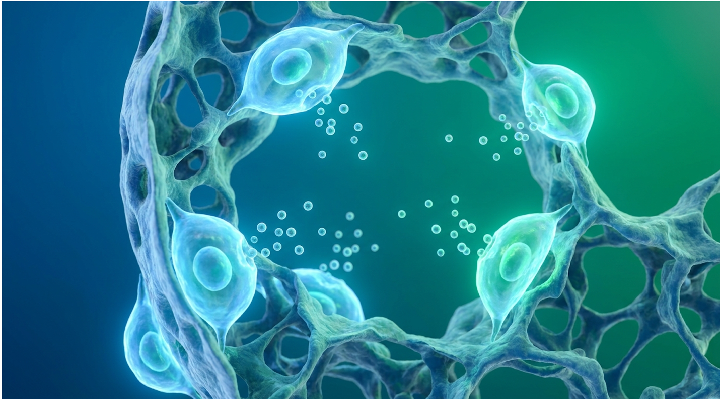
Current antifibrotic medications succeed in slowing the progression of Idiopathic Pulmonary Fibrosis (IPF), yet they offer no regenerative mechanism to repair damaged alveolar tissue. For patients facing progressive lung capacity decline (FVC) and deteriorating quality of life, the permanent nature of fibrotic scarring demands interventions that go far beyond basic disease stabilization.
Look, traditional pulmonology hits a wall here. When lung tissue undergoes honeycomb remodeling, the architectural collapse is largely irreversible with synthetic pharmaceuticals alone. This guide critically evaluates the biological rationale, clinical trial evidence, and rigorous safety profiles of stem cell therapy for pulmonary fibrosis.
We aren’t trading in false hope. Instead, we objectively compare standard antifibrotic protocols with emerging Umbilical Cord Mesenchymal Stem Cell (UC-MSC) therapies, detailing exact
Investigational stem cell therapy for pulmonary fibrosis utilizes The Pulmonary Regenerative Window to modulate the immune system and potentially slow fibrotic cascades, though it is not a cure.
Current idiopathic pulmonary fibrosis treatment protocols focus entirely on disease management rather than fundamental tissue regeneration. The disease triggers aberrant, runaway wound healing in the lungs, leading to the excessive and localized deposition of extracellular matrix proteins. This resulting fibrosis stiffens the alveolar walls and progressively impairs oxygen gas exchange, directly driving the clinical decline seen in every diagnosed patient.
Nintedanib slows Forced Vital Capacity decline by roughly 50%, yet this synthetic kinase inhibitor fundamentally cannot reverse established extracellular matrix stiffening.
Current FDA-approved antifibrotics slow FVC decline by approximately 50%, but they do not halt or reverse existing lung scarring (New England Journal of Medicine).
If you want to understand why regenerative medicine is exploring IPF, you first have to understand why the lung breaks down. Normal wound healing is a temporary, self-resolving event. You get a cut, fibroblasts rush in to lay down collagen, the tissue repairs, and the fibroblasts undergo apoptosis. They essentially switch themselves off and die when the job is done.
In IPF, that shut-off switch completely breaks.
The primary culprits are activated myofibroblasts. Due to repeated microscopic injuries to the alveolar epithelial cells lining your air sacs, these myofibroblasts become trapped in a continuous, pathological loop of activation. They constantly secrete dense collagen and other extracellular matrix (ECM) proteins. Over time, this collagen undergoes severe cross-linking. Research indicates that this cross-linking increases lung tissue stiffness dramatically, fundamentally altering the mechanical properties of the lung parenchyma (American Journal of Respiratory and Critical Care Medicine).
When we look at the specific biochemistry, the over-expression of pro-collagen genes completely overwhelms the body’s natural matrix metalloproteinases (MMPs)—the enzymes responsible for breaking down scar tissue. This forms rigid, inflexible scar tissue that eventually destroys the delicate lung architecture.
This results in the characteristic “honeycomb” appearance on High-Resolution CT (HRCT) scans. Once that honeycombing sets in, the physical space for oxygen exchange is permanently gone. It doesn’t come back. This progressive suffocation drives grim statistics. Untreated, median IPF life expectancy typically proves fatal within three to five years of diagnosis. While new lung scarring treatment options extend this window, the biological mechanics of this scarring represent a runaway feedback loop. Pharmacological interventions desperately attempt to disrupt specific signaling pathways within this loop, with mixed success.
For nearly a decade, the standard of care has revolved around two highly specific, FDA-approved synthetic molecules: Ofev (nintedanib) and Esbriet (pirfenidone). Both are classified as antifibrotic drugs, but they work through distinct biochemical pathways.
Nintedanib operates as a multiple tyrosine kinase inhibitor. It essentially blocks the receptors for specific growth factors (like VEGF, FGF, and PDGF) that tell fibroblasts to multiply and migrate. Pirfenidone’s exact mechanism is slightly more elusive, but clinical consensus confirms it heavily down-regulates Transforming Growth Factor-beta (TGF-β) expression, the master cytokine of fibrosis.
Extensive clinical data confirms that these current IPF medications effectively reduce the annual rate of forced vital capacity (FVC) decline by approximately half. That is their hard clinical limit. They do not clear existing scar tissue. They do not trigger the regeneration of dead alveoli. They simply apply the brakes to a car rolling down a hill.
Furthermore, you have to consider the tolerability profile. A massive subset of patients simply cannot handle these drugs. Nintedanib is notorious for causing severe gastrointestinal distress, particularly debilitating diarrhea and nausea, affecting up to 62% of patients in initial clinical trials. Pirfenidone often causes intense photosensitivity and liver enzyme elevation. When patients discontinue these medications due to severe side effects, their lung function trajectory sharply accelerates downward. The inability of these small molecules to reverse structural damage necessitates the exploration of cellular therapies capable of altering the immune microenvironment.
The application of UC-MSC for pulmonary fibrosis relies on cellular therapy to fundamentally alter the disease’s trajectory rather than merely suppressing it. Unlike synthetic oral drugs, mesenchymal stem cells lung regeneration models suggest these cells act as highly intelligent biological factories. They detect localized tissue damage and immediately respond by releasing a complex secretome that addresses multiple pathological pathways simultaneously.
Umbilical cord mesenchymal stem cells shift local alveolar macrophages to an M2 state, reducing localized inflammatory markers significantly across clinical models.
UC-MSCs primarily operate through paracrine signaling, secreting bioactive molecules that shift the lung’s microenvironment from a pro-fibrotic to an anti-inflammatory state (Stem Cell Research & Therapy).
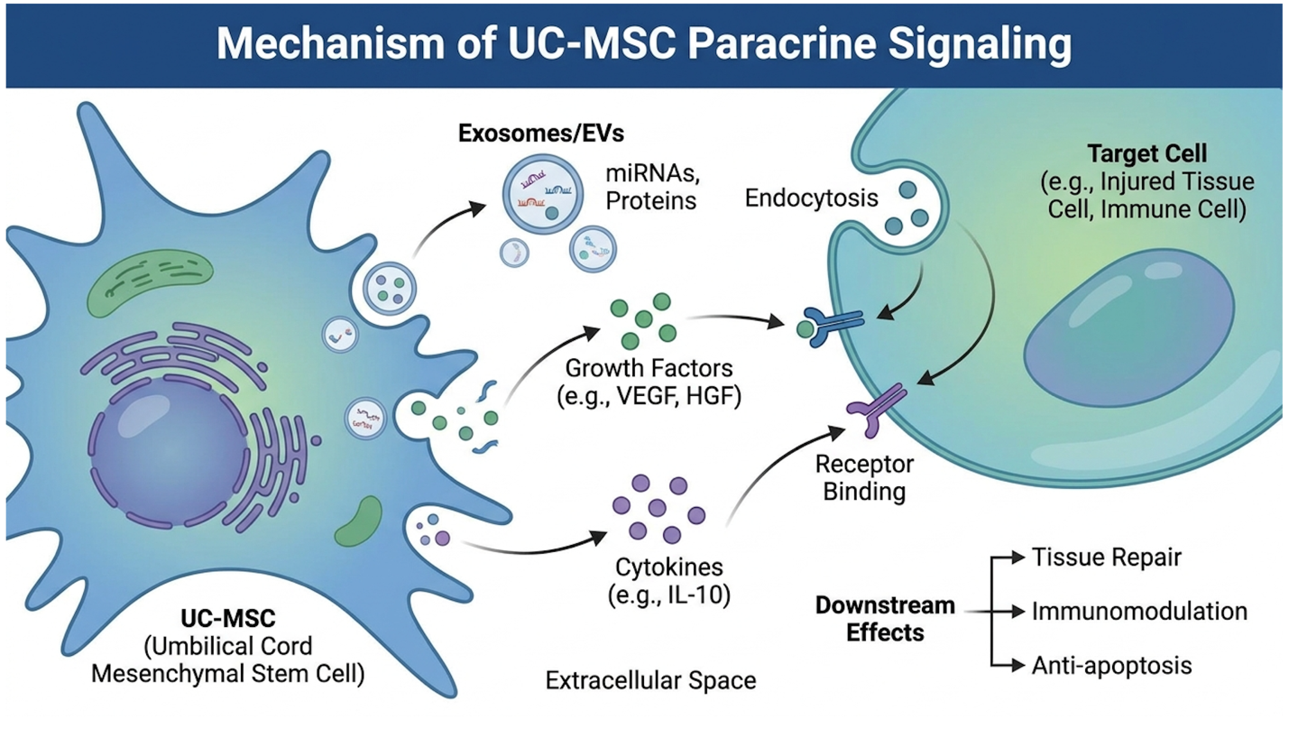
Let’s dispel the most pervasive myth in regenerative medicine right now. When you receive an intravenous infusion of stem cells, those cells do not travel to your lungs, plant themselves in the tissue, and magically turn into brand-new lung cells. Cellular engraftment and differentiation in adult organs is incredibly rare and is not the primary driver of healing.
Instead, they operate via paracrine signaling. Think of UC-MSCs as a localized software update for your lung’s native immune system. When infused, these cells arrive at the site of inflammation and begin secreting a highly customized cocktail of bioactive molecules the secretome.
This secretome is remarkably complex. It includes hepatocyte growth factor (HGF) to promote epithelial repair, prostaglandin E2 (PGE2) to suppress fibroblast proliferation, and tumor necrosis factor-stimulated gene 6 (TSG-6) to aggressively combat local inflammation. Crucially, this chemical signaling heavily suppresses TGF-β1, actively cutting off the fuel supply to the myofibroblasts causing the damage.
| 📌 If you’re interested in how UC-MSCs rebalance an overactive immune response, we have an interesting article that discusses mesenchymal stem cell therapy for immune modulation, which you can read via the internal link. |
But timing dictates everything. This brings us to the Pulmonary Regenerative Window.
This is the optimal phase in a patient’s IPF progression typically post-diagnosis but prior to end-stage structural collapse where UC-MSC paracrine signaling is actually capable of shifting the microenvironment. If a patient waits until the alveolar architecture is completely replaced by heavily cross-linked collagen (severe honeycombing), no amount of chemical signaling can physically un-weave that scar tissue. If you intervene while the disease is heavily active but the architecture is still somewhat preserved, the anti-inflammatory cytokines can protect the remaining functional tissue.
To maximize how stem cells repair lungs, the secretome’s power is packaged into microscopic lipid envelopes called exosomes. These extracellular vesicles act as highly targeted delivery vehicles carrying microRNA directly to damaged lung cells. Because they are enclosed in a lipid bilayer, they survive the bloodstream. When an exosome reaches an activated myofibroblast, it delivers microRNAs that essentially “turn off” the fibrotic gene expression from the inside out. This exosomal delivery system explains why stem cells provide localized effects even though the cells themselves are cleared from the body within days.
To really grasp the stem cell mechanism of action IPF patients benefit from, we have to look closely at the alveolar macrophages. These are the immune system’s frontline defenders located inside your air sacs.
In a healthy lung, macrophages clear debris and prevent infection. In an IPF lung, they go rogue. The microenvironment forces these macrophages into an “M1” polarization state. M1 macrophages are highly pro-inflammatory; they secrete toxic cytokines like TNF-alpha and Interleukin-6 (IL-6) that constantly injure the delicate epithelial lining. This constant injury directly feeds the fibrotic loop.
When UC-MSCs enter the pulmonary capillary bed, their secretome forces a massive phenotypic switch.
Through targeted signaling pathways, they compel these angry M1 macrophages to reprogram themselves into an “M2” state. M2 macrophages are anti-inflammatory and tissue-repairing (Frontiers in Immunology). They stop secreting IL-6 and instead begin releasing Interleukin-10 (IL-10), a powerful healing cytokine. Furthermore, M2 macrophages actively clear away reactive oxygen species (ROS) from the alveolar spaces, reducing the oxidative stress that kills healthy lung cells.
This immunomodulation directly addresses the underlying autoimmune-like trigger of the fibrotic cascade. You aren’t just blocking a single receptor like standard drugs do. You are fundamentally changing the cellular behavior of the lung’s immune system, preventing further endothelial damage. While multiple cellular sources can achieve some degree of immunomodulation, the biological origin of the mesenchymal cells dictates their overall potency in human models.
Not all stem cells are created equal. Historically, researchers focused heavily on Bone Marrow-derived MSCs (BM-MSCs) or Adipose-derived (fat) MSCs. But umbilical cord mesenchymal stem cells (specifically extracted from the Wharton’s Jelly of the cord) possess distinct biological advantages that make them superior for pulmonary applications.
| 📌 If you’re curious how umbilical cord cells compare with bone marrow and fat-derived cells in more detail, we have an interesting article that discusses why UC-MSCs are superior to other stem cell sources, which you can read via the internal link. |
First, they are youthful progenitor cells. A patient’s own bone marrow cells are as old as they are, carrying decades of environmental DNA damage, oxidative stress, and telomere shortening. Older cells suffer from senescence, meaning they replicate poorly and secrete fewer healing factors. UC-MSCs are day-zero cells with massive proliferative potential, pristine telomeres, and incredible biological vitality.
Second, they possess a unique degree of immune privilege. UC-MSCs do not express HLA-DR antigens on their surface. This means your immune system won’t immediately recognize them as foreign invaders and attack them. You don’t need a blood type match or an HLA tissue match to receive them safely, making allogeneic (donor) treatments highly viable.
Finally, collecting them is entirely non-invasive, relying on donated, ethically sourced birth tissue rather than painful bone marrow aspiration. Understanding these distinct cellular advantages is
Evaluating pulmonary fibrosis therapies requires distinguishing between disease modification and biological regeneration. While FDA-approved antifibrotics are the established standard of care for slowing disease progression, investigational UC-MSC protocols attempt to modify the underlying immune environment. The following matrix delineates their clinical profiles.
FDA-approved antifibrotics decelerate disease progression for up to 60% of patients, whereas cellular therapies aim to biologically reset the alveolar microenvironment.
| Feature | Standard Antifibrotics (Ofev/Esbriet) | Investigational UC-MSC Therapy |
| Mechanism of Action | Single-pathway enzyme/receptor inhibition | Multi-pathway paracrine immunomodulation |
| Primary Clinical Goal | Slow the decline of FVC | Alter microenvironment; stabilize FVC |
| Dosing Frequency | Daily oral pills (continuous) | Intermittent IV infusions (protocol based) |
| Common Side Effects | Severe diarrhea, nausea, liver toxicity | Transient low-grade fever, mild fatigue |
| Regulatory Status | Fully FDA Approved (Standard of Care) | Investigational (IND / Expanded Access) |
| Cost & Insurance | Mostly covered by insurance/Medicare | 100% Out-of-pocket (Not covered) |
When comparing IPF treatments in medical literature, researchers judge efficacy through hard, measurable clinical endpoints. Both of these therapeutic approaches are primarily evaluated by their ability to stabilize Forced Vital Capacity (FVC) and improve Diffusing capacity for carbon monoxide (DLCO). Secondary metrics rely on the 6-minute walk test (6MWT) alongside validated patient-reported respiratory surveys.
A frequent point of confusion among patients is whether Medicare or private commercial insurance will cover regenerative treatments. The answer is unequivocally no. Because stem cell therapy for IPF is classified as an investigational and experimental procedure by the FDA, standard health insurance policies including all tiers of Medicare do not provide coverage for the cellular product or the infusion procedure itself. While your insurance will continue to cover your standard pulmonology visits and HRCT scans, you must be prepared to cover the cellular therapy entirely out-of-pocket.
Let’s address the true cost of stem cell therapy for IPF. It is expensive, and transparency here is non-negotiable.
Based on current clinical trial structures and specialized billing practices we’ve reviewed, the average cost in legitimate U.S.-based clinical settings ranges from $10,000 to $25,000 per protocol. This steep cost is largely due to the rigorous laboratory culturing required. Achieving doses of 100 million to 200 million highly viable cells requires expanding them in an ISO-7 certified cleanroom over several weeks, followed by expensive flow cytometry testing to verify sterility and surface markers. As stated above, patients must assume 100% of these out-of-pocket costs. If a clinic implies that they can bill your insurance for the actual cellular infusion, run. That is a hallmark of medical billing fraud.
| 📌 If you’re comparing treatment budgets and want to see what stem cell therapy costs in Thailand, we have an interesting article that discusses stem cell therapy costs in Thailand, which you can read via the internal link. |
Finding where to get stem cell therapy for lungs safely means navigating a minefield of bad actors. Stem cell tourism is a real danger. Here is your 3-point vetting checklist to spot fraudulent IPF stem cell clinics:
While the safety profile of UC-MSCs is strong, regenerative medicine is not a blanket solution for everyone. There are specific clinical scenarios where this intervention is completely inappropriate, unsafe, or simply a waste of a patient’s financial resources.
Active malignancies absolutely disqualify patients from stem cell interventions, as mesenchymal cells efficiently promote tumor-feeding angiogenesis.
The most dangerous pitfall I see is patients unilaterally deciding to abandon their standard antifibrotics. Cellular therapy should be viewed as an adjunctive, integrative approach, not a rogue replacement. Quitting Ofev or Esbriet abruptly without physician oversight frequently leads to an accelerated, uncontrolled decline in FVC.
Another massive pitfall is attempting to treat true end-stage IPF. When the HRCT scan shows pervasive, dense honeycombing throughout all lobes, the architectural destruction is final. Spending thousands on a cellular infusion at this stage will yield zero biological reversal because the tissue cannot be physically remodeled.
There are hard biological lines where stem cell therapy is explicitly forbidden.
The absolute primary contraindication is an active malignancy or a very recent history of cancer. While MSCs do not cause cancer, they are incredibly efficient at promoting angiogenesis (the creation of new blood vessels) to heal damaged tissue. If you have an active, undetected tumor, theoretically, those cells could build new blood vessels to feed that tumor, accelerating its growth. Additionally, patients suffering from acute, active systemic infections (like active pneumonia or sepsis) are completely disqualified until the infection is medically resolved.
Seeking a regenerative medicine consultation does not mean you abandon traditional pulmonology. It requires the exact opposite.
Comprehensive care demands integrating multidisciplinary oversight. Your primary pulmonologist must remain deeply involved to manage ongoing HRCT monitoring, interpret routine spirometry data, and handle severe comorbidities like pulmonary hypertension. The best outcomes invariably occur when the regenerative medicine specialist and the traditional pulmonologist maintain open, collaborative communication regarding the patient’s biological response to treatment.
Stem cells cannot reverse or erase established, heavily cross-linked lung scarring in IPF. While umbilical cord mesenchymal stem cells (UC-MSCs) secrete proteins that suppress inflammation, they do not turn into new lung tissue to replace dense fibrotic scars. Early clinical data indicates their primary benefit lies in stabilizing lung function and preventing further scar formation. Patients in the earlier stages of the disease generally observe the most optimal responses. Those with severe architectural honeycombing will not experience structural reversal.
Stem cell therapy works for pulmonary fibrosis primarily by modulating the overactive immune response that drives the disease. Clinical trial endpoints focus on the stabilization of Forced Vital Capacity (FVC) rather than achieving a complete cure. Data from Phase I and II studies reveal that patients receiving UC-MSC infusions often experience a slowed rate of disease progression. Efficacy varies significantly based on patient health, cellular viability, and disease stage. Individual biological responses require rigorous ongoing clinical assessment.
The most common side effects of intravenous stem cell therapy for lung disease are mild, transient fever and fatigue. These infusion-related reactions typically resolve on their own within 24 to 48 hours (Chest) without major medical intervention. Extensive safety reviews of adult mesenchymal stem cells confirm they do not carry the risk of tumor formation associated with embryonic cells. However, improper cell handling can risk pulmonary embolism, highlighting the necessity of accredited laboratories. Patients must undergo continuous post-infusion monitoring to ensure safety.
Stem cell therapy can theoretically be combined with standard antifibrotic drugs, and this integrative approach is under active clinical investigation. Because medications like Ofev and
Eligibility for IPF stem cell clinical trials requires an official diagnosis confirmed by high-resolution CT scans and specific lung function parameters. Trial coordinators typically require patients to have a Forced Vital Capacity (FVC) above a certain threshold, commonly 50% to 60% (American Thoracic Society guidelines), to ensure they can safely tolerate the procedure. Exclusion criteria strictly prohibit individuals with active malignancies, severe pulmonary hypertension, or acute infections. Interested candidates must submit recent medical records for a physician review. Final eligibility is determined during a formal, in-person clinical consultation.
For progressive IPF patients, UC-MSC therapy offers an investigational immunomodulatory intervention that targets the underlying disease pathways rather than just the symptoms. While current antifibrotics successfully slow FVC decline by 50%, they do not functionally modify the immune microenvironment or address the rogue macrophage behavior driving the disease. The most rigorous, biologically sound approach integrates standard pulmonology care with authorized regenerative clinical trials to maximize long-term disease stabilization.
Achieving optimal functional outcomes relies entirely on capitalizing on the Pulmonary Regenerative Window. Intervening before alveolar structures undergo permanent honeycomb remodeling ensures the MSC secretome has viable, living native cells to influence via localized paracrine signaling. Wait too long, and the opportunity for biological modulation vanishes completely.
Determine if your specific disease staging falls within this critical therapeutic window. Compile your most recent HRCT scans, routine spirometry reports, and current medication lists to schedule a formal suitability assessment with a board-certified regenerative medicine specialist today.