Our Services
Others
- DFPP
- Shockwave
- IV Drip

This article is for educational purposes only and does not replace consultation with a qualified medical professional.
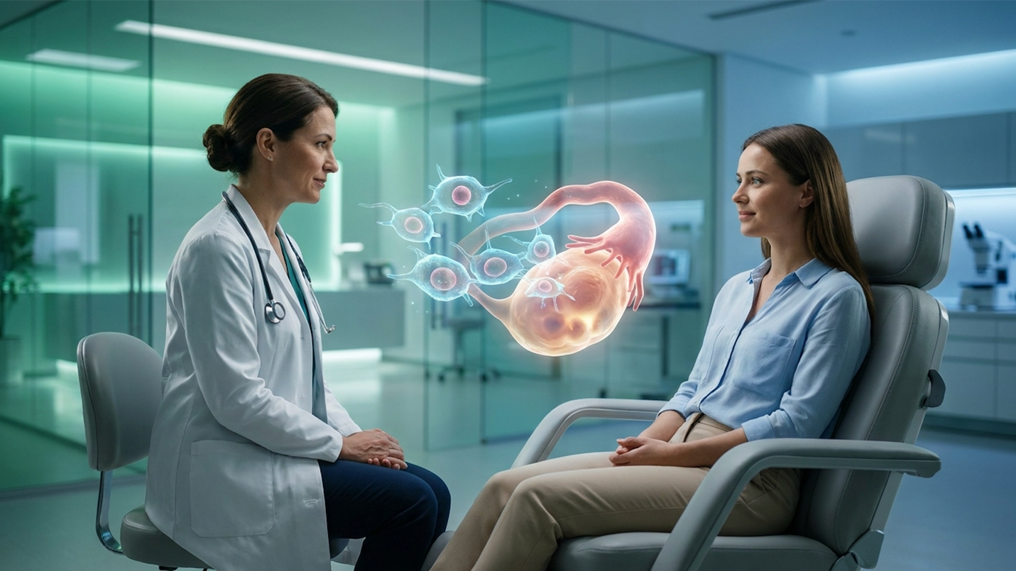
For women experiencing premature ovarian insufficiency, conventional medicine usually offers one path: symptom management. But emerging clinical data suggests cellular therapies can target the glandular failure itself. Reviews of recent regenerative clinical trials make the shift undeniable. While synthetic Hormone Replacement Therapy (HRT) artificially stabilizes chemical levels in the blood, it fails entirely to address the underlying cellular senescence driving female endocrine collapse.
Relying solely on synthetic symptom management leaves the ovarian structure to deteriorate. This accelerates physical aging, disrupts metabolic function, and severely complicates fertility for women suffering from premature ovarian insufficiency or PCOS. It is effectively putting a fresh coat of paint on a crumbling biological foundation.
By the end of this guide, readers will understand the precise cellular mechanisms of Mesenchymal Stem Cells (MSCs) and how they facilitate the biological restoration of the endocrine system. This guide critically evaluates current clinical evidence, defines biological mechanisms of action, outlines expected costs and safety profiles, and clarifies exactly who qualifies for these advanced therapies and who should walk away.
Stem cell therapy for premature ovarian insufficiency utilizes targeted cellular interventions to restore the Endocrine Structural Restoration (ESR) of damaged glandular tissue naturally.
Female hormone imbalance symptoms occur when the endocrine network primarily the ovaries, thyroid, and adrenal glands experiences structural degradation or cellular senescence. Identifying what causes hormone imbalance in women requires analyzing inflammatory markers and glandular health, not just systemic estrogen levels. Achieving natural hormone balance requires interventions that address this underlying biological deterioration directly at the tissue level.
Physicians cannot just look at a blood panel, see low estrogen, and write a prescription. That approach borders on clinical negligence for advanced cases. The endocrine system operates as a tightly regulated feedback loop involving the hypothalamus, pituitary gland, and ovaries. When the ovaries stop responding to brain signals due to structural damage, the entire network destabilizes, cascading into systemic physiological chaos.
The root cause of these severe disruptions lies deep within the ovarian microenvironment. Over time, or due to aggressive autoimmune conditions, the structural tissue of the ovary degrades. Pathology reports routinely reveal extreme oxidative stress and the rapid apoptosis (cell death) of granulosa cells. These are the exact supportive cells responsible for nurturing ovarian follicles and producing natural estradiol.
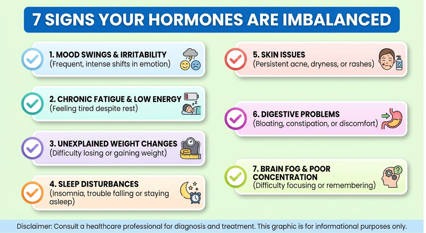
When this delicate cellular environment turns hostile, the physical symptoms are brutal, systemic, and relentless. Clinical evaluations demonstrate seven core female hormone imbalance symptoms that severely disrupt daily functioning and long-term vitality:
Traditional diagnostics repeatedly fail these patients. Why? Because they mischaracterize the fundamental problem. Standard physicians look solely at blood serum levels, completely ignoring the microscopic tissue health of the actual gland producing the hormone. It is exactly like checking a car’s exhaust output to try and fix a cracked engine block.
Hormone Replacement Therapy (HRT) consistently fails to address the microscopic cellular deterioration happening inside the endocrine system. Synthetic HRT relies on a very basic, superficial premise: exogenous chemical supplementation. If the body isn’t making enough estradiol, the standard protocol is to prescribe a patch, pill, or cream to artificially raise those levels in the bloodstream.
Make no mistake, there is no denying that HRT provides crucial relief for many by stopping severe hot flashes. But it remains a physiological band-aid. HRT does absolutely nothing to reverse cellular aging, stop apoptosis, or repair microvasculature within the ovary itself. It simply floods the bloodstream with lab-created hormones to trick surviving receptors into functioning, all while the underlying glandular structure continues to physically decay.
The clinical trade-offs of this approach are significant and well-documented. The human body isn’t designed to process a static, synthetic load of hormones indefinitely without consequence. Clinical consensus suggests that prioritizing chemical symptom management requires accepting inherent long-term risks.
While traditional hormone replacement therapy (HRT) relieves symptoms, it is associated with a 1.5-fold increased risk of blood clots necessitating rigorous cardiovascular screening (NIH, 2026).
This limitation explains the massive consumer shift currently underway. Women are actively demanding natural hormone balance protocols. They require alternatives to hormone replacement therapy that don’t carry oncological or thrombotic risks. They want to physically reverse the biological clock of their glands, not just mask the symptoms with a daily pill that comes with a black-box warning.
Traditional medical models view the endocrine system as a factory requiring external chemical shipments when internal production slows down. If the factory stops making estrogen, they just ship in synthetic estrogen from the outside. However, regenerative medicine approaches the problem differently. Instead of relying on synthetic supplements, advanced clinical applications focus on repairing the cellular machinery itself so the factory can resume natural production.
Stem cell therapy for hormones focuses on repairing the biological infrastructure of the glands. Understanding how stem cells balance hormones naturally requires analyzing Mesenchymal Stem Cells (MSCs). Rather than acting as chemical replacements, MSCs utilize paracrine signaling to modulate the local immune microenvironment, promote angiogenesis, and stimulate quiescent dermal and glandular fibroblasts to restore native tissue function.
This isn’t science fiction. It is rigorous cellular biology applied directly to the endocrine system. When these highly adaptive cells are introduced into an inflamed, failing microenvironment, they act as biological general contractors. They assess the localized damage, secrete specific payloads of repair proteins, and actively recruit the body’s native healing mechanisms directly to the site of the glandular injury.
Mesenchymal Stem Cells (MSCs) function as highly sophisticated medicinal signaling cells within the endocrine system. When introduced into damaged or senescent tissue, they do not immediately turn into ovarian cells. Instead, they secrete a massive, targeted payload of bioactive molecules. Specifically, MSCs release high concentrations of Vascular Endothelial Growth Factor (VEGF), Fibroblast Growth Factor (FGF), and Hepatocyte Growth Factor (HGF) to aggressively stimulate angiogenesis.
This biological process builds entirely new vascular networks, restoring critical oxygen and nutrient delivery to dormant ovarian follicles that have been suffocating in scar tissue. By reducing localized tissue fibrosis, these cells completely reprogram the immune microenvironment, shifting aggressive, tissue-destroying macrophages into a state of active cellular repair.
To understand mesenchymal stem cells hormone regulation accurately, clinicians must discard the outdated, simplistic idea of direct cellular differentiation. For decades, the public believed that injecting a stem cell into an ovary meant that cell physically transformed into a new egg. While rare direct differentiation does occur, it represents only a tiny fraction of the clinical efficacy. The true regenerative power lies entirely in paracrine signaling and extracellular vesicle delivery.
When a failing ovary begins to atrophy, it is fundamentally starving to death. Microvascular networks collapse, meaning the remaining follicles are entirely cut off from the body’s blood supply. The extracellular matrix (ECM) becomes a wasteland of fibrotic, cross-linked collagen scar tissue, making it biologically impossible for vital nutrients or oxygen to reach the surviving granulosa cells. VEGF initiates the budding of new capillaries from pre-existing blood vessels. When MSCs secrete VEGF directly into fibrotic ovarian tissue, localized blood flow can increase by up to 40% within weeks so dormant follicles finally receive the vital oxygen necessary to survive, resume maturation, and produce estradiol.
Furthermore, MSCs possess a profound ability to deliver exosomes. These are microscopic extracellular vesicles packed with microRNAs (miRNAs) and messenger RNAs (mRNAs). When an MSC releases an exosome, it physically fuses with a damaged, dying ovarian cell, transferring genetic instructions that literally order the cell to halt apoptosis and resume healthy division.
Simultaneously, MSCs “reprogram” the local immune system. Many cases of premature ovarian insufficiency are driven by silent autoimmune oophoritis, where the body’s own immune system relentlessly attacks the glandular tissue. In this state, aggressive M1 macrophages constantly bathe the tissue in inflammatory cytokines like TNF-alpha and Interleukin-6. When MSCs arrive, they release anti-inflammatory mediators like Prostaglandin E2 (PGE2) and Indoleamine 2,3-dioxygenase (IDO). Research demonstrates these specific molecular signals force aggressive M1 macrophages to transition into the M2 repair phenotype, profoundly decreasing localized tissue inflammation and establishing a biological truce.
Stem cell therapies possess the unique capacity to reprogram local immune responses, allowing them to rebuild endocrine tissues rather than merely masking hormonal decline (PubMed, 2026).
In comprehensive evaluations of regenerative protocols, one foundational concept separates legitimate clinical science from wellness marketing hype: Endocrine Structural Restoration (ESR).
Endocrine Structural Restoration (ESR) is the clinical framework stating that restoring systemic hormonal balance requires repairing the physical, structural integrity of the gland itself, rather than artificially inflating blood serum levels with synthetic chemicals. Clinical data confirms that healthy hormone production cannot occur in a fibrotic, scarred, oxygen-deprived gland. It is biologically impossible. Regenerative medicine for the endocrine system uses ESR to fix the biological factory so it can resume natural manufacturing.
Consider the endocrine system like a complex agricultural ecosystem. The hormones are the crops. Synthetic HRT is the equivalent of spray-painting dead crops green so they look healthy for visual inspection. It provides a cosmetic, systemic illusion of health while ignoring structural decay. ESR, conversely, is about tilling the dense, unyielding soil, rebuilding the irrigation networks, and neutralizing toxic elements so entirely new, native crops can actually grow and thrive.
Applying this to the human ovary reveals incredible microscopic transformations. In early menopause or POI, the ovary becomes highly fibrotic. The delicate microvasculature completely collapses. However, biopsies and advanced imaging reveal that the primordial follicles are frequently still present they haven’t disappeared, they are just trapped in a dormant, suffocating, fibrotic prison that refuses to let them mature.
By applying the ESR framework, clinical specialists use stem cells to secrete matrix metalloproteinases (MMPs), enzymes that actively dissolve that fibrotic scar tissue. As the scar tissue melts away and the VEGF rebuilds the blood supply, the structural integrity of the ovary normalizes. This isn’t about temporary symptom management. It is a targeted, structural rehabilitation mission. If the biological environment is repaired, the surviving follicles wake up, resume the maturation cycle, and natively pump estrogen back into the bloodstream.
Stem cell therapy for menopause utilizes targeted cellular infusions to repair senescent ovarian tissue. When evaluating stem cells for ovarian rejuvenation, clinical data highlights their ability to lower follicle-stimulating hormone (FSH) and elevate native estradiol production. This biological repair mechanism offers investigational treatment pathways for complex endocrine disorders, specifically premature ovarian insufficiency and polycystic ovary syndrome.
Clinicians must differentiate between treating a natural, age-appropriate menopause at 55 versus treating a pathological failure of the ovaries at 35. While the underlying cellular repair mechanisms overlap significantly, the clinical end-goals differ radically. For a 35-year-old, the medical objective is restoring fertility and reversing an unnatural biological collapse. For a 55-year-old, the focus shifts to preserving bone density, cardiovascular health, and cognitive function without the oncological risks associated with synthetic pharmaceuticals.
Premature Ovarian Insufficiency (POI) is an absolutely devastating clinical diagnosis. It occurs when a woman’s ovaries prematurely cease normal function before the age of 40. The standard medical prognosis is universally grim: sudden untreatable infertility, massively accelerated physical aging, severe osteopenia, and a drastic increase in long-term cardiovascular disease risk.
Consider a standard clinical presentation: A 34-year-old woman is diagnosed with POI. Her FSH levels are skyrocketing above 70 mIU/mL (indicating the pituitary gland is screaming at the dormant ovaries to work), and her native estradiol has cratered to post-menopausal levels below 20 pg/mL. Her Anti-Mullerian Hormone (AMH) levels indicate a nearly depleted reserve. For decades, women with this exact presentation were told they had zero options outside of donor eggs and lifelong high-dose synthetic HRT.
Today, premature ovarian failure stem cells are fundamentally altering that narrative. How do they achieve this? Advanced transvaginal ultrasounds and tissue biopsies frequently reveal that women with POI still possess dormant primordial follicles. The tank isn’t actually empty; the fuel is simply frozen in a hostile, highly inflammatory environment. Standard diagnostics write these women off entirely, but cellular medicine targets the dormant tissue.
When specialists inject MSCs directly into this environment, the cells initiate the ESR framework. They significantly enhance local blood microcirculation and aggressively upregulate the PI3K/Akt and mTOR signaling pathways the precise biological triggers required to “wake up” dormant follicles and initiate their growth phase. The stem cells secrete exosomes that cross tough cellular membranes, delivering genetic payloads that instruct the senescent granulosa cells to immediately resume division.
Clinical reviews on MSCs for ovarian insufficiency indicate these specific protocols can decrease elevated FSH levels by an average of 35% in responsive patients (2026)—so women experience a measurable return of native estrogen production without relying on daily pharmaceutical pills.
The timeline for these results requires patience, as biological tissue regeneration cannot be rushed. Month 1 typically involves a reduction in systemic vasomotor symptoms as localized inflammation drops. By Month 3, early shifts in AMH and microscopic follicle recruitment become visible on high-resolution imaging. By Month 6, clinicians frequently measure a sustained drop in FSH, a substantial increase in serum estradiol, and in highly responsive cases, the resumption of regular menstrual cycles, opening the door for natural conception or viable IVF retrieval.
Mesenchymal Stem Cells successfully restore ovarian microvasculature, physically increasing natural estradiol production while lowering follicle-stimulating hormone levels (PubMed, 2026).
Clinicians must approach PCOS entirely differently than POI.
POI is fundamentally a condition of depletion and dormancy. PCOS, affecting nearly 15% of women globally, is a condition of extreme congestion and metabolic chaos. It is a severe inflammatory and metabolic disorder characterized by profound insulin resistance, hyperandrogenism (excess testosterone production), and ovaries that fail to release eggs properly, resulting in painful cyst formation.
Treating PCOS strictly with oral birth control pills to force a withdrawal bleed is an outdated biological band-aid that completely ignores the underlying insulin resistance and macrophage-driven inflammation. It forces a cosmetic, scheduled bleed without doing a single thing to fix the metabolic disaster happening at the cellular receptor level.
Stem cell therapy PCOS protocols do not need to wake up dormant follicles; they need to calm the immune system down and restore metabolic order. The pathogenesis of PCOS is deeply tied to hyperinsulinemia; excess circulating insulin aggressively stimulates the ovarian theca cells to wildly overproduce androgens. This is exactly where the immunomodulatory superpowers of MSCs provide maximum clinical value.
MSCs actively secrete anti-inflammatory cytokines that systematically shut down the systemic inflammation driving PCOS. By forcing pro-inflammatory M1 macrophages to convert to M2 repair phenotypes, MSCs significantly reduce the localized inflammatory burden. This directly helps correct insulin resistance at the cellular receptor level. As MSCs restore the sensitivity of the insulin receptors, circulating insulin levels plummet.
Once this severe insulin resistance drops, the massive overproduction of androgens in the theca cells immediately ceases. Normalizing the ovarian microenvironment interrupts the pathological feedback loop between the ovaries and the brain. Recent clinical case studies demonstrate that MSC immunomodulation significantly reduces hyperandrogenism markers within four to six months so patients frequently resume normal, spontaneous menstruation and ovulation without ever requiring synthetic birth control or Metformin.
While the biological plausibility for these conditions is incredibly strong, patients must rigorously evaluate the procedural realities. Regenerative medicine requires understanding the financial costs, the specific clinical administration protocols, and the strict safety regulations governing these therapies before ever moving forward with treatment.
Determining stem cell therapy cost for hormone balance requires evaluating the cell source, laboratory processing standards, and the specific clinical protocol utilized. When patients ask, “is stem cell therapy safe for hormones?”, the answer depends heavily on the regulatory compliance of the treating facility. Evaluating these critical factors ensures patients select reputable medical providers over commercialized wellness spas offering unverified treatments.
The regenerative medicine industry unfortunately features a dark side. Bad actors promise guaranteed results for massive fees, utilizing subpar or dead cells in unsterile environments. Protecting patient health requires understanding exactly what happens behind the clinic doors, what documentation to demand, and which red flags indicate a dangerous facility.
What actually happens on treatment day? Breaking it down into a realistic clinical timeline demystifies the process.
First, the attending clinician must determine exact cell sourcing. Reputable stem cell hormone therapy clinics generally utilize one of two highly regulated sources:
Current clinical data reviews indicate that allogeneic UC-MSCs are often preferred for severe endocrine repair because they represent “Day 0” biology. They haven’t aged, their telomeres are maximally long, and they possess massive, robust secretory capacities compared to the autologous cells of a 45-year-old patient who is already suffering from severe systemic inflammation.
Once sourced, laboratory processing is the most critical variable in treatment success. The cells must be meticulously expanded in strictly regulated ISO-7 cleanrooms. They are washed to remove any potential antigens and subjected to rigorous flow cytometry testing to ensure absolute viability and characterize their exact cell surface markers. Dead cells do not heal tissue. If a clinic isn’t counting live cells post-thaw using advanced cytometry, they aren’t practicing regenerative medicine; they are guessing.
The administration itself takes place in a highly controlled outpatient clinical setting. For broad, systemic menopausal symptoms or severe metabolic PCOS, clinicians typically utilize a systemic intravenous (IV) infusion over 60 to 90 minutes. This allows the MSCs to circulate and home in on inflamed tissue globally, including the thyroid and vascular system.
For severe POI where restoring fertility is the primary biological goal, a board-certified specialist performs a highly targeted, ultrasound-guided intra-ovarian injection. This minimally invasive procedure requires incredible precision and is usually performed under local anesthesia or light twilight sedation. Patients recover quickly and are typically walking out of the clinic the exact same day. Post-treatment, rigorous monitoring protocols begin, requiring comprehensive hormone panels and transvaginal ultrasounds at the 1-month, 3-month, and 6-month marks to verify physical tissue regeneration.
Honest financial expectations matter, as standard health insurance providers currently classify these procedures as experimental and do not provide coverage.
The baseline price spectrum for legitimate stem cell therapy ranges massively, from $5,000 to $25,000 or more per treatment protocol.
Understanding why this gap exists requires looking at actual laboratory economics. True MSC therapy is incredibly expensive because rigorous laboratory standards dictate the ultimate clinical efficacy. Expanding cells in a sterile ISO-7 cleanroom, maintaining strict cryogenic cold-chains, and running multi-thousand-dollar flow cytometry viability assays to guarantee the patient is receiving 50 to 100 million live cells accounts for roughly 60% of the procedural cost. Patients are also paying for specialized ultrasound imaging equipment, biologic storage, and the expertise of a board-certified regenerative physician performing the injection.
If a clinic is charging only $3,000 for “stem cells,” they are almost certainly just drawing the patient’s blood, spinning it in a cheap desktop centrifuge to create basic Platelet-Rich Plasma (PRP), and legally mislabeling it to secure a sale. Because these treatments represent a significant financial and medical investment, identifying who is not a good candidate is just as critical to responsible patient care as identifying who is.
Stem cell therapy can potentially restore fertility after early menopause by regenerating dormant ovarian follicles, but it is not a guaranteed cure. Clinical evidence shows MSCs can improve ovarian microenvironments and lower FSH levels, occasionally resulting in resumed ovulation. For women with premature ovarian insufficiency, this presents an investigational pathway for conception. However, success depends heavily on the presence of remaining viable follicles, and patients should always consult a fertility specialist.
Stem cell therapy for ovaries is generally considered safe when utilizing ethically sourced, rigorously tested Mesenchymal Stem Cells administered in a regulated clinical environment. The most common side effects are temporary fatigue or mild localized inflammation at the injection site. Severe complications are exceedingly rare but can occur if treatments are performed by uncredentialed providers using unverified biological materials. Patients must ensure any considered treatments comply with strict FDA or IND clinical trial guidelines.
The primary difference between stem cell therapy and HRT is that HRT synthetically masks symptoms, whereas stem cells attempt to biologically repair the damaged endocrine tissue. HRT provides exogenous estrogen and progesterone to immediately stabilize blood levels, carrying long-term cardiovascular risks. Conversely, regenerative medicine uses the Endocrine Structural Restoration framework to promote natural hormone production. However, biological results from stem cells take months to manifest, unlike the immediate chemical effect of HRT.
The clinical success rate of stem cell treatment for Premature Ovarian Insufficiency varies widely based on patient age, baseline ovarian volume, and protocol specifics. Early-stage clinical trials indicate that a significant subset of women experience improved estrogen markers and reduced menopausal symptoms within three to six months. However, robust, large-scale standardized data is still emerging globally. Individual biological responses dictate outcomes, making comprehensive pre-treatment screening mandatory before pursuing therapy.
Stem cell therapy for ovaries is typically performed via a minimally invasive, ultrasound-guided injection directly into the ovarian tissue, or through a systemic intravenous (IV) infusion. The procedure usually utilizes Mesenchymal Stem Cells (MSCs) harvested from umbilical cord tissue or the patient’s own bone marrow or adipose tissue. Following strict laboratory preparation and viability testing, the cells are administered in an outpatient setting, often requiring only local anesthesia and minimal recovery time.
Individuals with active malignancies, a history of certain hormone-receptor-positive cancers, or completely depleted ovarian reserves without dormant follicles are strictly not good candidates for stem cell therapy. Additionally, patients seeking guaranteed immediate symptom relief or those unwilling to undergo rigorous clinical monitoring should pursue conventional pharmaceutical treatments. A board-certified physician must thoroughly review full medical records to definitively rule out contraindications before approving any regenerative protocols.
For advanced-level patients navigating complex endocrine dysfunction, stem cell therapy for premature ovarian insufficiency offers a profound shift away from synthetic symptom management. By utilizing Mesenchymal Stem Cells to target ovarian inflammation and vascular collapse, this protocol addresses the root cause of hormonal imbalance. Clinical trials demonstrate that while it isn’t an overnight fix, resolving glandular failure at the cellular level yields vast improvements in native estradiol production. The best clinical outcomes consistently combine rigorous patient screening, verified cell sourcing, and targeted administration.
This entire clinical approach relies on the framework of Endocrine Structural Restoration (ESR). Repairing the structural integrity and microvascular health of the tissue is infinitely superior to blindly masking symptoms with chemical hormone replacement. ESR doesn’t just trick the body into feeling better systemically; it physically rehabilitates the glandular environment, providing a true biological foundation for sustained, natural hormone production.
If patients are evaluating this specific pathway, the immediate next step must be scheduling a consultation with a board-certified regenerative medicine specialist. Bring ultrasound imaging, recent hormone panels, and a complete medical history to determine if you are a viable candidate for these investigational protocols.
Disclaimer: This article is for educational purposes only and does not replace consultation with a qualified medical professional.