Our Services
Others
- DFPP
- Shockwave
- IV Drip

This is the story of a patient who went through several lumbar spine surgeries and was left with chronic nerve injury, postoperative scar tissue, neuropathic pain, and weakness affecting his right hip and leg.
He shared that his first surgery caused significant nerve injury near the neural foramen. An emergency revision followed, and later a third operation involving posterior instrumentation, pedicle screws, and a foraminotomy was performed to create more space around the affected nerve.
That third surgery brought him substantial pain relief and helped him regain the ability to walk. Even so, chronic nerve pain, altered sensation, and concerns about postoperative scar tissue remained part of his daily life. Along the way, he also developed right greater trochanteric bursitis, likely related to repeated falls and altered movement during his long recovery.
The patient described a range of ongoing symptoms:
What he wanted to explore was whether a supportive, regenerative approach could help ease inflammation, support the tissue around his injured nerve, and improve the discomfort in his right hip.
The patient underwent three lumbar spine procedures. The first was followed, in his account, by significant nerve injury and severe weakness of the right hip and leg. A second, emergency revision was performed, but he felt the narrowing around the right neural foramen was not fully resolved. During the third surgery, posterior instrumentation and pedicle screws were placed, together with a foraminotomy to improve the passage around the affected nerve and this time, he reported meaningful pain relief and improved mobility, even though the neuropathic symptoms remained.
The images reviewed were postoperative lumbar flexion and extension X-rays, showing interbody fusion and posterior spinal instrumentation. These served as a general structural reference a formal radiological review would still be needed to fully assess alignment, movement, fusion status, hardware integrity, residual nerve compression, or postoperative scar tissue.
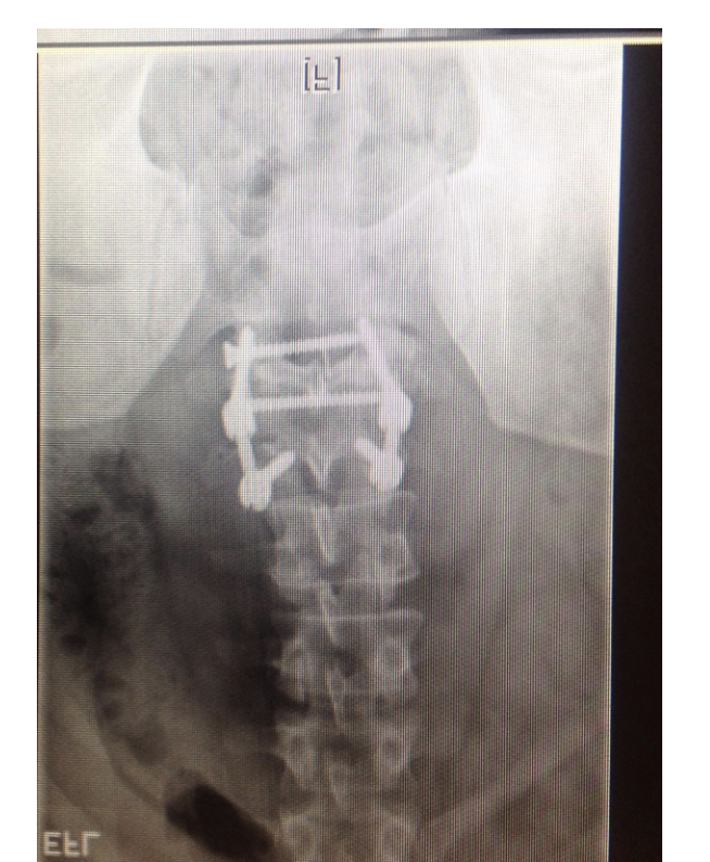
Figure 1: Lumbar flexion and extension radiographs demonstrating postoperative interbody fusion and posterior spinal instrumentation.
This case involved two main concerns: chronic neuropathic symptoms following nerve injury and lumbar spine surgery, and right greater trochanteric bursitis.
It’s important to be clear about what Cell Therapy can and can’t do here it cannot remove mature scar tissue, directly decompress a nerve, reposition spinal implants, or guarantee recovery from chronic nerve damage. Its role is supportive, and may include:
The patient received a targeted Cell Therapy programme, delivered by transforaminal epidural injection at the L4–L5 level, together with a local injection into the right greater trochanteric bursa. In the days that followed, he also received a Signature Vitamins Drip and an NAD+ intravenous infusion.
This programme was designed to support the biological environment around the affected nerve region and the inflamed soft tissue of the right hip not to eliminate scar tissue, fully repair the damaged nerve, or alter the existing spinal implants.
Specific cell doses and treatment dates are intentionally not disclosed here to protect the patient’s privacy.
From a clinical standpoint, this was never simply a case of persistent back pain after spinal fusion. His symptoms reflected a combination of previous nerve compression, possible direct nerve injury, postoperative fibrosis, chronic neuropathic pain, and altered movement built up over a long recovery.
The third surgery appears to have addressed the structural component well, improving the space around the nerve and adding spinal stability. But successful decompression doesn’t always mean complete neurological recovery a nerve that has been significantly injured can go on producing pain, numbness, weakness, or abnormal sensation even once the mechanical compression has been corrected.
His right greater trochanteric bursitis was considered as its own issue too, since altered gait, repeated falls, and compensatory loading can all contribute to hip pain independently of the spine.
In this case, Cell Therapy was positioned as supportive care rather than a way of reversing the earlier surgical injury the goal was to support the biological environment, help regulate inflammation, and complement his neurological and physical rehabilitation.
This is a story of chronic right-sided neuropathic pain following a complicated surgical journey, alongside right greater trochanteric bursitis. The patient regained his ability to walk after a successful third surgery, but continued to live with neuropathic pain and abnormal sensation.
He went on to receive a targeted Cell Therapy programme at the L4–L5 level and the right greater trochanteric bursa, followed by supportive intravenous therapy.
His care team was careful to frame this as part of a broader supportive care plan not a guarantee of nerve regeneration, scar-tissue removal, or reversal of his previous surgical complications. Ongoing follow-up continues to track his pain, mobility, muscle strength, neurological symptoms, hip discomfort, and overall quality of life.