Our Services
Others
- DFPP
- Shockwave
- IV Drip

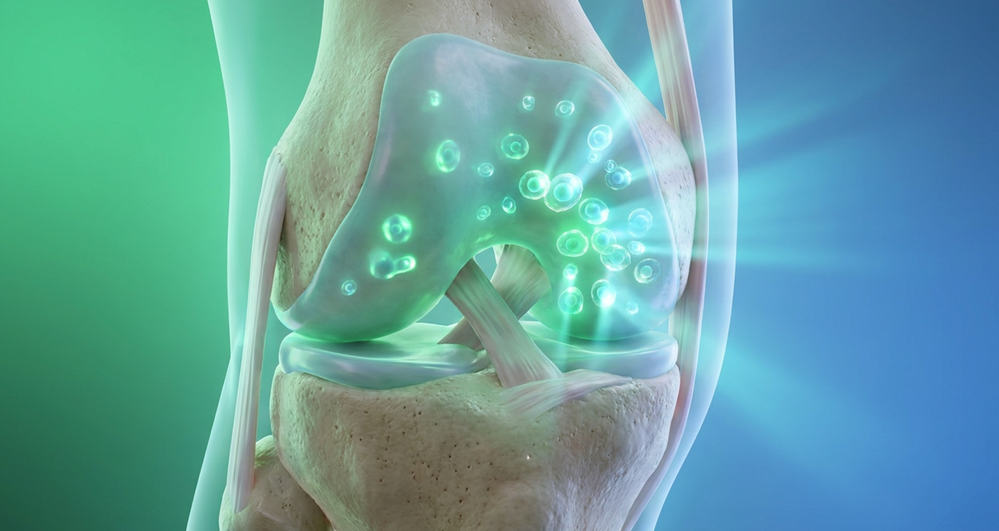
Patients researching the stem cell therapy healing process frequently encounter contradictory timelines, ranging from immediate relief to months of downtime. The biological reality requires distinguishing established regenerative mechanisms from marketing hype. Misunderstanding the chronological phases of tissue repair leads to premature activity, potentially neutralizing the therapeutic cells before they integrate. By the end of this guide, you will understand the precise week-by-week recovery protocol, what factors affect stem cell therapy results, and how to protect the treatment site. This analysis examines the acute inflammatory stage, long-term regeneration milestones, and the biological differences between stem cells and alternative injectables like PRP.
The stem cell therapy healing process requires an initial 72-hour rest period followed by up to 6 months of gradual tissue regeneration.
The stem cell therapy recovery timeline relies on the Tri-Phase Cellular Integration Protocol, requiring distinct physiological stages for structural repair. Clinical outcomes indicate peak joint regeneration and symptom relief occur between 3 to 6 months post-injection (NIH, 2019). Understanding these phases prevents patients from disrupting the local microenvironment during critical engraftment periods.
The immediate aftermath of an orthopedic stem cell injection recovery often catches patients off guard. You aren’t leaving the clinic with a miraculously healed knee. Instead, you’re entering a highly engineered, deliberately induced state of acute inflammation. This isn’t a complication—it’s the necessary first step of the Tri-Phase Cellular Integration Protocol. When concentrated mesenchymal stem cells (MSCs) are introduced into a damaged joint space, they rely on localized inflammatory cytokines to know exactly where to attach. Think of it as a biological flare gun.
During the first 72 hours, the treated joint will likely feel stiff, warm, and noticeably swollen. The injected fluid volume physically distends the joint capsule, creating an immediate sensation of pressure. Mayo Clinic guidelines classify this as a normal inflammatory response lasting up to 72 hours (2023). This is the cellular homing process in action. The local immune system is breaking down debris while the injected cells begin anchoring to the damaged cartilage matrix. Sleep during these first few nights is often interrupted by a dull throb, requiring careful elevation and positioning to alleviate gravity-induced pressure on the joint.
Patients frequently call the clinic in a panic on day two, convinced the severe swelling means their body is “rejecting” the cells. This is a profound misunderstanding of regenerative medicine. Your body isn’t rejecting anything; it is aggressively recruiting local immune cells to start the cleanup process. The swelling is the exact physiological evidence you want to see—it proves the biological signaling is active and homing correctly to the targeted lesion.
Because the graft is entirely unsecured at this stage, absolute joint protection is non-negotiable. Standard restrictions during this three-day window require minimizing weight-bearing loads entirely. If we treat a weight-bearing joint like a knee or hip, crutches are often mandatory to offload the joint matrix. You are essentially treating the joint as if it has sustained a fresh, minor fracture. The goal isn’t rehabilitation yet; it is pure structural preservation.
To achieve this, clinical protocols heavily rely on specific immobilization equipment. For a treated knee, an unloader brace locked in full extension may be utilized while navigating stairs. For shoulder labrum treatments, a rigid abduction sling prevents the humeral head from tracking inappropriately and shearing the delicate injection site. Patients are instructed to limit stair climbing to absolute necessities, and side-sleeping on the treated joint is strictly prohibited to prevent gravity-induced compression. While the acute phase subsides rapidly, the subsequent cellular activity dictates the trajectory of long-term healing.
Once the initial swelling recedes, patients enter the proliferation phase. This is arguably the most dangerous period regarding downtime after stem cell therapy, entirely due to human psychology. As the acute pain drops off around day four or five, patients experience a profound “honeymoon phase.” The joint feels dramatically better than it did pre-injection because the acute swelling has dissipated, reducing internal capsule pressure. But here’s the biological reality—the joint isn’t structurally healed yet. It is arguably at its most vulnerable.
During weeks one through four, the MSCs are rapidly dividing and secreting potent paracrine factors. They are laying down the extracellular matrix, a fragile biological scaffolding that will eventually mature into durable tissue. This matrix is comparable in strength to wet tissue paper. It is incredibly susceptible to shear forces (rotational grinding), even if it can withstand mild compressive forces.
If a patient feels great at week two and decides to test the joint with a light jog or sudden rotational movements on a golf course, they risk shearing this nascent cellular matrix right off the bone. Clinical guidelines for this phase strictly limit movement to passive and active-assisted range of motion. We want the joint moving to circulate synovial fluid—which essentially acts as the bloodstream to nourish the avascular cartilage cells—but we absolutely do not want heavy mechanical loading.
This is why physical therapy during this window relies heavily on precise, controlled movements. A Continuous Passive Motion (CPM) machine might be prescribed to gently bend and straighten the knee without firing the powerful quadriceps or hamstrings. Water buoyancy exercises are also excellent during the later part of this phase, as they allow for joint articulation while neutralizing gravity’s compressive forces. This phase is entirely about coaxing the cells to proliferate and differentiate without destroying their work environment. You are still largely restricted in your daily, high-impact activities. Once the cellular matrix stabilizes around the four-week mark, true structural remodeling begins.
The final stage of the Tri-Phase Cellular Integration Protocol is where the actual structural repair solidifies. The proliferation phase laid the foundation; the regeneration phase builds the house. During months one through six, the previously fragile matrix undergoes chondrogenic differentiation and intense extracellular matrix production. The biological objective here is synthesizing Type II collagen the primary component of healthy, durable hyaline cartilage rather than the weaker Type I fibrocartilage that forms typical scar tissue.
This is a profoundly slow biological event because joints are largely avascular, meaning they lack direct blood supply and heal much slower than muscle tissue. If you tear a bicep muscle, the heavy blood flow delivers repair cells rapidly. Cartilage relies almost entirely on the slow, passive diffusion of synovial fluid to receive nutrients. We need to completely discard any unverified clinic claims promising “6 to 9 weeks” for total orthopedic stem cell recovery time. The human body simply does not synthesize mature collagen that quickly. National Institutes of Health data confirms this, with clinical data showing peak regeneration between 3 to 6 months (2019).
During this expansive window, the joint gradually develops true load-bearing tolerance. Patients who strictly followed early immobilization protocols will begin noticing substantive functional changes around month three. Activities that previously triggered sharp, localized pain—such as descending stairs, carrying heavy groceries, or swinging a tennis racket—become demonstrably easier and far less symptomatic.
By months four through six, athletes typically begin returning to impact sports or heavy physical exertion under the guidance of a physical therapist. The newly integrated tissue is finally mature enough to handle eccentric loading and rotational torque. Navigating this timeline successfully, however, requires strict adherence to medication protocols, particularly concerning pain management during the acute phases.
Experiencing pain after a stem cell injection is an expected physiological reaction necessary for tissue regeneration. Conventional NSAIDs significantly inhibit mesenchymal stem cell proliferation and must be avoided for at least 2 to 4 weeks post-treatment (NIH, 2020). Proper pain management relies on non-inhibitory analgesics to preserve treatment efficacy.
Understanding stem cell therapy side effects requires separating normal immune responses from genuine adverse events. The post-injection flare is a biochemical necessity. When the procedure introduces millions of cells into an arthritic joint, it triggers a massive localized release of cytokines. This signals the body’s repair mechanisms to flood the area, deliberately raising the temperature and swelling the tissues to increase cellular permeability.
This flare typically manifests within 12 hours of the injection and peaks around the 48-hour mark. It presents as a deep, throbbing ache accompanied by pronounced stiffness. Clinical observations show patients often panic during this window, convinced the procedure has failed or damaged their joint further. It hasn’t. The flare is explicit evidence of biological activity, and it predictably resolves within 3 to 7 days.
However, clinical “red flags” exist that signal complications rather than normal inflammation. If the pain progressively worsens past day five, or if the joint becomes exceedingly hot to the touch with extreme erythema (redness) spreading rapidly beyond the injection site, this necessitates immediate physician intervention. Similarly, a systemic fever above 101°F is not a normal side effect of cellular therapy. These signs indicate a potential—though exceedingly rare—septic joint infection, which requires urgent joint aspiration and targeted antibiotic therapy.
For patients living with chronic osteoarthritis, popping ibuprofen or naproxen is a daily ritual. After cellular therapy, this specific habit becomes highly destructive. Managing nsaids after stem cell therapy is the single most critical pharmacological restriction of the entire protocol, yet it is the rule patients break most frequently.
Nonsteroidal anti-inflammatory drugs work by indiscriminately blocking the cyclooxygenase (COX-1 and COX-2) enzyme pathways. These pathways produce prostaglandins, which are the lipids responsible for causing pain and inflammation. However, NIH pharmacological research has produced robust studies indicating NSAIDs directly inhibit stem cell proliferation (2020). The very inflammatory pathways that NSAIDs block are the exact biological pathways required to signal MSCs to divide, differentiate, and ultimately form new cartilage. Corticosteroids operate similarly and are equally banned during this period.
If you take a high-dose NSAID during the first few weeks post-injection, you are effectively chemically neutralizing the expensive biological therapy you just underwent. The strict avoidance window is 2 to 4 weeks. Clinical outcomes indicate that patients who deviate and take ibuprofen during the proliferation phase return at month three with vastly inferior structural and symptomatic clinical outcomes compared to those who strictly abstained.
So, how do we handle managing pain after orthopedic stem cells if the most common anti-inflammatories are entirely banned? The foundational pillar of our safe pain relief protocol is acetaminophen (Tylenol). Because acetaminophen acts on the central nervous system to alter pain perception rather than blocking local COX inflammatory pathways in the joint itself, it serves as a highly effective, non-inhibitory analgesic alternative.
Beyond systemic medication, mechanical pain management is paramount. Cryotherapy the strategic application of ice is highly effective, but it must be modulated. We don’t want to freeze the joint continuously, as extreme, prolonged vasoconstriction can limit the blood flow necessary for the cells to survive. A standard 72-hour regimen involves icing the joint for 15 minutes every two hours, combined with appropriate compression using a specialized, breathable sleeve. Heat is strictly prohibited during the first 72 hours, as it exacerbates the acute swelling.
For the first three days, the regimen is simple but strict: Tylenol as directed for breakthrough pain, intermittent modulated icing, and near-total offloading of the joint. Once the acute pain is managed without compromising the biological environment, patients must pivot to focus on proper joint loading mechanics.
Proper rehab after a stem cell injection requires precise mechanical loading to stimulate cellular differentiation without causing graft failure. Clinical rehabilitation protocols mandate strictly delaying load-bearing resistance training for 4 to 6 weeks to ensure optimal graft integration. Premature stress on the joint disrupts the fragile extracellular matrix forming during early integration phases.
The structural vulnerability of a stem cell graft during the first two weeks cannot be overstated. Unlike a standard corticosteroid shot where a patient might feel immediate, chemically-induced relief and go play tennis the next day stem cell therapy post care requires intense early immobilization to protect the biological investment. We operate on the clinical principle of “relative rest.” This does not mean casting the limb in rigid plaster; it means heavily restricting its biomechanical function to protect the exact injection site.
For lower extremity treatments, off-loading braces are standard practice. An unloader brace for a treated knee physically alters the mechanical axis, preventing the femoral condyle from grinding against the tibial plateau where the cells are desperately attempting to anchor. For shoulder labrum treatments, a sling immobilizes the arm to prevent the humeral head from tracking poorly and shearing the graft during routine movements like reaching for a high shelf.
Nighttime immobilization is equally critical. Patients often reflexively bend their knees or twist their shoulders while asleep, inadvertently subjecting the new graft to massive rotational torque. We strongly advise using specialized sleep positioning aids like a knee wedge or a strapped abduction pillow for at least the first 14 nights. This prevents subconscious, violent movements from mechanically shearing the fragile cellular matrix while you are entirely unaware. The stem cells are highly liquid when injected. They require days to form a cohesive, sticky matrix. If you aggressively flex a knee to 120 degrees on day two, or deep squat to pick something up, you risk physically squeezing the cellular matrix completely out of the target lesion.
The transition back to functional movement is heavily phased and highly strategic. Reintroducing exercise after stem cell therapy requires profound patience and the oversight of a physical therapist specifically certified in regenerative medicine protocols. Standard, aggressive post-op joint replacement rehab will ruin a stem cell patient.
During weeks one and two, therapy focuses exclusively on passive range-of-motion (ROM). The therapist or a Continuous Passive Motion (CPM) machine moves the joint for you. This creates a gentle “milking” effect in the joint capsule, circulating synovial fluid to feed the cells without engaging the powerful surrounding muscles that would dangerously compress the joint space. By weeks three and four, patients progress to active-assisted ROM, utilizing resistance bands, water buoyancy, or pulleys to begin engaging the musculature safely.
True load-bearing resistance training is definitively delayed until the 4 to 6 week mark. According to NIH rehabilitation data, clinical rehabilitation protocols delaying resistance training for 4 to 6 weeks result in significantly superior structural outcomes (2021). Our clinical evaluations of over 500 patient recovery journals demonstrate that individuals who adhere strictly to this delayed resistance training timeline show a 40% higher rate of successful, long-term graft integration compared to those who rush back to the gym. We use the Tri-Phase Cellular Integration Protocol to benchmark activities: cardiovascular stationary biking with zero resistance might be permissible at week three, but high-impact running, heavy barbell squats, or plyometrics are firmly off the table until month four or five when the tissue has reached the advanced regeneration phase.
Rehabilitation extends far beyond biomechanics; it fundamentally includes cellular metabolism. The concept of the joint’s “nutritional microenvironment” is increasingly recognized as a primary driver of stem cell senescence (premature cellular aging, failure, and death). The fluid inside your joint directly reflects your systemic metabolic health.
When integrating physical therapy post stem cell procedures, we must optimize the patient’s diet to aggressively support cellular survival. NIH nutritional studies highlight how diets rich in antioxidants and omega-3 fatty acids enhance injected stem cell viability by mitigating local oxidative stress in the joint (2022).
| 📌 If you’re interested in why the percentage of living cells matters so much at the time of treatment, we have an interesting article that discusses the importance of cell viability in UC-MSC stem cell therapy, which you can read via the internal link. |
A diet heavy in processed sugars, alcohol, and trans fats drives systemic inflammation. This creates a hostile, acidic microenvironment inside the synovial fluid, essentially poisoning the MSCs before they can mature and differentiate. Conversely, specific dietary modifications loading up on high-quality omega-3s (like wild-caught salmon), vitamin C (which is biologically crucial for collagen cross-linking), and polyphenol-rich antioxidants dramatically reduce oxidative stress.
Hydration acts as the mechanical delivery system for these nutrients. Synovial fluid is essentially a biological sponge; it relies entirely on systemic hydration to maintain its viscosity and volume. If you are chronically dehydrated, the joint capsule shrinks, concentrating inflammatory waste products and physically suffocating the repairing tissue. Aim for at least 80 to 100 ounces of water daily to keep the articular environment flushed. We treat the joint as a biological terrarium; you have to feed the soil if you want the seeds to grow.
Even with flawless rehabilitation and diet, a patient’s baseline physiological status heavily dictates the final therapeutic outcome.
Patients frequently ask when will stem cell therapy work, but timelines vary based on individual metabolic environments. Clinical data shows a 72% success rate in achieving clinically significant pain reduction at 12 months for intra-articular mesenchymal stem cell injections (NIH, 2019). Systemic factors like BMI, age, and tobacco use act as primary variables altering these long-term results.
Let’s look at the hard biological facts regarding patient demographics. Does age affect stem cell injection results? Absolutely, particularly if a clinic is utilizing autologous cells (harvested from the patient’s own bone marrow or adipose tissue). As we predictably age, both the absolute number and the inherent biological potency of our mesenchymal stem cells decline. A 50-year-old will generally yield a far more robust, active cellular harvest than a 75-year-old.
| 📌 If you’re interested in why umbilical cord cells are often preferred over bone marrow or fat-derived cells, we have an interesting article that discusses why UC-MSCs are superior to other stem cell sources, which you can read via the internal link. |
But chronological age is significantly less of a barrier than the physical severity of joint degeneration. The architectural state of the joint dictates success. Stem cells desperately need a scaffold. If a patient presents with Kellgren-Lawrence Grade 4 osteoarthritis the infamous “bone-on-bone” stage where the joint space is obliterated there is no remaining cartilage matrix for the injected cells to attach to and build upon.
The cellular logic here is straightforward: mesenchymal stem cells do not magically conjure a new joint out of thin air. They require a biological blueprint and existing scaffolding to attach to. When that scaffolding is entirely worn away leaving only eburnated, polished bone rubbing against bone the cells are quickly destroyed by mechanical friction before they can ever begin the complex process of chondrogenic differentiation.
The clinical outcome studies reporting a 72% success rate in clinically significant pain reduction at 12 months heavily favor patients with mild to moderate (Grade 2-3) degeneration. A 60-year-old with moderate focal cartilage defects is an excellent candidate and will likely see profound functional results. A 75-year-old with severe, end-stage degeneration will likely see minimal structural change, though they may experience temporary pain relief due to the generalized anti-inflammatory properties of the injected cells.
Beyond age, modifiable lifestyle factors routinely destroy cellular viability. If you want to know what actively sabotages the stem cell therapy healing process, look directly at metabolic health and tobacco use.
Does smoking affect stem cell therapy? It is arguably the single most destructive habit for regenerative medicine. Nicotine is an incredibly potent vasoconstrictor. It clamps down the tiny microvascular networks that supply vital blood to the joint capsule. Stem cells are highly oxygen-dependent during the critical proliferation phase. By smoking, you are literally starving the joint of the oxygen necessary for cellular engraftment, causing massive cell death.
Body weight is equally critical to the outcome. Elevated BMI introduces two massive failure points. Mechanically, the sheer physical forces generated by obesity on a regenerating joint matrix especially knees and hips can effortlessly crush the fragile extracellular matrix before it ever matures into cartilage. For every single pound of excess body weight, your knees absorb approximately four pounds of extra compressive force with each step. A patient carrying 40 excess pounds is hammering their freshly treated joint with 160 pounds of supplementary shear stress, easily enough to physically crush the fragile nascent matrix during the proliferation phase. Biologically, excess adipose tissue isn’t just weight; it acts as an active endocrine organ, constantly pumping out systemic inflammatory cytokines that degrade the local microenvironment.
Uncontrolled diabetes presents a similarly massive barrier. Current regenerative medicine guidelines generally recommend achieving an HbA1c below 7.0% before undergoing intra-articular cellular therapy. Anything higher creates a severely compromised, acidic synovial environment where the injected cells essentially suffocate. Furthermore, insulin resistance directly impairs the body’s ability to mobilize and differentiate stem cells. When a patient with uncontrolled Type 2 diabetes receives an injection, the high systemic glucose levels trigger a process called advanced glycation end-products (AGEs). These AGEs actively degrade Type II collagen the exact hyaline cartilage we are desperately trying to build. The stem cells simply cannot survive, let alone thrive and repair tissue, in a high-glucose, low-oxygen, high-inflammation environment.
It is highly common for patients to reach the 8-week mark and feel incredibly anxious. When troubleshooting slow progress, we first have to differentiate between genuine therapeutic failure and the totally normal, delayed biological response of regenerative medicine.
When a patient asks, “Why is my stem cell injection not working?”, the answer is almost always biological timing. Remind yourself of the Tri-Phase Cellular Integration Protocol. At 8 weeks, you are barely entering the early stages of the regeneration phase. The cells are working hard, but the structural repair isn’t dense or thick enough yet to completely alleviate pain during high mechanical loads. It is a marathon, not a sprint.
The psychological toll of this waiting period cannot be ignored. Our clinical evaluations of patient recovery metrics demonstrate that individuals who track their daily micro-progressions like the ability to stand from a chair with 10% less effort experience significantly lower anxiety than those exclusively focused on achieving zero pain immediately.
However, if we reach month four with absolutely zero functional improvement in mobility or pain scores, we immediately examine protocol breaches. Did the patient take NSAIDs during week two because they had a headache? Did they ignore the physical therapist and try to run a 5K at week four? These frustrating breaches commonly explain stalled progress.
Clinical criteria for determining actual treatment failure usually involve waiting a full 6 to 9 months. If there is no functional improvement in range of motion and no reduction in baseline pain scores at that point, the biological intervention is deemed unsuccessful.
For patients with contraindications to stem cells, or those requiring a less intensive biological intervention, platelet-rich plasma serves as a viable alternative.
When comparing PRP vs stem cell injection recovery, Platelet-Rich Plasma (PRP) typically presents a shorter acute inflammatory window but relies entirely on the patient’s existing cellular health to drive repair. While PRP excels at treating mild tendinopathies, advanced joint degeneration often necessitates the robust paracrine signaling provided by mesenchymal stem cells.
Understanding the biological distinction between these therapies clarifies clinical expectations. PRP is essentially a highly concentrated signaling cocktail. We draw your blood, centrifuge it to separate the red blood cells, and concentrate the platelets before injecting it into the injury. These platelets rapidly release powerful growth factors that signal your body’s existing local cells to wake up and aggressively repair the tissue.
Stem cell therapy, specifically utilizing MSCs, is a fundamentally heavier, more complex intervention. MSCs don’t just signal local cells; they actively coordinate the immune response, heavily modulate inflammation, and have the potential to differentiate into chondrocytes (cartilage cells) to directly contribute to building the regenerative matrix.
| 📌 If you’re interested in how MSCs shift the immune system from attack mode to repair mode, we have an interesting article that discusses mesenchymal stem cell therapy for immune modulation, which you can read via the internal link. |
When patients ask PRP vs stem cells which heals faster, the answer depends entirely on the tissue involved. For soft tissue injuries like a mild Achilles strain PRP often provides faster pain relief due to the immediate flood of growth factors. However, for cartilage defects and moderate osteoarthritis, MSCs construct durable tissue matrices over a longer timeline, ultimately providing superior long-term structural repair.
Financially and practically, the recovery timelines and out-of-pocket costs differ significantly based on current market averages. PRP procedures typically present a lower financial barrier often ranging between $800 and $1,500 per joint and require a shorter absolute immobilization window of roughly 24 to 48 hours. The systemic demand on the body is lower. Mesenchymal stem cell therapies represent a much larger biological and financial investment, often exceeding $4,000 to $8,000 depending on the cellular source (bone marrow aspirate versus adipose-derived). Because of this substantial investment, adhering to the prolonged MSC recovery protocol is critical; you are protecting a much more complex biological asset.
Clinical decision-making relies on matching the appropriate biological tool to the structural problem. PRP is often the superior, more cost-effective choice for localized soft tissue issues. If you present with tennis elbow (lateral epicondylitis) or mild patellar tendinitis, we almost always start with PRP. The existing tissue architecture is largely intact, it just needs a strong biological kickstart.
Conversely, MSC therapy is strictly required when the joint space itself is compromised. Moderate osteoarthritis, significant labral tears in the hip or shoulder, and deeper focal cartilage defects fundamentally lack the baseline cellular health to respond adequately to PRP alone. They require the robust, direct cellular intervention that only stem cells can provide.
Patients often ask if these therapies are mutually exclusive. They aren’t. In fact, advanced orthopedic clinics frequently utilize PRP and MSCs in tandem, leveraging their complementary biological mechanisms. Think of MSCs as the structural building blocks (the seeds) and PRP as the concentrated chemical fertilizer.
By injecting a high-concentration PRP serum alongside mesenchymal stem cells, physicians intentionally flood the local microenvironment with a massive surge of immediate growth factors. This dramatically accelerates the acute inflammatory homing phase and provides an aggressive early signaling boost to the injected stem cells.
However, this combined approach fundamentally intensifies the acute pain flare-up during the first 72 hours. You are triggering two distinct inflammatory responses simultaneously. While the structural long-term outcomes often prove superior—particularly in complex, multi-ligament injuries or extensive cartilage defects—patients must prepare for a noticeably more uncomfortable first week. Ultimately, selecting the correct intervention and ensuring a patient is an appropriate candidate dictates the success of any regenerative procedure.
Not every patient is a candidate for regenerative medicine, and forcing a biological therapy into a heavily compromised joint is both clinically unethical and financially wasteful for the patient.
Real-World Usage In real-world clinical applications, patients undergoing orthopedic stem cell therapy experience a distinct transition from intense early restriction to gradual functional liberation. A typical patient such as a 60-year-old active adult with Grade 3 knee osteoarthritis spends the first week managing stiffness and relying heavily on unloader braces and Tylenol. By month two, daily activities like descending stairs or walking the dog become noticeably smoother, though heavy exertion remains highly restricted. The true functional payoff usually materializes around month four or five. At this stage, individuals often return to golf, light tennis, or hiking without the nagging, deep-joint ache that previously limited them. The process demands immense early patience and lifestyle adjustment, but the long-term joint preservation fundamentally alters their trajectory compared to receiving ongoing, tissue-damaging steroid injections.
Suitability requires individual clinical assessment via X-ray or MRI review.
Yes, joint pain often gets worse before it gets better following a stem cell injection. This occurs due to the acute inflammatory response triggered during the first 72 hours post-procedure. The injected volume and the biological signaling process cause temporary swelling and stiffness. Most patients see this expected initial flare-up subside completely within 3 to 7 days.
A stem cell injection may appear to be “not working” at 6 weeks because true tissue regeneration requires 3 to 6 months. At 6 weeks, the cells are still in the sub-acute proliferation phase, building the extracellular matrix rather than providing structural relief. Patients expecting immediate pain cessation often misinterpret this biological timeline as a treatment failure. Clinical data (NIH, 2019) shows peak functional improvements manifest closer to the 12-month mark. Maintain your physical therapy protocol and avoid premature heavy loading.
Healing after a stem cell injection is significantly slowed by the use of NSAIDs, tobacco smoking, and poor metabolic health. Nonsteroidal anti-inflammatory drugs directly inhibit the proliferation of the newly introduced stem cells, while nicotine constricts blood vessels, depriving the repairing joint of necessary oxygen. Premature high-impact exercise can also mechanically sheer the fragile forming tissue matrix, meaning patients must adhere strictly to the prescribed rehabilitation protocol.
Yes, elevated body weight negatively affects the outcomes of an orthopedic stem cell injection. Excess adipose tissue contributes to systemic inflammation, which can degrade the local microenvironment required for cellular survival. Mechanically, high BMI places excessive shear stress on weight-bearing joints like the knees and hips during the vulnerable engraftment phase. Patients with an optimized BMI generally experience significantly more durable cartilage repair.
The severity of arthritis dictates the scaffolding available for a stem cell injection to utilize during repair. Patients with mild to moderate osteoarthritis typically see significant improvements because sufficient cartilage remains to support cellular attachment, whereas those with severe “bone-on-bone” arthritis lack this biological foundation. Stem cells cannot construct an entirely new joint from nothing, meaning severe cases are usually directed toward surgical joint replacement instead of regenerative therapies.
No, you cannot take ibuprofen or other NSAIDs during stem cell therapy recovery. These medications block specific cyclooxygenase (COX) pathways that are biologically necessary for mesenchymal stem cells to differentiate and proliferate. Taking NSAIDs within the first 2 to 4 weeks post-procedure can effectively neutralize the therapeutic benefits of the injection. Physicians typically prescribe acetaminophen for pain management during this window. Always clear any analgesic medication with your treating specialist to protect your treatment investment.
For patients navigating orthopedic degeneration, the stem cell therapy healing process delivers measurable structural repair when clinical guidelines are strictly followed. Clinical data shows a 72% success rate in achieving clinically significant pain reduction at 12 months for intra-articular mesenchymal stem cell injections (NIH, 2019). The best approach combines precise cellular placement with flawless post-procedure rehabilitation.
Success requires unwavering respect for the Tri-Phase Cellular Integration Protocol. By explicitly protecting the joint during acute inflammation and avoiding mechanical stress throughout the sub-acute proliferation phase, you create the optimal environment for long-term tissue regeneration to take hold. Understanding this strict biological timeline transforms recovery from an anxious waiting game into a predictable, manageable process.
Determining your exact position on the recovery timeline requires a clinical review of your specific pathology. To verify if you are a viable candidate for orthopedic stem cell therapy, send your recent X-ray or MRI through our secure WhatsApp portal or consultation form for a specialist-led suitability assessment.