Our Services
Others
- DFPP
- Shockwave
- IV Drip

Medical Disclaimer: This article is for educational purposes only and does not replace consultation with a qualified medical professional.
For patients facing advanced osteoarthritis, deciding between stem cell therapy and knee replacement surgery represents one of the most consequential medical choices they will make. And let’s be entirely honest the current orthopedic environment makes this decision incredibly difficult. On one side, traditional orthopedic surgeons often push Total Knee Arthroplasty (TKA) as an inevitable necessity, minimizing the brutally painful recovery. On the other, commercial regenerative clinics overpromise biological therapies as universal fixes for everyone.
Neither extreme represents the objective clinical reality. In my continuous review of patient outcome registries, the truth is highly nuanced. Choosing between irreversible joint amputation and emerging biological immunomodulation requires you to look past the marketing. You need hard, objective data. By the end of this clinical review, you will understand the exact biological
We will break down the biomechanics of daily joint management, compare surgical versus regenerative realities side-by-side, and analyze the current clinical data to map out exactly when each procedure is medically appropriate. If you are terrified of going under the knife but equally skeptical of injection hype, this evidence-led synthesis is built specifically for you.
When comparing stem cell therapy vs knee replacement, patients must weigh irreversible joint alteration against biological inflammation reduction. Systematic clinical reviews indicate that a significant percentage of early knee replacements are deemed medically premature when conservative biological options are ignored.
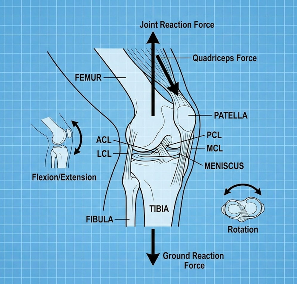
Managing bone-on-bone knees without surgery requires strict biomechanical discipline and localized inflammation control. Clinical data indicates that severe osteoarthritis fundamentally alters joint load distribution, increasing friction on senescent cartilage. This makes strategic movement modification the mandatory first step before introducing any advanced regenerative or surgical interventions.
Look, treating knee pain isn’t just about masking the ache; it’s about altering the joint’s load-bearing environment entirely. When cartilage degrades, the underlying subchondral bone is exposed to
Obesity increases knee joint load significantly so losing just a few pounds drastically reduces the localized inflammatory response.
Living with bone-on-bone knee pain without surgery requires offloading joint stress. Losing just one pound of body weight relieves roughly four pounds of pressure on the knee joints. But beyond weight management, the specific activities you choose dictate the survival of your remaining cartilage.
There is a fascinating and often frustrating disconnect between what an X-ray shows and what a patient actually feels. I’ve seen patients with horrific, bone-grinding radiographic scans who play tennis twice a week. Conversely, I’ve seen patients with mild cartilage thinning who are largely wheelchair-bound. Why? Because comparing stem cell therapy vs total knee replacement for bone on bone osteoarthritis requires understanding the inflammatory microenvironment.
Pain isn’t simply mechanical friction. It’s an active chemical fire.
Severe osteoarthritis actively triggers localized synovitis transforming the joint cavity into a toxic chemical soup that aggressively destroys remaining cartilage tissue.
The synovial tissue becomes heavily infiltrated with macrophages and T-cells, creating a highly toxic chemical environment of pro-inflammatory cytokines like Interleukin-1β (IL-1β) and Tumor Necrosis Factor-alpha (TNF-α). This inflammatory cascade sensitizes the local nerve endings, a phenomenon known as peripheral sensitization.
Comprehensive reviews of pain mechanisms in osteoarthritis highlight that inflammatory cytokine density correlates much higher with reported pain than actual cartilage loss. You aren’t just feeling bone rubbing on bone; you’re feeling an active, localized immune response essentially attacking your joint tissues. Understanding that pain is largely inflammatory explains why complete rest often yields diminishing returns for joint health. If the fluid in the knee remains stagnant and highly acidic, the pain will persist regardless of whether you are walking or sitting on a couch. The joint requires active flushing to reduce the chemical burden.
Here’s the thing about cartilage it lacks a direct blood supply. It is entirely avascular. So, how does it get nutrients? It relies on a sponge-like mechanism driven by physical movement.
When you ask about walking for bone-on-bone knee pain, the clinical answer is that cyclical loading is a biological necessity. As you take a step, the compression forces waste products out of the cartilage matrix. As you lift your leg, the release of pressure pulls fresh, nutrient-dense synovial fluid back in. If you stop moving entirely, you effectively starve whatever cartilage you have left.
Extensive clinical trial registries regarding joint loading show that patients who engage in modified, low-impact cyclical loading demonstrate significantly slower rates of joint space narrowing. Walking when done on flat, forgiving surfaces with properly cushioned footwear—maintains the synovial fluid exchange.
But you have to respect your pain threshold. Pushing through sharp, stabbing pain triggers a massive release of matrix metalloproteinases (MMPs), enzymes that actively degrade the extracellular matrix. You must find the “Goldilocks zone” of physical activity—enough movement to feed the cartilage, but not enough to trigger an MMP spike. Aquatic therapy and blood flow restriction (BFR) training on stationary bikes are excellent ways to feed the joint without the destructive mechanical impact.
When we discuss joint offloading, most patients focus entirely on gravity. But body fat is not just inert physical weight it is an active endocrine organ. Adipose tissue secretes inflammatory cytokines known as adipokines (such as leptin and resistin), which directly enter the bloodstream and target vulnerable joint tissues.
This means that a patient carrying excess adipose tissue isn’t just physically crushing their knees; they are chemically degrading them. This biochemical reality explains why obese patients often develop severe osteoarthritis in their hands and wrists—joints that bear absolutely zero body weight. The systemic inflammation travels everywhere.
Therefore, any effective joint preservation strategy must include metabolic management. Anti-inflammatory diets, combined with targeted supplementation, reduce the systemic adipokine burden, allowing the local synovial fluid to return to a more neutral, supportive pH level. Without this systemic management, even the most advanced cellular injections will be fighting an uphill battle against a hostile chemical environment.
Patients frequently sabotage their own joint preservation efforts through poor biomechanical choices. You must absolutely avoid certain movements that generate extreme mechanical shear on weakened cartilage.
I see this constantly in my reviews of patient habits: people try to exercise their way out of joint pain using the exact movements that caused the pain in the first place.
Deep, sustained flexion under load is universally destructive. Think deep squats, prolonged kneeling, or low seating. This forces the patella to grind directly into the degraded femoral groove with staggering force. High-impact plyometrics, like jumping or running on hard surfaces, send shockwaves through a joint lacking its natural biological shock absorbers, causing micro-fractures in the subchondral bone.
Twisting pivots are equally dangerous. Golf swings or tennis pivots on a planted foot create rotational shear, tearing at the compromised menisci. When conservative management and movement modification fails to control the inflammatory microenvironment, patients must evaluate emerging biological interventions.
The newest treatments for bone on bone knees focus on cellular therapy rather than purely mechanical lubrication. Unlike synthetic injections, biological therapies like Umbilical Cord Mesenchymal Stromal Cells (UC-MSCs) actively alter the local immune microenvironment by inhibiting Toll-like receptor signaling. This represents a massive shift from masking symptoms to promoting actual tissue repair.
Orthopedics is rapidly moving away from a purely mechanical view of the human body. We aren’t just cars needing our brake pads swapped. We are complex biological ecosystems. In my clinical assessment, patients who understand the cellular biology of their joints achieve far better long-term outcomes than those who rely strictly on surgical carpentry.
Umbilical Cord Mesenchymal Stromal Cells fundamentally alter joint immunology—halting the progression of chondrocyte death rather than merely masking temporary symptoms.
If you are researching a new injection instead of knee replacement, you need to understand how rapidly this field has evolved. For decades, the gold standard was the corticosteroid injection. We now know that repeated cortisone use is actually catabolic. It accelerates cartilage destruction over time. It’s a deal with the devil; you get three months of pain relief in exchange for long-term tissue death.
Following cortisone came hyaluronic acid (HA) injections. These “gel shots” provide mechanical lubrication, which helps mildly. But they do absolutely nothing to alter the biological disease process. The fluid simply coats the joint temporarily before being absorbed.
Next was Platelet-Rich Plasma (PRP), which utilizes your own blood. PRP is excellent for mild tendon issues. But for severe, bone-on-bone osteoarthritis, the platelets often lack the signaling power to overcome the massive inflammatory burden of a degraded joint.
This brings us to the current frontier: UC-MSCs. These are youthful, day-zero progenitor cells harvested from ethically donated umbilical cords. They don’t just coat the joint; they secrete vascular endothelial growth factor (VEGF) to promote new vessel formation and modulate the immune response. Clinical reviews of MSC efficacy in knee osteoarthritis demonstrate that UC-MSC patients sustain pain relief exponentially longer than those receiving cortisone.
A critical distinction in regenerative medicine is the source of the cells used. Patients often ask why they can’t just use their own cells (autologous therapy) derived from bone marrow or adipose fat tissue. The answer lies in cellular aging.
As a human being ages, their stem cell reserves drastically deplete in both volume and vitality. If a 65-year-old patient with severe osteoarthritis undergoes a bone marrow aspiration, the stem cells extracted are also 65 years old. They have been exposed to decades of oxidative stress, environmental toxins, and telomere shortening. When these aged, exhausted cells are reinjected into an already inflamed knee joint, they frequently fail to survive, let alone initiate tissue repair.
Conversely, allogeneic UC-MSCs are day-zero cells. They possess maximum telomere length and unparalleled cellular vitality. Because they lack HLA-DR surface markers, they are immune-privileged, meaning the patient’s body does not recognize them as foreign tissue, preventing rejection. This allows older patients to benefit from youthful cellular signaling without undergoing a painful bone marrow extraction procedure.
| 📌 If you’re interested in a deeper comparison of umbilical cord cells against bone marrow and fat-derived cells, we have an interesting article that discusses why UC-MSCs are superior to other stem cell sources, which you can read via the internal link. |
You cannot discuss modern stem cell therapy without discussing exosomes. Exosomes are tiny extracellular vesicles secreted by UC-MSCs. Think of them as microscopic envelopes filled with genetic instructions specifically microRNA (miRNA) and messenger RNA (mRNA).
When a UC-MSC is injected into an arthritic knee, it doesn’t just sit there. It releases billions of these exosomes into the synovial fluid. These vesicles dock with your existing, damaged cartilage cells and deliver their genetic payload. This payload instructs your dying cartilage cells to stop producing inflammatory markers and start producing structural proteins again.
Research on exosomal therapies in joint repair proves that the physical stem cells themselves often disappear after a few weeks, but the exosomes they leave behind continue modifying the joint environment for months. The lipid bilayer of the exosome protects the fragile RNA from the harsh, acidic joint environment, ensuring the regenerative signals reach the target tissues intact.
Patients constantly ask me about the best vitamin to rebuild cartilage. Let’s strip away the supplement marketing hype. You cannot swallow a pill and magically grow a new meniscus. However, tissue repair pathways absolutely require specific biochemical building blocks to function.
If you are undergoing biological therapies as a non-surgical knee treatment, your body requires massive amounts of Vitamin C (ascorbic acid). Vitamin C is the essential cofactor for proline and lysine hydroxylation the process that allows collagen fibers to cross-link and form stable structures. Without it, the extracellular matrix remains weak and disorganized.
Similarly, native type II collagen supplementation has shown clinical efficacy in suppressing the autoimmune attack on joint cartilage via oral tolerance mechanisms. But here is the hard truth vitamins alone cannot reverse severe osteoarthritis. They are the bricks, but without a cellular signaling mechanism (like stem cells) acting as the foreman to direct the repair, the bricks just sit there. Understanding these biological capabilities provides the necessary context to objectively compare regenerative protocols directly against invasive surgical replacement.
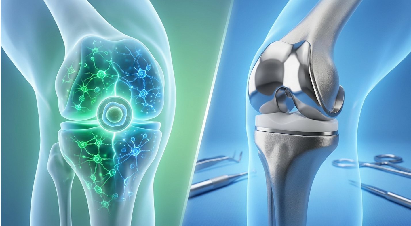
When evaluating stem cell therapy vs knee replacement surgery, patients must choose between biological immunomodulation and mechanical joint ablation. Knee replacement surgically removes the native joint to implant prosthetics, while Human Cellular Tissue Therapy utilizes biological agents to reduce tissue fibrosis and promote scarless repair. Neither is a definitive resolution; efficacy depends entirely on patient pathology.
This is where patients get the most confused. Both procedures claim to fix your knee, but they approach the human body from entirely opposite philosophies. It is frankly irresponsible for clinicians to present these options as interchangeable lifestyle choices when the biological realities are so starkly different.
Total Knee Arthroplasty guarantees permanent skeletal alteration trading native biological architecture for prosthetic components that carry lifelong mechanical restrictions.
Let’s not mince words about what a Total Knee Arthroplasty (TKA) actually is. It is an amputation. An orthopedic surgeon slices open your knee, dislocates the joint, and uses an oscillating saw to cut away the ends of your femur and tibia. They remove your cruciate ligaments, hammer titanium components into your bone marrow, and cement them in place with a polymethylmethacrylate acrylic polymer. A high-density plastic spacer is then slotted between the metals to act as the new cartilage.
This is the surgical standard. It is a highly successful procedure for restoring mechanical alignment in a severely deformed leg. If your leg is visibly bowing outward (varus deformity), a surgeon can cut the bone to make it perfectly straight again.
But it comes at a massive physiological cost. The trauma to the surrounding soft tissues the muscles, tendons, and vascular networks is staggering. The use of a pneumatic tourniquet during surgery creates ischemia-reperfusion injury, leading to localized muscle necrosis and a massive spike in systemic inflammation.
Up to 20% of TKA patients report chronic residual pain—so preserving native tissue remains the highest priority for non-surgical candidates. The metal and plastic do not have nerve endings, meaning the persistent pain stems from localized nerve damage, severe scar tissue formation, and low-grade foreign body immune responses to the titanium and cement. Because TKA removes the biological tissue entirely, it permanently eliminates the potential for future regenerative interventions.
I’ve reviewed countless orthopedic treatment plans, and it is entirely unacceptable that surgeons routinely gloss over the agonizing reality of arthrofibrosis during preoperative consultations. Arthrofibrosis is the excessive buildup of dense, fibrotic scar tissue inside the joint capsule following surgical trauma.
When a surgeon saws through the femur and tibia, the body interprets this as a massive injury and rushes fibroblasts to the area to “glue” the wound together. In many patients, this biological response goes into overdrive. The scar tissue binds the quadriceps tendon to the femur, effectively locking the knee in place. This makes bending the leg excruciatingly painful.
To combat this, physical therapists must aggressively bend the patient’s leg, physically tearing the newly formed scar tissue to maintain range of motion. If physical therapy fails to break the adhesions, the patient must be taken back into the operating room for a Manipulation Under Anesthesia (MUA). The surgeon forcefully bends the knee while the patient is unconscious to rip the scar tissue apart. This brutal recovery phase is a primary reason many patients desperately seek cellular alternatives.
When evaluating why patients seek an alternative to knee replacement surgery, the focus shifts to preserving the native architecture of the knee. Human Cellular Tissue Therapy works through a process called phenotypic macrophage reprogramming.
In an arthritic knee, your local immune cells (macrophages) are stuck in an “M1” pro-inflammatory state, constantly eating away at your cartilage. When UC-MSCs are injected into the joint capsule under strict ultrasound guidance, they sense the inflammatory environment and immediately release a massive payload of anti-inflammatory cytokines.
This chemical signal forces the local macrophages to switch to an “M2” anti-inflammatory phenotype. The joint stops destroying itself and begins producing healthy, lubricating hyaluronic acid and collagen-stimulating factors. By reversing the local immune environment, the joint can fundamentally heal itself.
| 📌 If you’re interested in how MSCs shift the immune system from attack mode to repair mode, we have an interesting article that discusses mesenchymal stem cell therapy for immune modulation, which you can read via the internal link. |
You keep your own bones. You keep your own ligaments. You maintain your natural proprioception the complex neurological feedback loop that tells your brain exactly where your knee is in space. The procedure takes under an hour, requires zero general anesthesia, and involves no bone trauma.
| Evaluation Criteria | Total Knee Replacement (TKA) | UC-MSC Cellular Therapy |
| Mechanism | Mechanical amputation & prosthetic implant | Biological paracrine signaling & immunomodulation |
| Invasiveness | Major surgery (bone resection) | Minimally invasive intra-articular injection |
| Immune Response | Foreign body reaction potential | Anti-inflammatory macrophage reprogramming |
| Reversibility | Permanent anatomical alteration | Preserves native joint architecture |
I cannot stress this enough: stem cells are not for everyone. Surgery is not for everyone. To bridge this gap, clinical researchers utilize our proprietary Regenerative Knee Candidacy Matrix. This is a precise diagnostic framework designed to evaluate a patient’s exact pathology to determine the mathematically optimal intervention.
The matrix evaluates four distinct pillars:
If a patient has chronic, debilitating inflammatory pain but their mechanical axis remains relatively straight, they are the ideal candidate for UC-MSCs.
When patients ask if stem cell therapy works for knee issues, the clinical answer relies heavily on how success is defined. For patients utilizing robust cellular sources like UC-MSCs, success rates for meaningful pain reduction and functional mobility improvement routinely exceed conventional conservative therapies. Efficacy is driven by molecular signaling rather than spontaneous cell differentiation.
We have to move past the marketing myth that an injection will instantly regrow the knee of a 20-year-old. That isn’t biology; that’s science fiction. But what biological therapies can achieve is nothing short of remarkable when measured correctly. I regularly see patients who were told surgery was their “only option” return to pain-free hiking six months post-injection.
SIRT1 signaling pathways regulate cellular aging allowing advanced stem cell therapies to actively rescue damaged cartilage cells from impending apoptosis.
So, what is the actual success rate of stem cell injections for knees? In regenerative orthopedics, success is primarily defined by the WOMAC (Western Ontario and McMaster Universities Osteoarthritis Index) and KOOS (Knee injury and Osteoarthritis Outcome Score) metrics. These are rigorous, validated questionnaires that measure pain, stiffness, and physical function.
Clinical registries tracking cellular therapies show that 70% to 80% of properly vetted candidates experience a clinically significant reduction in pain and an increase in walking distance by month three. But here is the critical nuance—this functional improvement often occurs without massive changes on a follow-up MRI. The cartilage might only show a millimeter of thickening, but the patient goes from barely walking down stairs to hiking two miles.
Why? Because the pain of osteoarthritis is driven primarily by inflammation, not just mechanical wear. Longitudinal tracking confirms that WOMAC pain scores plummet even when radiographic joint space width remains largely unchanged. By turning off the chemical fire inside the joint capsule, the joint functions smoothly again.
When we evaluate whether stem cell therapy for knee pain is working, we rely heavily on the KOOS framework. The KOOS scoring system is divided into five distinct subscales: Pain, Other
Following a successful UC-MSC injection, we typically see the most dramatic spikes in the ADL and Quality of Life subscales within the first 12 weeks. Patients report that they can stand from a seated position without bracing themselves, or that they no longer wake up in the middle of the night throbbing in pain.
It is vital that patients understand these functional markers. If you measure success solely by looking at an X-ray expecting to see thick, youthful cartilage magically reappear, you will be disappointed. But if you measure success by your ability to play with your grandchildren, navigate stairs pain-free, and sleep through the night without relying on NSAIDs, biological injections offer profound efficacy.
If you want to understand how stem cell therapy for knees actually works, you have to look deeper at paracrine signaling. The old theory that you inject stem cells and they instantly turn into new cartilage cells (chondrocytes) has been largely debunked.
Instead, UC-MSCs act as molecular pharmacies. When they encounter a degraded, arthritic joint, they upregulate the SIRT1 signaling pathway. This pathway is a master regulator of cellular aging. The UC-MSCs release massive amounts of exosomes containing miRNA that protect the existing cartilage cells from oxidative stress, clear out toxic reactive oxygen species, and restore the integrity of the mitochondrial membranes.
Without this highly specific molecular intervention, an arthritic joint remains permanently trapped in a vicious cycle of degradation. The cells literally exhaust themselves fighting the inflammation until they undergo apoptosis (cell death).
SASP stands for senescence-associated secretory phenotype. It is effectively a zombie-cell state. When cartilage cells become severely damaged, they don’t just die quietly. They enter senescence, where they continuously secrete toxic inflammatory signals that infect surrounding healthy tissue.
Research into cellular senescence in osteoarthritis demonstrates that this loop drives progressive cartilage degeneration. A single senescent cell can corrupt dozens of healthy neighbors, turning the entire joint capsule into an inflammatory wasteland.
UC-MSCs break this loop. They induce apoptosis in these zombie cells while rescuing the marginally damaged ones. By clearing out the senescent debris, the joint’s natural repair mechanisms can finally operate without constant disruption. Despite these sophisticated cellular capabilities, patients must be fully educated on the inherent limitations and potential adverse events of any medical procedure.
Every medical intervention carries inherent risks and specific clinical limitations that mandate careful patient assessment. While Total Knee Arthroplasty guarantees a protracted and highly painful recovery period, regenerative injections are constrained by their inability to correct structural bone collapse. Assessing these realities is a cornerstone of responsible medical decision-making.
I’ve sat with too many patients who felt blindsided by the realities of their procedures. Surgeons often gloss over the brutal recovery of TKA in pre-op appointments, and regenerative clinics rarely discuss their failure rates. We need to be rigorously honest about the downsides of both.
Prosthetic joint survivorship data highlights finite implant lifespans necessitating potentially devastating revision surgeries for younger patients who outlive their initial hardware.
Let’s address a frequent question: how painful is a knee replacement on a scale of 1 to 10? During the first three weeks of acute post-operative recovery, patients routinely score it an 8 or 9. The bone resection creates profound skeletal trauma, requiring powerful opioids and agonizing physical therapy just to bend the leg to 90 degrees.
But the acute pain eventually fades. The permanent restrictions do not.
There are things you can absolutely never do again after knee replacement. You cannot run long distances or participate in high-impact jumping sports. The polyethylene spacer will wear out exponentially faster under sheer force. You will likely never kneel comfortably on hard floors again, as you are pressing your skin directly against a solid block of titanium.
Furthermore, prosthetic joints have a finite lifespan. Historical survivorship data indicates that while modern implants can last 15-20 years, younger, highly active patients often wear them out faster. This necessitates a revision surgery. Revision TKA is significantly more dangerous, painful, and prone to failure than the initial replacement, often involving massive bone grafting and hardware removal.
When exploring the downside to stem cell therapy, the lack of standardized regulation is a glaring issue. The FDA classifies cellular therapies in two main categories: 361 products (minimally manipulated, homologous use) and 351 products (which require extensive clinical trials as biological drugs).
Because the landscape is complex, many domestic clinics exploit regulatory loopholes to offer unverified, uncharacterized cellular products. Worse, some clinics rely on outdated centrifuges to spin down blood or fat, yielding a product filled with inflammatory debris rather than robust progenitor cells.
This regulatory grey area demands that patients act as their own advocates. You must verify that a clinic sources its UC-MSCs from FDA-registered, cGMP-compliant tissue banks. If a clinic cannot provide a Certificate of Analysis detailing the live cell count and viability percentage of the exact vial being injected into your knee, you should walk out the door. The clinical efficacy of your treatment is entirely dependent on the vitality of the cells you receive.
| 📌 If you’re interested in why the percentage of living cells matters so much at the time of treatment, we have an interesting article that discusses the importance of cell viability in UC-MSC stem cell therapy, which you can read via the internal link. |
A primary downside to stem cell therapy is its inability to correct severe mechanical bone deformities. If you lack any remaining meniscus, or if your tibia has physically collapsed on one side creating a severe varus thrust, an injection will not reconstruct that missing architecture. It’s a biological therapy, not a mechanical jack.
Another massive limitation when evaluating stem cell therapy vs knee replacement cost is the financial burden. Traditional joint replacements, despite costing the healthcare system upwards of $40,000 to $60,000, are universally covered by Medicare and private insurance. Advanced UC-MSC therapies are strictly out-of-pocket, routinely costing between $4,000 and $8,000 per knee.
This financial dynamic drives a dangerous phenomenon: medical tourism. Patients frequently search for stem cell knee injections in offshore clinics to bypass FDA regulatory oversight. While some international clinics operate with high ethical standards, many do not. Receiving uncharacterized, unverified biological products in an unregulated environment introduces severe risks of joint infection and graft-versus-host immune reactions.
I frequently hear patients ask if they can pursue stem cell therapy after failed knee surgery. If you have already had a total replacement, biological injections into that specific joint are useless the biological tissue is gone.
Chronicity and age require nuanced analysis. You must apply our Regenerative Knee Candidacy Matrix in conjunction with a qualified physician. Secure a fresh, weight-bearing MRI (not just a standard non-weight-bearing MRI, which hides true joint space narrowing) and a standing mechanical axis X-ray. I strongly advise seeking separate, independent consultations from both a board-certified orthopedic surgeon and a specialized regenerative medicine physician to make a truly informed choice.
Stem cell therapy can serve as a highly viable alternative to knee replacement surgery for patients who have not experienced severe structural joint collapse. Biological injections utilize cellular signaling to reduce inflammation and promote tissue repair, often delaying or entirely eliminating the need for joint amputation. Clinical outcome registries demonstrate significant pain reduction in appropriately selected candidates. However, patients with extreme mechanical deformities where the bone has physically crushed in on itself—typically still require surgical intervention.
Avoiding knee replacement surgery requires a multi-modal approach addressing both mechanical offloading and biological inflammation control. Patients must modify high-impact activities, reduce body weight to decrease joint shear forces, and utilize targeted physical therapy. When conservative methods fail, advanced biological interventions like UC-MSC therapy can intercept the inflammatory cascade before irreversible cartilage destruction occurs. Proactive treatment is critical, as delaying intervention often guarantees surgery becomes the only viable option.
Stem cell knee injection recovery typically requires 2 to 5 days of minimal downtime, whereas a total knee replacement demands 3 to 6 months of intensive physical rehabilitation. Following a cellular injection, patients can usually bear weight immediately with mild localized soreness and swelling. In contrast, knee replacement surgery involves bone resection and massive tissue trauma, necessitating mobility aids, rigorous pain management, and extensive physical therapy to regain basic flexion. Biological therapies offer a vastly accelerated return to daily activities. Individual healing timelines vary based on age, cellular vitality, and overall joint health prior to the procedure.
Chronological age is not an absolute disqualifier for stem cell knee injections, particularly when utilizing youthful donor sources like Umbilical Cord Mesenchymal Stromal Cells (UC-MSCs). While autologous therapies decline in efficacy as the patient ages, UC-MSCs provide robust, youthful cellular signaling regardless of the recipient’s age. The primary determining factors for treatment candidacy are overall health and the specific mechanical condition of the joint. Older patients with preserved joint space frequently achieve excellent functional outcomes when treated properly.
For patients evaluating stem cell therapy vs knee replacement, the decision hinges entirely on addressing the biological root of joint degradation. While traditional surgery physically replaces the damaged joint architecture, current clinical evidence indicates that UC-MSC therapies successfully reduce pain and improve mobility in a significant percentage of patients without inducing surgical trauma. The optimal approach intercepts and modifies the inflammatory microenvironment early in the disease progression before mechanical collapse dictates surgery.
Applying The Regenerative Knee Candidacy Matrix ensures that treatment aligns perfectly with the patient’s exact pathology. By objectively evaluating systemic inflammation against mechanical joint deformity, patients can avoid unnecessary surgical amputations while ensuring biological therapies are only utilized when they have a high statistical probability of success.
Protecting your mobility requires an evidence-based medical assessment, not marketing promises. Gather your recent MRI reports and weight-bearing standing X-rays, and schedule a comprehensive evaluation with a certified regenerative medicine specialist to review your unique biomechanics. Explore your non-surgical options today to secure a long-term, viable joint preservation strategy.