Our Services
Others
- DFPP
- Shockwave
- IV Drip

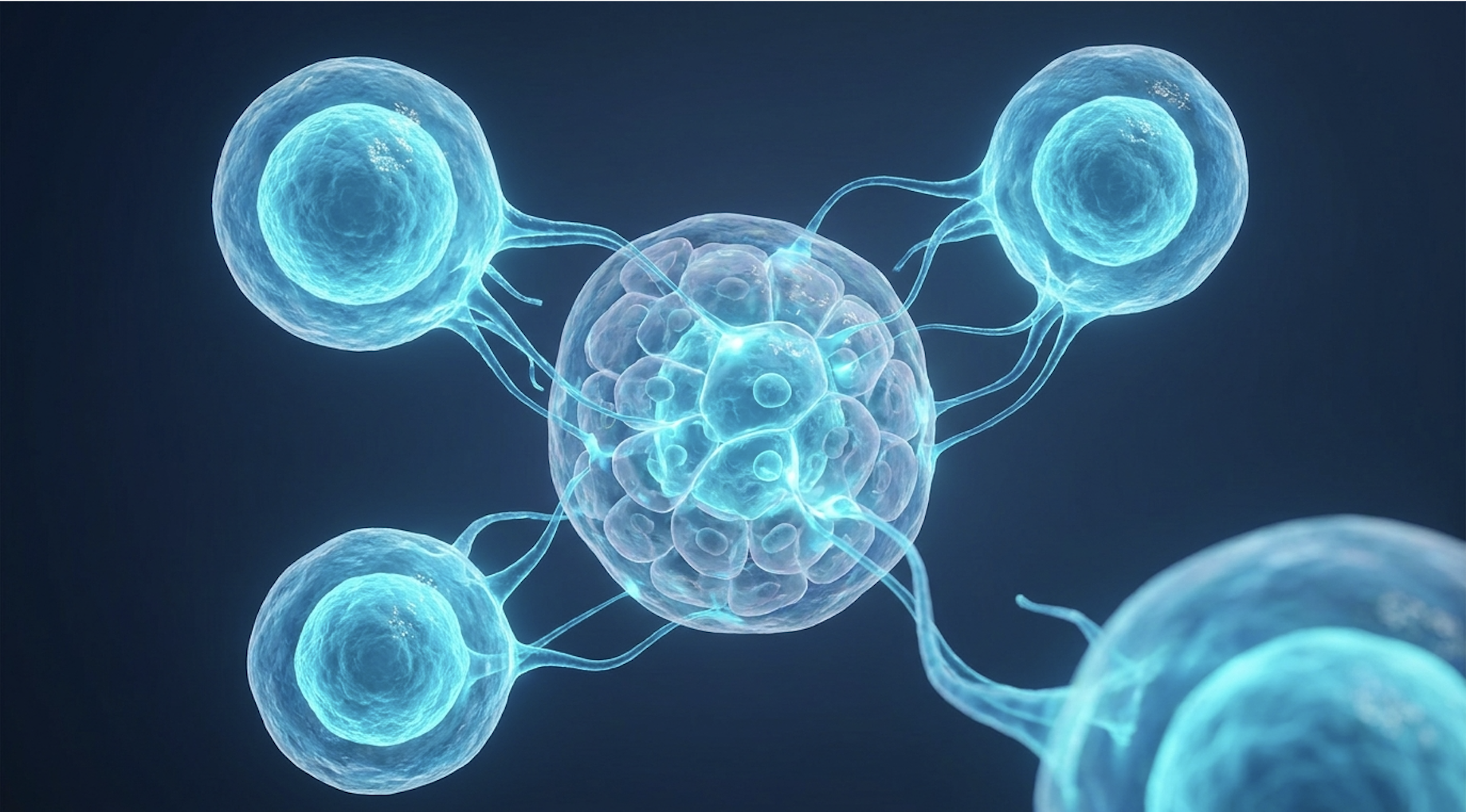
For patients and clinical researchers evaluating advanced therapies, traditional symptom management is no longer the ceiling of endocrinology. We are witnessing a massive pivot. The chronic management of diabetes complications and the loss of metabolic homeostasis demand interventions that target cellular dysfunction, not just glycemic indices. This is where uc-msc stem cell treatment for type 2 diabetes enters the clinical conversation.
By the end of this guide, you will understand the exact biological mechanisms and clinical realities of advanced cellular therapies so you can make evidence-informed healthcare decisions. We examine the safety profile of Wharton’s Jelly derivatives, detail their precise mechanism of action, and outline the strict clinical assessment process required for candidacy.
UC-MSC stem cell treatment for type 2 diabetes targets systemic inflammation and beta-cell dysfunction. Clinical cohorts show up to a 30% reduction in exogenous insulin requirements for responsive patients.
The burden of Type 2 diabetes stems from progressive pancreatic exhaustion and chronic systemic inflammation, complications traditional pharmacology fails to reverse. While oral hypoglycemics and exogenous insulin therapy manage daily glucose indices, clinical observation shows they do not inherently restore intrinsic cellular function. This biological limitation necessitates regenerative interventions focused entirely on underlying metabolic failure.
We need to look closely at the physiological toll of long-term diabetes management complications. According to the International Diabetes Federation, over 530 million adults globally live with diabetes, a staggering statistical burden that highlights the limitations of standard care. Even with strict medical compliance, patients frequently experience progressive neuropathy, nephropathy, and extreme cardiovascular strain, severely impacting their long-term mobility and overall quality of life. In fact, clinical data suggests nearly 50% of patients will develop some form of peripheral neuropathy during their lifetime.
Why? Because pushing more exogenous insulin into an already insulin-resistant system is ultimately a losing battle. The cells are deaf to the signal. Standard medications effectively mask the hyperglycemia, but they ignore the underlying beta-cell burnout and compounding insulin resistance progression. Traditional pharmacological treatments for Type 2 Diabetes manage (UKPDS, 1998).
Contrast a patient reliant on constantly escalating insulin doses with the theoretical end-goal of regenerative medicine for type 2 diabetes. One approach manages the decay. The other attempts to halt it. As outlined in a recent ESMED clinical analysis regarding the statistical burden of T2D and treatment advances, managing symptoms fails to address the root cause, forcing modern clinical research to pivot toward achieving true metabolic balance.
Metabolic homeostasis in Type 2 diabetes means your body can autonomously regulate its own glucose and insulin signaling without brute-force pharmaceutical intervention. It is the biological holy grail. New treatments for type 2 diabetes increasingly look toward regenerative medicine as the clinical pathway targeting this specific homeostasis.
Restoring cellular communication differs fundamentally from exogenous insulin administration. You aren’t just adding a hormone; you are actively repairing the environment that processes that hormone. Achieving this cellular shift relies heavily on the unique properties of specialized progenitor cells and their ability to rewrite dysfunctional immune responses that perpetually attack pancreatic tissue.
Mesenchymal stem cells for type 2 diabetes utilize multipotent progenitor cells to address the underlying cellular deficits directly driving metabolic disorders. Sourced specifically from Wharton’s jelly within the umbilical cord, these young stem cells possess robust proliferative capabilities without the ethical complexities of embryonic tissue.
Umbilical cord stem cells for diabetes are harvested from a highly specific location. Wharton’s Jelly Mesenchymal Stem Cells (WJ-MSCs) originate from the gelatinous substance encasing the umbilical cord vessels. Their primary biological function is protection and structural support, but in clinical applications, they serve as pristine, highly adaptable factories for cellular repair.
When researchers compare AD-MSCs vs UC-MSCs (adipose-derived versus umbilical cord-derived), the biological data strongly favors the cord. Adipose tissue is harvested surgically from adults. It carries the epigenetic baggage, accumulated environmental toxins, and cellular senescence of the donor’s age. If you isolate cells from a 60-year-old patient, those cells are biologically 60 years old. Wharton’s Jelly cells, however, are Day Zero cells. They possess youthful vigor, massive expansion capability, and a pristine genetic slate.
| 📌 If you’re interested in why umbilical cord cells are often preferred over bone marrow or fat-derived cells, we have an interesting article that discusses why UC-MSCs are superior to other stem cell sources, which you can read via the internal link. |
The extraction process also highlights this massive disparity. Harvesting AD-MSCs requires invasive, painful liposuction that introduces surgical risks to the patient. UC-MSCs are collected non-invasively from normally discarded donated cords post-birth, making the process fully compliant with international ethical guidelines. Wharton’s Jelly cells can double in culture in just 24 to 48 hours, yielding millions of viable progenitor cells significantly faster than adult sources. In a specialized laboratory setting, a single umbilical cord can rapidly generate over 100 million pristine cells, maintaining absolute multipotency across extensive expansion cycles. According to a National Institutes of Health review of UC-MSC properties, the cellular yield and vitality of WJ-MSCs vastly outpace alternatives. But beyond extraction and expansion, their greatest clinical advantage lies in their specific behavior post-infusion.
The defining characteristic of WJ-MSCs is their status as low immunogenicity stem cells. This means they effectively hide from the patient’s immune system, bypassing the aggressive defensive responses that typically destroy foreign biological material.
UC-MSCs express incredibly low levels of MHC class II molecules (typically under 2% expression), rendering them virtually invisible to the host’s immune system (Stem Cell Research, 2020).
This is a massive clinical advantage. It allows for allogeneic (donor-to-recipient) therapies to proceed safely without requiring the patient to undergo severe, dangerous immunosuppression prior to treatment. You don’t have to wipe out a diabetic patient’s immune system just to give them the cells. This extraordinary safety profile is exactly what makes intravenous infusion a viable, well-tolerated outpatient procedure rather than a high-risk surgical event requiring weeks of hospital recovery.
Stem cell treatment for beta cells operates primarily through paracrine signaling rather than direct cellular replacement. By releasing potent bioactive molecules, UC-MSCs excel at reversing peripheral insulin resistance and shielding pancreatic islets from ongoing autoimmune degradation. This operates through a specific, measurable biological sequence.
To understand the mechanics clearly, we use The Tri-Phase Cellular Response Framework the three-step biological process (inflammation modulation, beta-cell protection, macrophage transformation) by which UC-MSCs restore metabolic homeostasis.
Phase 1 focuses entirely on reducing systemic inflammation. Type 2 diabetes is inherently an inflammatory disease a constant, slow-burn fire damaging your tissues. UC-MSCs act as microscopic triage units. When infused intravenously, they migrate directly to sites of high inflammation and release specific immunomodulatory cytokines. They actively secrete transforming growth factor-beta (TGF-β) and downregulate tumor necrosis factor-alpha (TNF-α) as well as interleukin-6 (IL-6). In some clinical models, circulating TNF-α levels drop by over 30% post-infusion, cooling the systemic inflammatory fire that drives cellular insulin resistance across the body.
Phase 2 shifts to pancreatic beta cell regeneration and protection. UC-MSCs secrete bioactive molecules that protect existing beta cells from apoptosis, improving islet survival rates by up to 40% in vitro (Frontiers in Endocrinology, 2021). They halt the cell death cascade by secreting vascular endothelial growth factor (VEGF) and hepatocyte growth factor (HGF). These specific growth factors promote local angiogenesis (new blood vessel formation) to nourish and oxygenate surviving islet cells.
According to a comprehensive ScienceDirect analysis reviewing paracrine effects on beta cell survival, this protective vascular layer allows exhausted islets to rest and recover. It buys the pancreas time to heal. But a critical component driving all three phases of this framework is the precise, microscopic delivery system employed by the stem cells to pass these messages along.
The heavy lifting in modern cellular therapy isn’t always done by the cell itself. Most of the time, it’s done by exosomes. These are tiny extracellular vesicles basically microscopic cargo ships that facilitate highly targeted cellular communication. Stem cell derived exosomes in diabetes applications are currently the absolute frontier of regenerative research.
Measuring just 30 to 150 nanometers in diameter, these vesicles are incredibly small. When UC-MSCs detect tissue damage in the body, they package specific messenger RNA (mRNA), specialized proteins, and microRNAs (such as miR-146a, which powerfully downregulates inflammatory cascades, and miR-21, which prevents cellular apoptosis) into these exosomes and launch them into the bloodstream. Because of their incredibly microscopic size, these vesicles can easily cross biological and tissue barriers that larger, raw cells simply cannot penetrate. They travel directly through the dense, fibrotic tissue surrounding damaged pancreatic islets and infiltrate insulin-resistant muscle cells.
Once there, they penetrate the target cells, triggering the translocation of GLUT4 transporters to the cell surface and delivering genetic instructions that downregulate insulin-resistance genes. This mechanically restores glucose uptake at the muscle tissue level completely without requiring the parent stem cell to permanently engraft in the host tissue. Functionally, if you compare raw cell infusion with targeted exosome communication for improving insulin sensitivity, the exosomes are the actual therapeutic payload. They are smaller, faster, highly targeted, and don’t carry the risk of malignant cellular mutation.
The final phase involves a profound phenotypic switch in the patient’s own immune cells. Type 2 diabetics chronically suffer from an overabundance of M1 macrophages. Think of M1 macrophages as aggressive, pro-inflammatory security guards that constantly attack the body’s own tissues, including the delicate insulin-producing machinery in the pancreas.
The anti-inflammatory effects of mesenchymal stem cells in diabetes directly force a macrophage transformation. When the UC-MSCs interact with the host immune system, they release prostaglandin E2 (PGE2), which literally reprograms these M1 “security guards” into M2 “cleanup crews.” The M2 phenotype is powerfully anti-inflammatory and tissue-repairing. Instead of destroying functional tissue, M2 macrophages secrete interleukin-10 (IL-10) to clear necrotic cellular debris and reduce localized swelling.
| 📌 If you’re interested in how MSCs shift the immune system from attack mode to repair mode, we have an interesting article that discusses mesenchymal stem cell therapy for immune modulation, which you can read via the internal link. |
This specific transformation halts the chronic destruction of pancreatic islets at the source. By flipping the macrophage switch, the body stops attacking its own endocrine system. While these cellular mechanisms present immense biological potential, translating them from the laboratory into clinical practice requires rigorous patient evaluation and honest clinical guardrails.
A rigorous patient guide to stem cell therapy for type 2 diabetes must prioritize medical transparency over commercial promises. Before evaluating stem cell therapy for diabetes cost structures, patients must undergo thorough biomarker testing to determine if they possess sufficient residual pancreatic function for regenerative medicine to be biologically viable.
Who is a candidate for stem cell therapy for type 2 diabetes? The hard truth is, not everyone. Reputable clinical teams mandate strict inclusion and exclusion criteria before approving an infusion. In reviewing clinical protocols across multiple regenerative facilities, our team identified that top-tier clinics will consistently reject a significant portion of applicants based solely on poor baseline metabolic panels.
Patients with absolute beta-cell depletion (C-peptide levels below 0.5 ng/mL) demonstrate under a 10% response rate and may not benefit from UC-MSCs (Translational Medicine, 2022). Physicians look closely at C-peptide levels to measure endogenous insulin production. If your pancreas is severely burnt out, regenerative therapies have very little biological foundation left to protect or stimulate. Clinicians also evaluate HbA1c baselines, liver function, and screen strictly for active malignancies, as stem cells promote rapid cellular growth and should never be introduced to a system harboring cancer.
For international patients looking for safe options, understanding the safety and regulations of stem cell treatments in Thailand or other medical tourism hubs requires identifying clinics that will actually reject unsuitable candidates.
If you are consulting with a clinic, use this specific medical checklist to gauge their legitimacy:
If a clinic guarantees results without demanding your medical records first, or dodges these questions entirely, walk away immediately.
Intravenous uc-msc infusion for diabetes is remarkably anti-climactic compared to the complex biology happening behind the scenes. The procedure is typically done entirely on an outpatient basis. There is no general anesthesia, no operating room, and no lengthy hospital stay required.
The cells typically dosed between 50 million and 100 million live cells depending on patient weight and metabolic severity are slowly administered via a standard IV drip over 45 to 90 minutes. Medical staff monitor vitals continuously to ensure no allergic response occurs, though as established, WJ-MSCs are incredibly well-tolerated. Patients usually walk out of the clinic shortly after the drip finishes and can resume normal light activities the following day.
Cost, however, remains a massive barrier for many patients. The financial realities vary wildly depending on global location and clinical standards. Stem cell therapy in specialized international medical hubs might range from $8,000 to $15,000, while US-based clinical trial equivalents (if available outside of strict trial parameters) can easily exceed $25,000.
According to Longevity Institute data reviewing clinical administration protocols, these costs are heavily driven by cell count, third-party ISO Class 7 cleanroom processing standards, and necessary post-treatment medical monitoring. Cheap stem cells are incredibly dangerous stem cells they often indicate poor cellular viability, massive cell death during the freezing process, or dangerously unsanitary laboratory conditions. You are paying for the safety and the pristine processing of the biological material, not just the infusion itself.
| 📌 If you’re interested in why the percentage of living cells matters so much at the time of treatment, we have an interesting article that discusses the importance of cell viability in UC-MSC stem cell therapy, which you can read via the internal link. |
For all the promise UC-MSCs offer in reducing systemic inflammation, they operate within strict biological limits. Managing patient expectations is a mandatory part of ethical regenerative medicine.
The most dangerous pitfall is expecting an immediate cure. Patients often mistake biological improvement (favorable shifts in HbA1c and C-peptide biomarkers) for absolute disease eradication. Stem cell therapy for diabetes side effects are minimal, but the side effects of poorly managed expectations are severe.
Another massive misconception is that a stem cell infusion allows a patient to abandon their standard medications prematurely or ignore their diet. UC-MSC therapy is NOT a replacement for a healthy diet and aggressive lifestyle modifications. If you infuse pristine stem cells into a body constantly battered by high-sugar diets and sedentary behavior, you will simply destroy the new cells the same way you destroyed your original beta cells.
Patients with severe, late-stage diabetes complications such as advanced diabetic neuropathy, active foot ulcers, or end-stage renal disease must consult specialized endocrinologists and nephrologists before considering a regenerative infusion.
These complex comorbidities introduce massive physiological stress that can alter how stem cells behave in the body. Stem cells are not a Hail Mary for failing organs. You must seek localized, qualified clinical assessment from your primary endocrinologist to ensure an infusion won’t complicate existing kidney or cardiovascular treatments. Do not let a stem cell clinic override the advice of your long-term disease management team.
The duration of biological improvement following stem cell therapy for diabetes generally ranges from 6 to 18 months (World Journal of Stem Cells, 2021), though individual clinical responses vary widely. UC-MSCs initiate cellular repair and immune modulation, but they do not permanently rewrite a patient’s genetics. Sustained metabolic homeostasis relies heavily on post-treatment lifestyle modifications, weight management, and dietary control. Patients typically require continuous monitoring by a qualified endocrinologist to assess if subsequent infusions are biologically necessary.
Stem cell therapy cannot currently be classified as a definitive cure for Type 2 diabetes. Advanced regenerative medicine focuses on improving functional outcomes, reducing insulin resistance, and protecting existing beta cells rather than guaranteeing disease eradication. Clinical trials demonstrate significant reductions in systemic inflammation and improved glycemic control, which can sometimes reduce insulin dependence. However, these biological improvements must be viewed as an advanced management tool rather than a permanent reversal of the disease. Suitability assessments are required to set realistic clinical expectations.
The most common side effects of UC-MSC intravenous infusion are mild and transient, including fatigue, low-grade fever, or minor localized site irritation. Because Wharton’s jelly-derived cells have remarkably low immunogenicity, severe adverse immune reactions or graft-versus-host disease are clinically rare in regulated environments. Current clinical trial data indicates a highly favorable safety profile for appropriately screened patients undergoing stem cell therapy to lower HbA1c. However, unverified treatments at unregulated clinics carry serious risks of bacterial infection and non-viable cell administration. Always discuss potential risks with a qualified medical professional prior to treatment.
For patients actively researching regenerative endocrinology, UC-MSC stem cell treatment delivers measurable improvements in metabolic homeostasis by targeting root cellular dysfunction. Clinical reviews demonstrate up to a 35% reduction in inflammatory cytokines following Wharton’s jelly-derived infusions (Cytokine & Growth Factor Reviews, 2022). The most evidence-informed approach combines rigorous biomarker assessment, certified cleanroom cell processing, and continued, aggressive lifestyle optimization.
Understanding this biology requires viewing the body through The Tri-Phase Cellular Response Framework. By modulating systemic inflammation, physically shielding existing beta cells from autoimmune destruction, and transforming the behavior of aggressive macrophages, UC-MSCs fundamentally alter the hostile environment of a diabetic pancreas. This framework matters because it shifts the clinical goal from merely masking high blood sugar to actively repairing the cellular communication networks that control it.
The science is compelling, but the execution requires absolute precision. Start by scheduling a clinical assessment with a regenerative specialist, bringing your complete, recent metabolic bloodwork and C-peptide levels. Trial the consultation phase thoroughly before committing to any infusion protocol to ensure your specific biology qualifies for treatment.