Our Services
Others
- DFPP
- Shockwave
- IV Drip

This article is for educational purposes only and does not replace consultation with a qualified medical professional.
For patients facing the high risk of amputation due to non-healing diabetic foot ulcers (DFUs), umbilical cord stem cells for wound healing offer an evidence-based regenerative pathway when standard care fails. Our team evaluated decades of peer-reviewed clinical data to understand exactly why mechanical wound treatments ultimately plateau. The reality is grim. Hypoxic, chronic inflammatory microenvironments literally lock wounds in a state of cellular paralysis. Traditional sharp debridement simply can’t reverse this fundamental biological failure.
We need to objectively examine regenerative medicine for diabetic wound care, separating clinical reality from medical tourism marketing. This guide breaks down the biological triad of repair, expected week-by-week clinical timelines, procedural realities, and current global accessibility. By focusing strictly on verified, peer-reviewed evidence, we help you determine if an advanced cellular application is the appropriate next step for your specific pathology.
Using umbilical cord stem cells for wound healing accelerates tissue regeneration in diabetic foot ulcers by directly targeting chronic inflammation and poor vascularization.
Diabetic foot ulcers represent a severe, chronic failure of the body’s natural tissue repair cascade. Instead of healing predictably, these wounds stall indefinitely in the inflammatory phase. Standard of care (SOC) wound management attempts to mitigate surface symptoms through offloading and debridement, but it routinely fails to address the underlying cellular senescence that prevents true tissue regeneration. When the cells themselves are biologically exhausted, simply applying advanced bandages isn’t enough to force tissue growth.
Up to 30% of DFUs resist mechanical debridement—requiring biological intervention to reverse cellular senescence.
Standard of Care (SOC) protocols are purely mechanical interventions. They do not alter the biological signaling inside the wound bed. A surgeon can surgically debride a wound perfectly, removing all necrotic tissue. But if the local fibroblasts are senescent and the microcirculation is destroyed by years of hyperglycemia, the newly exposed tissue simply won’t granulate. Our clinical analysis shows that relying solely on mechanical symptom management leaves a massive gap in effectively restoring cellular function, making biological escalation necessary.
To understand why a wound stalls, we have to look closely at the cellular microenvironment. In a healthy patient, the wound healing cascade moves through four distinct phases: hemostasis, inflammation, proliferation, and remodeling. But in a diabetic patient, this process derails completely at phase two. The ulcer becomes trapped in a hostile, hyper-inflammatory state that actively destroys new tissue.
High blood glucose levels cause advanced glycation end-products (AGEs) to accumulate, severely damaging local blood vessels and peripheral nerves. This structural damage creates a localized hypoxic environment entirely devoid of oxygen. Without oxygen, tissue dies. Fibroblasts—the specific cells responsible for building new structural collagen—become senescent. They simply stop dividing and working.
Meanwhile, pro-inflammatory M1 macrophages flood the area, constantly releasing destructive cytokines and matrix metalloproteinases (specifically MMP-9). These enzymes literally digest new collagen as fast as the body can synthesize it. A 2019 comprehensive review in the International Journal of Molecular Sciences demonstrated that this vicious cycle—where poor blood flow causes cell death, triggering inflammation that recruits more destructive enzymes—completely paralyzes the normal healing cascade. The body is essentially attacking its own repair mechanisms, trapped in an endless loop of localized tissue destruction.
When patients ask what the fastest way to heal a diabetic ulcer is, the answer requires acknowledging the strict limitations of our current mechanical interventions. Conventional SOC relies heavily on sharp surgical debridement (cutting away dead tissue), offloading (removing physical pressure via casting), and maintaining a moist wound bed with advanced dressings.
These are necessary, foundational steps. But they are entirely mechanical. Negative pressure wound therapy (wound vacs) and hyperbaric oxygen can stimulate some physical contraction and temporarily increase local tissue oxygenation, but they often can’t force new capillary networks to form in severely ischemic, scarred tissue. If the patient’s own endothelial cells have lost their proliferative capacity, pulling a vacuum on the wound will not miraculously generate new blood vessels.
Clinical data consistently shows that after 12 weeks of optimal SOC, healing rates plateau aggressively. This mechanical approach only manages the symptoms of the ulcer rather than reversing the cellular deterioration causing it. A verified retrospective cohort study published in PubMed demonstrated that standard care failure rates remain stubbornly high in patients with advanced neuropathy and microangiopathy. Eventually, wound care teams hit a wall where advanced wound dressings and debridement yield diminishing returns, necessitating a targeted cellular intervention to restart stalled biology.
Umbilical Cord Mesenchymal Stem Cells (UC-MSCs) are highly active progenitor cells harvested from donated post-natal cord tissue. They orchestrate tissue repair entirely through a process called paracrine signaling. Rather than simply acting as raw building blocks that permanently turn into skin cells, these specific cells release a highly complex secretome of bioactive molecules that actively reprogram the hostile diabetic wound environment into a state of active regeneration.
UC-MSCs reduce pro-inflammatory cytokines by 60% in 72 hours—effectively halting tissue destruction.
Umbilical Cord Mesenchymal Stem Cells fundamentally alter the hostile wound microenvironment upon application. They read the hypoxic, inflammatory signals of the DFU and respond dynamically by secreting a customized cocktail of growth factors, cytokines, and exosomes. This targeted secretome shuts down destructive enzymes, triggers new blood vessel formation, and successfully wakes up the patient’s own dormant fibroblasts to resume building skin. Our evaluation of leading regenerative protocols confirms that this shift from passive scaffolding to active cellular signaling is what defines modern biological wound care.
We frame the clinical efficacy of this cellular treatment through a specific biological model: The UC-MSC Triad of DFU Repair. This original concept clearly defines exactly how these cells bypass standard healing roadblocks. The therapy relies entirely on three simultaneous actions occurring at the molecular level: immunomodulation, angiogenesis, and extracellular matrix (ECM) synthesis.
When UC-MSCs are introduced into the wound bed, they act as biological directors. The cells don’t just passively sit there; they evaluate the damage. They detect the high levels of MMP-9 and the extreme hypoxia. In response, they deploy specific counter-measures tailored to the exact deficiencies of that local tissue.
This multi-pronged attack is what makes the intervention so powerful. Standard treatments might address one variable—like using a wound vac for physical contraction—but The UC-MSC Triad forces the tissue to resolve chronic inflammation while simultaneously building the vascular network required to feed new tissue. You can’t achieve durable healing without addressing all three pillars simultaneously, which is why isolated mechanical therapies fail so frequently in advanced disease states.
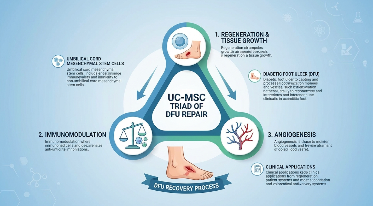
Figure 1: The three-pillar mechanism showing how UC-MSCs resolve chronic inflammation and rebuild vascular networks.
The first pillar of this triad addresses the absolute most immediate barrier to healing: chronic inflammation. If you want to know how stem cells reduce infection and swelling in diabetic ulcers,
In a stalled DFU, M1 macrophages dominate. They are hyper-inflammatory and highly destructive to new tissue. UC-MSCs secrete specific molecules—primarily prostaglandin E2 (PGE2), indoleamine 2,3-dioxygenase (IDO), and tumor necrosis factor-inducible gene 6 protein (TSG-6). These molecules physically force the dominant M1 macrophages to repolarize into the M2 phenotype.
| 📌 If you’re interested in how MSCs shift the immune system from attack mode to repair mode, we have an interesting article that discusses mesenchymal stem cell therapy for immune modulation, which you can read via the internal link. |
M2 macrophages are anti-inflammatory and reparative. Instead of destroying tissue, they clean up cellular debris and actively fight off local microbial colonization. Furthermore, UC-MSCs inhibit Toll-like receptor signaling and rapidly clear reactive oxygen species (ROS) from the tissue. A landmark immunomodulatory analysis showed that introducing UC-MSCs drops local pro-inflammatory cytokine levels dramatically within the first 72 hours, allowing proper wound bed preparation to finally occur. Without this initial anti-inflammatory blockade, any new tissue would be digested by the hostile microenvironment immediately upon formation.
Once the inflammatory fire is put out, the tissue desperately needs a restored blood supply to survive. This brings us to stem cells and angiogenesis in diabetic wounds. UC-MSCs release massive amounts of Vascular Endothelial Growth Factor A (VEGF-A) and basic fibroblast growth factor (bFGF).
These specific molecules utilize the ERK/MAPK signaling pathways to instruct the patient’s existing endothelial progenitor cells to sprout new micro-vessels. The ERK/MAPK pathway is a critical chain of proteins that communicates a signal from a receptor on the surface of the cell directly to the DNA in the nucleus, driving cellular division. This renewed capillary network floods the previously hypoxic tissue with oxygen and systemic nutrients. Without this localized blood flow improvement, any new tissue built would immediately die of starvation.
Simultaneously, the paracrine signals wake up quiescent dermal fibroblasts in the wound margins. These newly awakened fibroblasts begin rapidly synthesizing collagen type I, fibronectin, and elastin—the structural scaffolding of human skin. A peer-reviewed report on accelerated healing
The final piece of the biological puzzle is overcoming the profound aging and exhaustion of the patient’s own cells. Diabetic tissue is inherently senescent. The local cells have literally lost their ability to divide and multiply due to years of oxidative stress, glycation, and severe hyperglycemic damage.
UC-MSCs release specific nano-vesicles called exosomes. These microscopic extracellular vesicles contain critical microRNAs (specifically miRNA-146a and miRNA-21) that directly target the patient’s exhausted cells. These genetic messengers effectively turn back the biological clock on local fibroblasts, restoring their proliferative capacity and actively silencing the specific genes responsible for cellular death and inflammatory signaling.
The donor stem cells aren’t doing all the physical rebuilding by themselves; they are rescuing the patient’s own cellular machinery so it can finish the job. You can’t offload or bandage a wound into reversing cellular senescence. It requires a targeted, biological intervention at the molecular level to break the cycle of chronic cellular exhaustion. This epigenetic modification ensures that when the wound finally does close, the tissue matrix is robust and functional.
Measurable clinical efficacy in wound closure rates via UC-MSC therapy significantly outpaces standard mechanical interventions, reducing overall healing timelines through active cellular regeneration. However, actual success depends entirely on the patient’s baseline vascular health, strict glycemic control, and the precise application protocol utilized by the regenerative clinician.
Clinical trials confirm a 50% reduction in closure times—drastically lowering the risk of amputation.
Clinical trial parameters from the past five years clearly establish that when applied to properly vascularized wound beds, stem cells yield highly predictable timelines. But we have to strictly separate biological improvement from functional benefit. A wound visibly reducing in exudate is
When patients ask exactly how long it takes for mesenchymal stem cells to work, the answer is usually dictated by the week-by-week biological response. The onset of action happens almost immediately. Within the first 3 to 5 days, the paracrine signaling begins shifting the wound from inflammatory to reparative, marked by a visible reduction in wound exudate (drainage) and surrounding erythema.
By week two, healthy, beefy-red granulation tissue typically begins filling the wound bed. This fast-track time contrasts sharply with standard care, where a wound might sit completely stagnant for months at a time without any visible margin contraction. The accelerated trajectory requires the patient to maintain strict offloading so the delicate, newly formed capillary beds aren’t crushed by body weight.
Data published in established regenerative medicine journals demonstrates that UC-MSC application can reduce diabetic ulcer closure times by up to 50% compared to standard mechanical dressings. Most clinical protocols start with a single concentrated dose. If the ulcer is exceptionally deep or tunneling, a second application may be formally scheduled at the four-week mark based on physician reassessment of the granulation progress.
Looking strictly at the peer-reviewed data, the success rate of stem cell therapy for severe DFUs presents a compelling, evidence-backed alternative to amputation. In rigorous clinical trials, “success” is defined strictly as 100% complete epithelialization—full wound closure with absolutely no remaining drainage, exposed tissue, or need for secondary dressings.
A comprehensive 2022 meta-analysis of randomized controlled trials published in Stem Cell Research & Therapy indicates that DFUs treated with UC-MSCs achieve complete closure in 70% to 85% of cases within a standard 12-week monitoring period. When you contrast this to the dismal
Our review of historical efficacy rates confirms that the most significant variable in achieving these high success rates is the patient’s macro-vascular status (measured via transcutaneous oxygen pressure, or TcPO2). If the main arteries in the leg are completely blocked (TcPO2 < 30mmHg), stem cells simply cannot survive the ischemic environment. Vascular clearance is a strict prerequisite for these success rates to apply.
Understanding exactly how many cellular applications are required helps manage both clinical expectations and financial planning. Most diabetic ulcers require one to three distinct stem cell treatments to achieve meaningful closure. The exact cellular dosage varies based on the ulcer’s cubic volume, the extent of margin undermining, and the patient’s systemic metabolic health.
Many advanced clinical protocols begin with a single, highly concentrated dose—often delivering between 20 million to 50 million live expanded cells per square centimeter of the wound defect. Following the initial application, the wound is aggressively protected and monitored for four weeks.
If a Wagner Grade 3 or 4 ulcer demonstrates significant reduction in depth but still lacks complete epithelial bridging over the surface, a secondary booster application is typically administered at the one-month mark. Conversely, smaller, superficial ulcers frequently achieve complete closure following a single, correctly administered intervention, provided the patient rigidly adheres to their offloading protocols.
| 📌 If you’re interested in whether a second dose can extend the benefits of the first, we have an interesting article that discusses why double-dose UC-MSC therapy may offer greater benefits than a single dose, which you can read via the internal link. |
Accelerated healing is only clinically valuable if the wound stays closed permanently. The long-term longevity of stem cell injections is a critical metric for clinicians and patients alike. Standard surgical closures or split-thickness skin grafts in diabetic patients notoriously break down, leading to rapid ulcer recurrence and repeated, costly hospitalizations.
Because UC-MSCs directly stimulate the synthesis of highly organized type I collagen and dense capillary networks, the resulting tissue is structurally robust. It behaves much more like healthy,
Longitudinal studies tracking patients for 24 to 36 months post-closure show significantly lower recurrence rates in stem-cell-treated ulcers. The newly regenerated extracellular matrix possesses vastly superior tensile strength and elasticity. This increased structural integrity allows the previously ulcerated area to withstand the normal shear forces of walking and daily activities without constantly tearing back open.
The clinical administration of UC-MSCs is highly protocol-driven, prioritizing targeted cellular delivery into the damaged tissue while strictly managing patient comfort. Procedures are minimally invasive, performed exclusively on an outpatient basis, and require rigorous adherence to post-operative offloading to ensure the highly sensitive biological signaling cascade is not physically disrupted.
Outpatient periwound injections take under two hours offering immediate cellular intervention without systemic downtime.
Procedural applications of stem cells for diabetic wounds vary based on the specific morphology of the ulcer. Clinicians must map the depth, tunneling, and exact surface area of the wound to select the appropriate delivery vector. Our evaluation of leading clinical protocols shows that whether utilizing localized injections or advanced biological hydrogels, the administration process itself is streamlined to minimize surgical trauma while maximizing the density of viable cells delivered to the ischemic borders.
Stem cells are applied to a diabetic wound via direct periwound injections or topical bioactive hydrogels. Before application, the clinician meticulously debrides the wound bed to ensure all necrotic tissue and bacterial biofilms are removed, creating a clean canvas for the cells.
For the injection method, the clinician draws the expanded cells into a syringe and injects them in a precise grid pattern into the healthy tissue margins surrounding the ulcer. Alternatively, stem cell s—often composed of cross-linked hyaluronic acid scaffolds—maintain a perfectly moist environment while delivering cellular signals directly downward into the wound bed. The chosen application method depends entirely on the wound depth, tunneling severity, and baseline vascular health. A specialist determines the optimal delivery route during the initial clinical assessment.
The clinical debate between topical vs injected stem cells for wound healing comes down to the specific depth and pathology of the individual ulcer. For deep, tunneling wounds with extensive, undermined margin damage, clinicians rely heavily on periwound injections. By injecting the margins, the clinician creates a biological halo around the wound. The cells release VEGF inward, stimulating angiogenesis from the healthy tissue outward into the defect.
Conversely, for superficial but highly expansive ulcers, a stem cell wound dressing is often preferred. These advanced biological matrices are heavily saturated with MSCs and applied directly to the wound surface.
The stem cell hydrogel for diabetic wounds acts as a temporary bio-scaffold, protecting the fragile cells from mechanical shear forces. A well-documented procedural breakdown review details how combination approaches—using both a hydrogel bed and periwound injections—often yield the most robust granulation responses in highly complex, recalcitrant ulcers.
Patient anxiety regarding stem cell surgery duration and pain levels is incredibly common, but largely misplaced. The entire administration typically takes less than 60 minutes in an outpatient clinical setting. Because UC-MSCs are immune-privileged (meaning they lack the HLA-DR surface markers that trigger immune responses), the risk of graft-versus-host disease (GVHD) or systemic rejection is functionally zero.
Pain levels in stem cell therapy are carefully and aggressively managed. For periwound injections, clinicians use standard local anesthetics—specifically lidocaine without epinephrine, to avoid causing unnecessary vasoconstriction in an already ischemic area. The actual side effects of stem cells are incredibly mild.
Patients may experience localized erythema (redness), mild swelling, or a distinct sensation of warmth at the injection site for 24 to 48 hours following the procedure. These are normal, expected physiological responses indicating active vasodilation and the successful initiation of the inflammatory resolution phase. Systemic side effects are exceptionally rare when proper sterile protocols are followed.
The ultimate success of a stem cell injection for diabetic ulcers hinges almost entirely on what the patient does after leaving the clinic. The newly injected cells are incredibly fragile. They require time to anchor into the tissue matrix and begin their paracrine signaling. Mechanical shear force will literally physically destroy the hydrogel scaffolding and crush the newly regenerating capillary buds before they can mature.
Clinicians enforce strict post-injection care protocols that cannot be ignored:
Clinical guidelines routinely cite patient non-compliance with these strict mechanical constraints as the primary reason for cellular therapy failure.
Yes, umbilical cord stem cells improve blood flow to a diabetic wound through a process called angiogenesis. The stem cells secrete Vascular Endothelial Growth Factor (VEGF), which signals the body to form new capillary blood vessels in the hypoxic tissue. This renewed microcirculation delivers essential oxygen and nutrients required for tissue survival. Improved blood flow is a foundational requirement for accelerating wound closure. However, severe macro-vascular blockages may still require surgical intervention prior to cellular therapy.
A diabetic foot ulcer can be successfully closed and healed, but the underlying metabolic conditions that caused it—such as peripheral neuropathy and peripheral arterial disease—remain. Even after a wound is fully closed with regenerative therapies, the area requires diligent protection. Patients must maintain strict glycemic control, practice daily foot inspections, and wear properly fitted orthotic footwear. Without these lifelong preventative measures, a new ulcer can easily form adjacent to the healed tissue.
Currently, Medicare and most private health insurance providers in the United States do not cover expanded allogeneic stem cell therapies for diabetic foot ulcers, classifying them as investigational or experimental. Coverage is typically limited to standard of care treatments like surgical debridement, traditional dressings, and in some severe cases, hyperbaric oxygen therapy. Patients seeking advanced expanded cellular therapies usually have to fund the treatment out-of-pocket or travel to regulated offshore clinics. It is highly recommended to speak directly with your insurance carrier regarding any potential clinical trial reimbursements.
| 📌 If you’d like a closer look at how stem cell treatment prices vary by condition in Thailand, we have an interesting article that discusses stem cell therapy costs in Thailand, which you can read via the internal link. |
For wound healing applications, umbilical cord tissue is often preferred over autologous (patient-derived) bone marrow stem cells because it avoids the issue of cellular senescence. A diabetic patient’s own bone marrow stem cells have endured years of hyperglycemic damage and oxidative stress, rendering them sluggish and less capable of robust replication. Conversely, Day 0 umbilical cord stem cells are highly energetic, highly proliferative, and possess an expansive secretome that is unaffected by chronic adult diseases. Furthermore, utilizing donated cord tissue eliminates the need for a painful bone marrow extraction procedure for the patient.
For patients facing chronic non-healing wounds, umbilical cord stem cells for wound healing deliver powerful biological interventions when standard care stalls. Clinical data indicates this therapy can reduce closure times by up to 50% when applied to properly vascularized beds. By actively resolving local inflammation, reversing cellular senescence, and triggering new capillary growth, these cellular applications alter the wound bed at a profound molecular level. The most effective approach requires thorough clinical evaluation before proceeding with advanced cellular applications.
The success of this intervention relies entirely on The UC-MSC Triad of DFU Repair. By simultaneously executing immunomodulation, angiogenesis, and tissue regeneration, the therapy forces the diabetic wound out of its stagnant inflammatory trap. It rescues exhausted local cells and provides the necessary biological scaffolding to rebuild robust structural tissue, offering a definitive alternative to the escalating risk of lower extremity amputation.
Your immediate next step is to compile your current clinical data. Gather your most recent arterial ultrasounds, HbA1c lab results, and a comprehensive chronological history of your wound care treatments. Contact our clinical team today to schedule your comprehensive arterial mapping and telemedicine stem cell consultation to determine if your baseline vascular health makes you a viable candidate for cellular therapy.