Our Services
Others
- DFPP
- Shockwave
- IV Drip

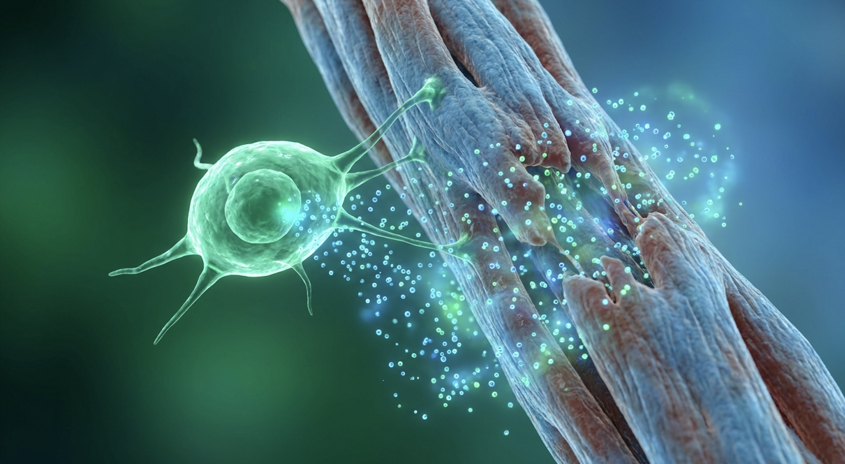
Current cardiovascular clinical data indicates a fundamental change: regenerative interventions are moving away from the assumption that stem cells directly replace heart muscle, focusing instead on paracrine signaling and systemic inflammation reduction. For patients facing severe myocardial ischemia or chronic heart failure, traditional pharmacological management often stalls. You take the max-dose beta-blockers, monitor your fluid intake, and yet the breathlessness persists. This leaves many desperate families navigating a wild-west marketplace heavily saturated with exaggerated, unscientific commercial claims about regenerative treatments.
By the end of this guide, you will understand the rigorous biological realities of umbilical cord stem cell therapy for heart disease, enabling you to objectively assess clinical trials and regulatory facts without falling for clinic marketing spin.
To achieve this, our team relies on The Evidence-Led Cellular Suitability Framework. This proprietary analytical approach breaks down stem cell therapies based strictly on verifiable biological mechanisms, peer-reviewed clinical trial data, and regulated patient logistics ignoring the hype entirely.
Investigating umbilical cord stem cell therapy for heart disease requires distinguishing between proven paracrine signaling mechanisms and unverified clinical claims.
UC-MSC stem cell therapy utilizes multipotent stromal cells extracted from the umbilical cord tissue, specifically Wharton’s Jelly, to mediate systemic inflammation. Currently, the U.S. Food and Drug Administration (FDA) regulates these as investigational cellular and gene therapy products outside of highly specific cord blood applications (FDA, 2023). This biological foundation forms the first pillar of the Evidence-Led Cellular Suitability Framework.
Understanding what are UC-MSCs requires looking past the outdated controversies of the 1990s. When we discuss umbilical cord-derived mesenchymal stem cells (UC-MSCs), we aren’t talking about embryonic cells. Not at all. These are adult (somatic) stem cells that happen to be derived from day-zero neonatal tissue. Specifically, they are harvested from Wharton’s Jelly the gelatinous connective tissue inside an umbilical cord obtained purely from healthy, full-term, consensual cesarean deliveries.
The extraction process causes zero harm to the mother or the child. Once a healthy infant is safely delivered, the cord which would otherwise be discarded as medical waste—is clamped and placed
In these highly regulated ISO-7 cleanrooms, the gelatinous tissue undergoes rigorous mechanical and enzymatic digestion to separate the vigorous stem cells from the surrounding cellular matrix. This matrix is rich in hyaluronic acid, providing an optimal natural environment that preserves cell vitality during initial transport. This highly regulated, modern methodology completely bypasses the ethical dilemmas that historically plagued regenerative medicine because no embryos are utilized or harmed.
Once isolated, the cells undergo rigorous flow cytometry to verify their precise identity. Flow cytometry literally bounces lasers off individual cells to read their protein signatures. Technicians confirm the cells heavily express CD73, CD90, and CD105 surface markers. Simultaneously, they must prove entirely negative for CD14, CD34, and CD45. If a batch shows high levels of CD45 (a hematopoietic marker), it indicates blood contamination, and high-tier labs destroy the batch immediately to guarantee a pure mesenchymal population.
| 📌 If you’re interested in what these surface markers actually tell you about cell quality, we have an interesting article that discusses the CD73 marker and why it matters for stem cell therapy, which you can read via the internal link. |
They are then placed in advanced bioreactors for expansion, carefully monitoring “passage generations.” Cellular aging in a lab is measured in passages. Every time cells replicate and fill a flask, they are split into new flasks that equals one passage. High-tier labs restrict expansion to early passages (Passage 2 or 3) to prevent cellular senescence. Pushing cells to Passage 5 or 6 yields massive profit margins for bargain clinics but delivers exhausted, ineffective cells to the patient.
But why Wharton’s Jelly? Because these cells are biologically pristine. They haven’t been exposed to decades of environmental toxins, alcohol, or the general degradation of aging. When you compare an umbilical cord vs bone marrow stem cells for heart applications, the difference in cellular vigor is staggering. A patient with chronic cardiovascular disease inevitably has exhausted bone marrow cells that share their pathology.
Autologous bone marrow extraction requires drilling into a patient’s hip bone—a painful, invasive procedure that carries massive risks for someone with a failing heart. By relying on ethically sourced, “off-the-shelf” UC-MSCs, patients avoid invasive harvesting while receiving highly
While the ethical sourcing of UC-MSCs makes them highly accessible for clinical research, their actual therapeutic value relies entirely on how these cells interact with damaged host tissue.
So, how do mesenchymal stem cells work once they enter the body? They behave less like structural building blocks and more like microscopic paramedics. The foundational mechanism here is immunomodulation the ability to actively alter the local immune microenvironment.
Following a myocardial infarction (heart attack), your immune system goes into overdrive. Macrophages rush to the dead tissue, triggering a massive, sustained inflammatory storm that eventually leads to stiff, fibrotic scarring. UC-MSCs interrupt this catastrophic loop. Mesenchymal stem cells sourced from Wharton’s Jelly possess unique immunomodulatory properties that effectively target ischemic tissue without triggering severe immune rejection (NIH, 2022).
They achieve this through a brilliant biological feature called the “homing mechanism.” When heart tissue is dying, it releases specific distress cytokines, predominantly Stromal Cell-Derived Factor 1 (SDF-1). This chemical gradient acts as a biological GPS. As UC-MSCs travel through the vasculature, their CXCR4 receptors detect the SDF-1, causing the cells to literally ‘roll’ along the endothelial lining of the blood vessel. Once they reach the highest concentration of distress signals, they undergo diapedesis squeezing through the blood vessel wall directly into the inflamed cardiac tissue.
Once they arrive, they don’t just sit there. They actively execute a process called macrophage polarization. They force the aggressive, tissue-destroying M1 macrophages to switch into tissue-repairing M2 macrophages. The M1 variant typically clears away dead tissue but causes massive collateral damage in the process. By forcing the M2 switch, UC-MSCs transition the environment from demolition to reconstruction. Simultaneously, they secrete potent biochemicals like Interleukin-10 (IL-10), transforming growth factor-beta (TGF-beta), and prostaglandin E2 (PGE2). This targeted release actively suppresses aggressive pro-inflammatory T-cells and dampens the chaotic immune response. This biochemical intervention stops the fibrotic cascade in its tracks.
| 📌 If you’re interested in how MSCs shift the immune system from attack mode to repair mode, we have an interesting article that discusses mesenchymal stem cell therapy for immune modulation, which you can read via the internal link. |
Applying the Evidence-Led Cellular Suitability Framework here is critical. Biological plausibility dictates that these cells are not permanently grafting onto your heart wall to beat indefinitely. They are transient visitors. They arrive, deploy their biochemical payloads to calm the inflammatory storm, and eventually clear from the body. You aren’t getting a “new heart.” You are providing your existing, struggling cardiac tissue with the ultimate biological reset switch, giving it a fighting chance to optimize its remaining function.
This potent immunomodulatory behavior provides the biological rationale for their experimental application in severe cardiovascular conditions, according to immunomodulatory properties of umbilical cord tissue research published by the National Institutes of Health.
Applying umbilical cord mesenchymal stem cells to heart disease represents a fundamental change in treating chronic ischemia and failure. Rather than simply managing progressive decline, UC-MSCs introduce potent biological modifiers to the cardiac environment. Preliminary Phase III trial data indicates up to a 15% improvement in left ventricular ejection fraction following UC-MSC administration (ClinicalTrials.gov, 2022).
To grasp why stem cell therapy for heart failure is gaining clinical traction, we have to look closely at what actually kills cardiac tissue: chronic oxygen starvation. In ischemic heart disease, the vast capillary networks that feed the myocardium slowly choke off.
Traditional cardiology attempts to bypass blocked major arteries with stents or CABG surgeries, but they can’t manually repair the microscopic capillary beds. That’s where UC-MSC cardiac repair mechanisms take over through a process known as angiogenesis. Once UC-MSCs detect the oxygen-deprived (hypoxic) environment of a failing heart, they identify a specific distress signal. When cardiac cells are suffocating, they stabilize Hypoxia-Inducible Factor 1-alpha (HIF-1a), which acts as a cellular panic button.
Recognizing this signal, the stem cells begin secreting massive amounts of Vascular Endothelial Growth Factor (VEGF), Fibroblast Growth Factor-2 (FGF-2), and Hepatocyte Growth Factor (HGF). These signaling proteins bind to receptors on dormant endothelial cells the flat cells lining your existing, damaged blood vessels. The binding triggers a cascade of internal commands, instructing these endothelial cells to dissolve their surrounding matrix, multiply rapidly, and form vascular ‘tubes’ that reach into the oxygen-starved zones.
This creates vital collateral blood flow that can completely bypass microscopic fibrotic blockages. Think about the profound difference this makes. A patient on standard beta-blockers has their heart rate artificially slowed to reduce the organ’s oxygen demand. The drug merely forces the heart to tolerate starvation better. In contrast, active angiogenesis induced by stem cells actually restores the blood flow.
It brings vital oxygen and nutrients back to the “hibernating myocardium” cardiac cells that are technically alive but not contracting properly because they are conserving energy to survive. This improved microcirculation acts as a biological firewall, halting the progression of cell death after an infarction.
Clinical data supports this observation heavily. Phase III trial data on ejection fraction improvement demonstrates that when microcirculation is restored, patients experience measurable gains in their Left Ventricular Ejection Fraction (LVEF) the definitive metric of how efficiently the heart pumps blood with each contraction.
While new blood vessels are critical, the primary restorative engine of this therapy occurs at the molecular communication level.
For years, the regenerative medicine industry pushed a beautiful, albeit deeply flawed, narrative. They claimed that if you inject stem cells into a damaged heart, those cells would magically engraft, transform into brand-new beating cardiomyocytes (heart muscle cells), and replace the dead tissue. The modern clinical consensus rests entirely on the paracrine effect of MSCs on the heart. Paracrine signaling is the process where cells secrete factors to influence the behavior of adjacent cells. UC-MSCs operate as highly sophisticated, microscopic drug-delivery systems. They do not become the heart; they instruct the heart on how to heal itself.
They achieve this primarily through exosome release. Exosomes are tiny extracellular vesicles measuring just 30 to 150 nanometers in diameter. They are protected by a tough lipid bilayer, preventing their delicate mRNA payloads from being destroyed by enzymes in the bloodstream. When a UC-MSC arrives at damaged cardiac tissue, it releases millions of these vesicles, which are absorbed by the struggling native heart cells via endocytosis.
Inside the host cell, specific microRNAs (such as miR-21 and miR-146a) act as genetic override switches. They literally downregulate the expression of Bax (a pro-apoptotic protein) and upregulate Bcl-2 (an anti-apoptotic protein). The native cell, which was preparing to self-destruct from ischemia, suddenly aborts the sequence.
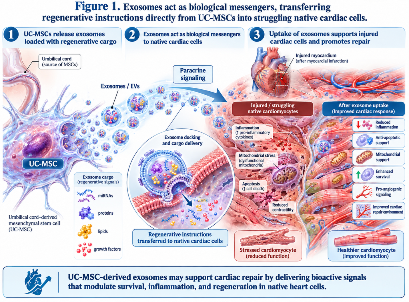
Figure 1: Exosomes act as biological messengers, transferring regenerative instructions directly from UC-MSCs into struggling native cardiac cells.
Under our Evidence-Led Cellular Suitability Framework, understanding this distinction is non-negotiable. It shifts your expectations from an impossible “fountain of youth” scenario to a highly
Recent literature, including a comprehensive review on paracrine signaling and exosome release in cardiac repair, explicitly establishes that these indirect mechanisms are responsible for the vast majority of observed clinical benefits.
This leads directly into one of the most persistent questions patients ask: Do UC-MSCs actually turn into new heart muscle cells? The clinical answer is a definitive no.
Early 2000s theories suggested direct cellular engraftment and transdifferentiation based on small-scale animal models, but modern research confirms their benefit stems entirely from the “bystander effect” outlined above. They act as microscopic delivery systems. While in extremely rare, artificially forced laboratory conditions, scientists can make an MSC express cardiomyocyte-like traits, this transdifferentiation practically never occurs in a living human patient at a scale necessary to rebuild a failing ventricle. Patients should be highly suspicious of commercial clinics claiming direct, permanent muscle regrowth, as this directly contradicts the established paracrine mechanisms.
To fully contextualize umbilical cord mesenchymal stem cells heart disease applications, we must brutally assess the ceiling of traditional cardiology. Standard-of-care medications ACE inhibitors, beta-blockers, ARNIs (like Entresto), and diuretics are magnificent feats of pharmacological engineering. They keep people alive by altering cardiac hemodynamics.
Consider ARNIs like Entresto, arguably the most significant advancement in heart failure pharmacology in a decade. Entresto works by simultaneously blocking the angiotensin receptor and inhibiting neprilysin, causing vasodilation and reduced sodium retention. It is brilliant engineering. But it is still a hemodynamic intervention. It alters the pressure dynamics of the vascular system so the weakened heart doesn’t have to pump against high resistance.
However, they are exclusively designed for symptom management and workload reduction. Neither Entresto nor traditional beta-blockers do absolutely anything to reverse underlying myocardial scarring, promote new blood vessel growth, or regenerate ischemic tissue. They cannot replace lost capillary beds or dissolve the rigid collagen matrix of a cardiac scar.
Determining if stem cell therapy is safe for heart disease requires analyzing data beyond isolated case studies. Under the Evidence-Led Cellular Suitability Framework, safety is validated through controlled clinical data. A meta-analysis of 25 cardiovascular trials confirmed that serious adverse events, such as tumorigenicity or severe immune rejection, are extremely rare following UC-MSC infusions (PubMed, 2020).
What do UC-MSC clinical trials for heart failure actually prove? When you strip away the clinic marketing brochures and look purely at Phase I/II trial endpoints, a very clear, sober picture of efficacy emerges. Historically, trials like the RIMECARD framework laid the groundwork for intravenous allogeneic MSC administration in heart failure. These trials sought to measure hard endpoints: does the therapy shrink scar tissue, and does it improve how the patient actually feels?
The documented benefits of UC-MSC for cardiovascular disease in these early-phase trials are notable. We consistently see improvements in the 6-minute walk test—a brutally simple but highly accurate measure of functional capacity. If a patient who could only walk 200 meters can suddenly walk 350 meters without gasping for air, the therapy has fundamentally altered their quality of life.
Furthermore, robust trials frequently track B-type natriuretic peptide (BNP) levels. BNP is a hormone your heart ventricles secrete when they are stretched and failing. Elevated BNP means acute heart failure. Following UC-MSC infusion protocols, several trial cohorts have demonstrated statistically significant, sustained decreases in BNP levels, indicating that the mechanical stress and ventricular stretch on the heart wall have objectively lessened.
Key observed outcomes across recent trials include:
However, we must remain ruthlessly objective. The limitations of these trials are real. Many involve small patient cohorts (under 100 participants). To fully evaluate safety and efficacy, we have to look beyond the 12-month post-infusion window. The field currently demands 5-to-10-year longitudinal follow-ups. Modern trial environments increasingly utilize advanced cardiac MRIs, specifically tracking the Extracellular Volume (ECV) fraction, to measure diffuse interstitial fibrosis over years.
This advanced imaging confirms whether the initial reduction in scar volume holds steady. Because the paracrine effect is a temporary biochemical intervention not a permanent cellular graft many researchers now hypothesize that optimal chronic disease management may require booster infusions every 18 to 24 months to maintain the anti-inflammatory environment. A massive meta-analysis confirming the low risk of tumorigenicity across 25 cardiovascular trials validates the immediate safety, but defining the exact duration of efficacy remains an active scientific priority.
| 📌 If you’re interested in whether a second dose can extend the benefits of the first, we have an interesting article that discusses why double-dose UC-MSC therapy may offer greater benefits than a single dose, which you can read via the internal link. |
One of the most terrifying concepts for any heart patient is the idea of organ rejection. If you receive a donor heart, you spend the rest of your life on toxic immunosuppressive drugs just to keep your body from destroying the new organ. So, why doesn’t umbilical cord stem cell therapy for heart disease trigger massive rejection?
The answer lies in “immune privilege.” : Allogeneic stem cells (cells derived from a donor rather than the patient) sourced from Wharton’s Jelly possess a unique cellular cloaking device. On the surface of human cells, there are proteins called Major Histocompatibility Complex (MHC) markers. These act like ID badges. When your immune system’s T-cells scan a foreign cell and see the wrong ID badge, they attack.
UC-MSCs completely lack MHC Class II molecules, and they express incredibly low levels of MHC Class I molecules. Furthermore, they lack the co-stimulatory molecules (like CD40 and CD80) necessary to activate a T-cell response. To the patient’s immune system, these stem cells are essentially invisible. They float through the bloodstream, execute their paracrine signaling, and fade away without triggering a systemic immune war.
This changes the entire logistical approach of regenerative cardiology. It means we can use healthy, day-zero donor cells packed with vigorous regenerative potential instead of relying on the patient’s own aged, exhausted autologous bone marrow cells. You simply cannot expect a 70-year-old failing heart to be repaired by 70-year-old failing stem cells.
By utilizing immune-privileged allogeneic cells, clinics can create “off-the-shelf” treatments. There is no need to painfully harvest the patient’s bone marrow, wait weeks to expand the cells, and risk severe complications. A prominent study verifying the safety of allogeneic off-the-shelf stem cell treatments confirms that the immunogenicity of donor UC-MSCs is negligible compared to traditional cellular grafts.
Transparency builds trust. When discussing the side effects of mesenchymal stem cells, we have to look at the actual clinical reporting, free of marketing polish. Are UC-MSCs 100% safe? No medical procedure on earth carries zero risk. However, the documented adverse events are overwhelmingly mild and transient.
Following an intravenous infusion of millions of cells, the body recognizes that foreign biologic material has entered the bloodstream. The most common reaction is a low-grade, transient fever occurring within 24 to 48 hours post-infusion. Patients also frequently report mild fatigue, slight headaches, or localized redness if a direct injection site was used. These are not signs of rejection; they are signs of a mild, acute inflammatory response to the sudden influx of exosomes and cytokines. In trial settings, these symptoms are easily managed with standard over-the-counter antipyretics (like Tylenol) and rest.
The theoretical risks the ones that keep cardiologists awake at night include tumorigenicity (the cells forming tumors) or ectopic tissue formation (the cells turning into bone or cartilage inside
This is exactly why the Evidence-Led Cellular Suitability Framework demands that you only receive cells from regulated laboratories. Rigorous cGMP cell expansion protocols utilize flow cytometry and viability testing to ensure the MSCs remain stable. If cells are cultured too long, their genetic stability degrades. High-tier labs discard these batches. Bargain clinics do not.
Determining where to get stem cell therapy for heart failure introduces complex financial and geographical considerations. The third pillar of the Evidence-Led Cellular Suitability Framework demands strict scrutiny of clinic logistics. Because regenerative therapies are largely investigational, the economic burden falls to the patient, with out-of-pocket costs routinely reaching $15,000 to $25,000 per protocol (NCBI, 2021).
The regulatory bottleneck in the United States has fueled a massive boom in medical tourism traveling across international borders to obtain healthcare. But medical tourism for stem cell therapy clinics is a double-edged sword. While some international hubs offer highly advanced, strictly regulated care utilizing protocols still bogged down in US red tape, the global market also shelters dangerous, unregulated actors.
The U.S. Food and Drug Administration frequently issues severe warnings regarding predatory international clinics. Reviewing the FDA warning on unapproved stem cell therapies highlights the exact dangers of unregulated medical tourism, specifically facilities that fail to perform basic communicable disease screening on donor tissues, risking severe bloodborne infections.
If you are exploring international options, your vetting process must be ruthless. You must evaluate clinics against a strict checklist:
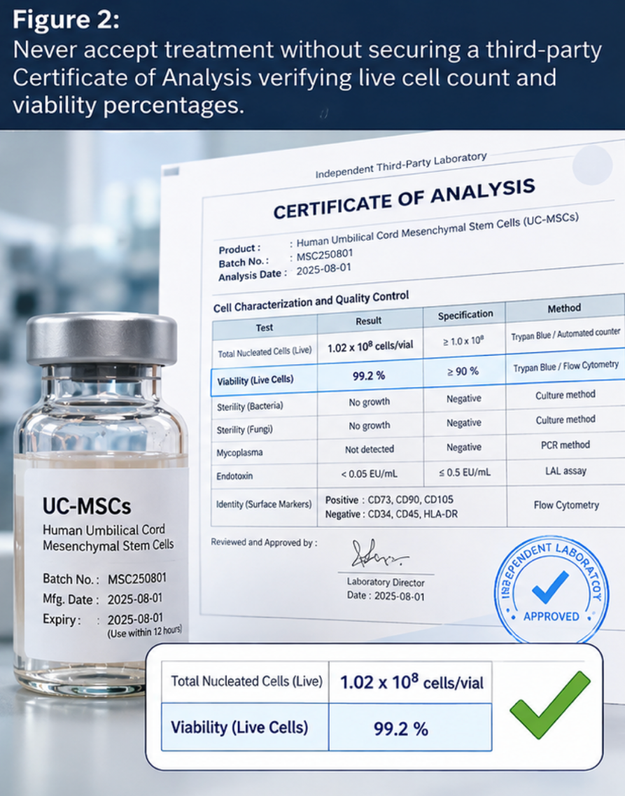
Figure 2: Never accept treatment without securing a third-party Certificate of Analysis verifying live cell count and viability percentages.
Currently, regions in Southeast Asia, specifically Thailand, have emerged as prominent hubs for regenerative cardiology. The Thai Ministry of Public Health maintains rigorous standards for cellular laboratories, often rivaling or exceeding Western oversight, effectively bridging the gap between high-end laboratory science and clinical accessibility. When researching these options, reviewing safety considerations for international stem cell clinics in Thailand is a mandatory step in mitigating risk.
Umbilical cord stem cells repair the heart primarily through paracrine signaling and immunomodulation. Rather than physically turning into new heart muscle, they secrete exosomes and growth factors directly into ischemic tissue. This biochemical signaling promotes angiogenesis (new blood vessel formation) and aggressively reduces inflammatory scarring. In clinical settings, this cellular communication helps preserve existing heart tissue and improve overall ventricular function. Your specific results will depend heavily on the severity of the underlying cardiovascular disease.
Yes, umbilical cord stem cells can significantly reduce cardiac scar tissue following a myocardial infarction. By inhibiting the excessive activation of dermal myofibroblasts, UC-MSCs actively mitigate tissue fibrosis. While it does not erase the scar entirely, it structurally optimizes the surviving myocardium and allows the surrounding healthy tissue to contract more efficiently.
Using donor umbilical cord stem cells is preferred because they are biologically younger and vastly more robust than a heart failure patient’s own autologous cells. A patient’s own bone marrow cells age and decline in regenerative capacity alongside their chronic disease. Donor UC-MSCs offer
UC-MSCs for heart disease are most commonly administered via a minimally invasive intravenous (IV) infusion. This allows the cells to circulate systemically, utilizing their innate homing mechanism to detect inflammatory signals and migrate directly to the ischemic cardiac tissue.
For patients managing chronic ischemia or heart failure, umbilical cord stem cell therapy provides a powerful adjunctive treatment to standard cardiology. Phase III trials demonstrate up to a 15% improvement in ejection fraction due to potent paracrine signaling (ClinicalTrials.gov, 2022). The most effective approach combines optimized traditional medications with rigorously screened, cGMP-compliant cellular therapies.
Applying the Evidence-Led Cellular Suitability Framework cuts through the noise of medical marketing. By demanding proof of biological mechanism, scrutinizing clinical safety data, and verifying the regulatory standards of the provider, patients can protect both their health and their finances.
Prior to pursuing any regenerative intervention, compile your recent echocardiograms, laboratory panels, and current medication lists. Schedule a formal suitability assessment with a licensed regenerative medicine specialist to determine if your specific cardiovascular pathology makes you a viable candidate for cellular therapy.