Our Services
Others
- DFPP
- Shockwave
- IV Drip

This article is for educational purposes only and does not replace consultation with a qualified medical professional.
For patients with advanced joint degeneration who have exhausted cortisone injections and physical therapy, the decision often comes down to joint replacement or regenerative medicine. Clinical observations reveal a frustrating reality patients endlessly cycling through temporary fixes, watching their mobility slowly degrade. Stem cell therapy for joints represents a profound biological shift away from this endless symptom masking.
Rather than artificially dampening pain while the cartilage continues to disintegrate, modern cellular therapies target the underlying microenvironment. By the end of this guide, you’ll understand the exact cellular mechanisms of tissue matrix regeneration, moving past the marketing hype. This guide analyzes the clinical science, recovery timelines, and exact financial costs of modern regenerative orthopedics so you can make an evidence-informed decision regarding your treatment suitability.
Stem cell therapy for joints utilizes targeted cellular therapies to alter the joint microenvironment, potentially supporting extracellular matrix regeneration.
What is joint stem cell therapy? It is an evidence-informed medical intervention where targeted cellular derivatives primarily Mesenchymal Stromal Cells (MSCs) are introduced into a degenerated joint to alter the local immune microenvironment. Unlike corticosteroids that mask pain, joint stem cell therapy introduces progenitor cells that secrete bioactive molecules to stimulate tissue-repairing pathways (National Institutes of Health, 2022). This isn’t magic. It’s applied cellular biology.
When discussing stem cell therapy for joint degeneration, the medical community is almost always referring to Umbilical Cord Mesenchymal Stromal Cells (UC-MSCs). These aren’t embryonic cells. They are an abundant, ethically sourced population of active progenitor cells known for their incredible paracrine signaling capabilities. Their primary regenerative ability doesn’t come from transforming into cartilage cells directly. Instead, they act as cellular directors.
| 📌 If you’re interested in why umbilical cord cells are often preferred over bone marrow or fat-derived cells, we have an interesting article that discusses why UC-MSCs are superior to other stem cell sources, which you can read via the internal link. |
UC-MSC therapy relies on the specific bioactive molecules these cells secrete—epidermal, vascular, and basic fibroblast growth factors. Think of it like comparing a superficial chemical peel to a deep cellular repair protocol in dermatology. Orthopedic applications similarly aim to reverse cellular senescence rather than just mask biological deterioration. When researching the safety of stem cell therapy in Thailand or vetting domestic clinics, understanding this paracrine mechanism is non-negotiable. Furthermore, when patients consider joint injections stem cells offer a uniquely dynamic biological response that static treatments simply cannot match.
Evaluating stem cells versus cortisone and hyaluronic acid injections reveals a stark contrast in medical philosophy. Corticosteroids, traditional anti-inflammatory injections that mask symptoms, provide temporary, relatively cosmetic improvements. They shut off the pain alarm. But the house is still burning down.
Clinical observations indicate that steroids carry severe adverse consequences. Long-term clinical trials demonstrate that repeated intra-articular corticosteroid injections actually accelerate joint space narrowing and cartilage volume loss compared to saline injections (JAMA, 2017). You get less pain today, but you guarantee more structural degradation tomorrow. Continuous clinical review has led advanced orthopedic practices to move away from these destructive interventions. Instead, they favor biological therapies utilizing umbilical cord stem cells for joint degeneration, aiming to halt the breakdown rather than just silence the symptoms. This biological shift is driven by a highly specific cellular cascade, governed by the body’s macrophage population.
Understanding how stem cells regenerate cartilage requires looking far beyond simple cellular replacement. The primary mechanism for rebuilding tissue matrix relies on the complex secretomes released by UC-MSCs. These bioactive molecules enhance local blood microcirculation, deliver vital nutrients to senescent tissues, and initiate a cascade of biological repair signals throughout the damaged joint capsule. It’s a highly orchestrated sequence of events, and mesenchymal stem cells joint repair capabilities are entirely dependent on these precise chemical signals.
Before any physical tissue rebuilding can happen, the joint’s toxic environment must change. Enter The Macrophage Reprogramming Switch the specific mechanism by which UC-MSCs alter the joint microenvironment from a tissue-destructive state to a tissue-repairing one. Macrophages, the immune cells responsible for regulating inflammation, typically get stuck in a pro-inflammatory “M1” state in degenerated joints.
UC-MSCs perform phenotypic macrophage reprogramming, forcing a biological shift to the anti-inflammatory “M2” population. At the molecular level, UC-MSCs inhibit the signaling of Toll-like receptors while promoting the secretion of protective proteins (Frontiers in Immunology, 2021). They also activate the SIRT1 pathway, which protects against oxidative stress and restores the integrity of mitochondrial membranes. Research shows this shift occurs rapidly, often within 72 hours of administration. Reducing excessive tissue fibrosis is the mandatory first step. Once the local immune microenvironment is stabilized, the biological focus shifts to structural repair.
| 📌 If you’re interested in how MSCs shift the immune system from attack mode to repair mode, we have an interesting article that discusses mesenchymal stem cell therapy for immune modulation, which you can read via the internal link. |
With the inflammation neutralized, the physical rebuilding of joint structure begins. UC-MSCs stimulate quiescent dermal fibroblasts to synthesize essential extracellular matrix proteins, including collagen type I, fibronectin, and elastin (Stem Cell Research & Therapy, 2023). This Extracellular Matrix (ECM) is the structural protein scaffolding of the joint.
Furthermore, MSC exosomes for cartilage repair carry specific microRNAs that downregulate cartilage-destroying enzymes. Vascular endothelial growth factor secreted by the stem cells stimulates local endothelial cells to promote new vessel formation (angiogenesis) by activating the ERK MAPK signaling pathway.
As the anatomical diagram above illustrates, this complex matrix provides the physical scaffolding necessary to cushion the joint, mimicking the natural biological structure that was lost. While these molecular pathways are scientifically established, their translation into meaningful functional outcomes depends heavily on the specific joint condition being treated.
Determining the best conditions for stem cell therapy requires a rigorous, objective suitability assessment. While the biological plausibility of cellular repair is high, the clinical efficacy varies dramatically by diagnosis. From widespread degenerative disease to localized tears, evidence-informed practice demands matching the pathology with realistic functional outcomes. Which joints can be treated with stem cell therapy? Almost any major synovial joint knees, hips, shoulders, and ankles but the severity of the damage dictates the success rate.
When analyzing stem cells for synovial inflammation (synovitis) and osteoarthritis, success heavily depends on the disease stage. What happens to the cartilage matrix in osteoarthritis? It gradually dehydrates, fragments, and wears away. In cases of mild to moderate OA (Kellgren-Lawrence grades 2 and 3), evidence-informed stem cell applications have demonstrated measurable improvements in joint mobility and pain reduction within a 6-month clinical monitoring period (Journal of Clinical Medicine, 2022).
But let’s address the reality of bone-on-bone knee conditions objectively. If the joint space is completely obliterated and severe mechanical deformity exists (grade 4), cellular therapy cannot reverse the structural collapse. It might modulate pain via The Macrophage Reprogramming Switch, but it will absolutely not regrow a completely missing joint. Early to moderate OA presents the optimal microenvironment for extracellular matrix scaffolding to succeed. Contrasting experimental claims of “curing” OA with the clinical reality of delaying joint replacement builds trust and sets accurate expectations for prospective patients.
Beyond systemic degenerative diseases, isolated structural injuries present a different clinical profile. Stem cell therapy for a meniscus tear or severe ligamentous injury operates under challenging biological constraints. Meniscal tissue has a notoriously poor blood supply, specifically in the inner avascular “white zone.”
Standard physiological healing here is incredibly difficult, which is exactly why the vascular endothelial growth factor secreted by UC-MSCs becomes critical. By triggering angiogenesis, stem cells bring new blood vessels to a starved area. The joint regeneration success rate for these isolated soft-tissue applications is compelling when patients are properly screened. Recent clinical
Patients frequently ask how long it takes for stem cells to rebuild cartilage following a treatment protocol. It is vital to separate the minimally invasive joint stem cell procedure from the prolonged biological remodeling phase. A scientifically accurate stem cell injection recovery time acknowledges that cellular regeneration is a months-long marathon, not a days-long sprint. You aren’t getting a magical fix; you’re initiating a physiological rebuild.
The day-of treatment experience is surprisingly straightforward. The procedure is an outpatient process completed in a single visit. However, precision is everything. Advanced clinics utilize exact imaging guidance either high-resolution ultrasound or fluoroscopy to ensure the UC-MSCs are delivered directly into the damaged joint capsule. Blind injections simply don’t cut it. Ultrasound-guided intra-articular injections ensure near-perfect delivery accuracy, whereas unguided injections frequently miss the synovial space entirely.
Local anesthesia handles the discomfort, making the injection process itself similar to a standard joint aspiration. Unlike surgical interventions that require elongated recovery periods and carry severe immune system risks, this biological therapy aims to mitigate excessive activation of dermal myofibroblasts. The goal is scarless local healing. Once the cells are delivered, the body enters a highly orchestrated, phased recovery timeline.
While the joint stem cell procedure is completed in a single outpatient visit, the biological process of cartilage matrix regeneration with stem cells requires 3 to 6 months of continuous cellular activity.
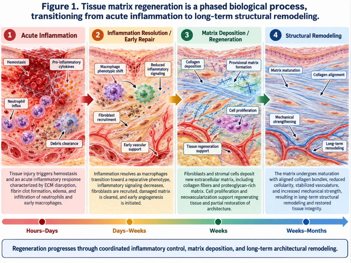
Figure 1: Tissue matrix regeneration is a phased biological process, transitioning from acute inflammation to long-term structural remodeling.
Here is the standard recovery progression:
Post stem cell therapy care explicitly requires targeted physical therapy. Proper mechanical loading is mandatory to align the new tissue matrix correctly. Factoring in this extensive rehabilitation timeline is essential when weighing the financial investment of the procedure against traditional orthopedic alternatives.
| 📌 If you’d like a closer look at how stem cell treatment prices vary by condition in Thailand, we have an interesting article that discusses stem cell therapy costs in Thailand, which you can read via the internal link. |
Stem cells repair damaged joints by secreting bioactive molecules that initiate the macrophage reprogramming switch, altering the joint environment from inflammatory to tissue-repairing. These secretomes actively stimulate local fibroblasts to synthesize new extracellular matrix proteins like collagen and elastin. Furthermore, they promote the formation of new blood vessels, delivering vital nutrients to starved, senescent tissues. For example, in mild osteoarthritis, this paracrine process can significantly improve joint lubrication and mobility.
Yes, targeted cellular therapy can help rebuild cartilage matrix in documented cases of mild to moderate joint degeneration. Umbilical cord mesenchymal stromal cells provide the necessary growth factors to prompt the body’s native cells to produce structural scaffolding. However, stem cells absolutely cannot regenerate a fully destroyed joint where severe bone-on-bone contact has caused mechanical deformity.
Repeated cortisone shots are clinically shown to accelerate cartilage damage when utilized excessively over long periods. While corticosteroids provide rapid, localized improvement by chemically masking acute inflammation, they concurrently inhibit the synthesis of native cartilage proteins. Studies indicate that patients receiving multiple intra-articular steroid injections exhibit much higher rates of joint volume loss over time. Consequently, many modern orthopedic specialists now limit cortisone injections to rare, acute flare-ups. Patients requiring long-term management are increasingly evaluated for safer biological alternatives.
Stem cell therapy can successfully delay joint replacement for several years in properly selected clinical candidates. By reducing excessive tissue fibrosis and restoring the integrity of the joint’s microenvironment, cellular therapy mitigates the biological deterioration that ultimately necessitates surgery. Many patients experience enough functional improvement and pain reduction to postpone invasive arthroplasty indefinitely. However, patients with advanced structural collapse remain immediate surgical candidates regardless of injection therapy.
For patients facing advanced orthopedic decline, stem cell therapy for joints offers an evidence-informed alternative to endless symptom masking and highly invasive surgery. By utilizing UC-MSCs, this biological therapy stimulates quiescent fibroblasts to synthesize essential extracellular matrix proteins, actively fighting tissue degradation. The most effective clinical approach combines rigorous patient suitability assessments with targeted cellular administration, followed by mandatory post-treatment physical therapy to load the new tissue properly.
The success of this intervention relies entirely on the Macrophage Reprogramming Switch the biological mechanism by which UC-MSCs alter the joint from a tissue-destructive state to a tissue-repairing environment. Understanding this molecular process allows patients to look beyond the aggressive marketing hype of miraculous cures and appreciate the realistic, phased timeline required for genuine cellular rejuvenation.
Before committing to any regenerative or surgical procedure, consult with a qualified orthopedic biologic specialist to evaluate your recent MRI imaging for a comprehensive suitability assessment.