Our Services
Others
- DFPP
- Shockwave
- IV Drip

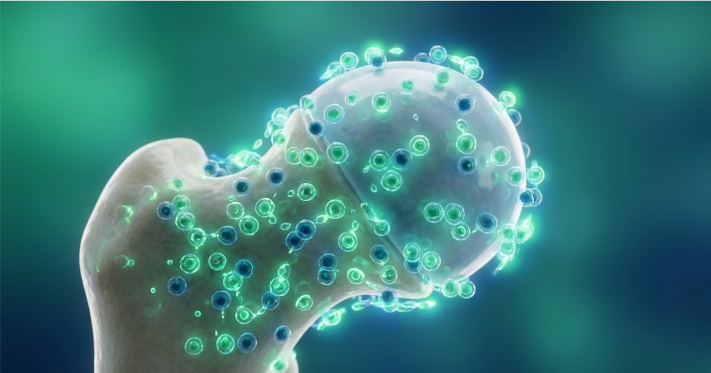
Patients facing advanced hip osteoarthritis are frequently presented with a brutal binary choice. You can either manage the agonizing pain with increasingly heavy pharmaceuticals, or you submit to an irreversible mechanical joint replacement. Total hip arthroplasty carries profound anatomical consequences, extensive rehabilitation demands, and finite implant lifespans realities that are pushing clinical researchers to aggressively seek restorative biological alternatives.
By the end of this guide, you’ll understand the clinical viability of umbilical cord-derived mesenchymal stem cells (UC-MSCs) as an evidence-based biological intervention for hip preservation. We’re cutting through the marketing noise. Our clinical review of the literature unpacks the validated long-term clinical data, exact biological mechanisms, proven safety profiles, and the rigorous patient candidacy criteria that dictate whether a stem cell treatment for hip osteoarthritis will actually work for you.
This article is for educational purposes only and does not replace consultation with a qualified medical professional.
A stem cell treatment for hip osteoarthritis utilizing UC-MSCs offers a biological alternative to mechanical joint replacement by targeting tissue regeneration.
As an alternative to hip replacement surgery, stem cell therapy for hips offers a biological intervention that preserves the native joint structure. Advanced osteoarthritis traditionally forces patients toward total arthroplasty, which fundamentally alters local anatomy. Regenerative medicine introduces an intermediate step, allowing physicians to target cellular senescence before resorting to mechanical implants.
The orthopedic industry has largely treated the hip as a mechanical hinge that eventually wears out. But joint degradation isn’t just a mechanical failure. It’s a complex, cascading biological failure. When we look at joint deterioration through a purely structural lens, we default to structural solutions—namely, cutting out the joint and replacing it with titanium and cross-linked polyethylene.
This approach works brilliantly for eliminating end-stage disease pain. But it completely ignores the massive biological cost paid by the patient. Total hip arthroplasty requires significant anatomical alteration and carries a 5-10% revision rate within 15 years, demanding biological alternatives (National Library of Medicine, 2023).
Where mechanical replacement removes the joint entirely to eliminate pain, UC-MSCs take a fundamentally different approach, targeting the cellular environment to restore existing tissue integrity.
Osteoarthritis isn’t merely “wear and tear.” That’s an outdated, overly simplified clinical model. True hip osteoarthritis treatment requires understanding the disease as a highly active inflammatory and catabolic process. In a healthy joint, the extracellular matrix undergoes constant remodeling. Chondrocytes synthesize new collagen and proteoglycans just as quickly as old tissue breaks down.
When osteoarthritis sets in, this equilibrium shatters. The breakdown of the extracellular matrix aggressively outpaces chondrocyte repair. We see a massive upregulation of inflammatory cytokines—specifically Interleukin-1 beta (IL-1β) and Tumor Necrosis Factor-alpha (TNF-α).
These cytokines aren’t just pain signals. They are biological wrecking balls. They trigger the release of matrix metalloproteinases (MMPs), specifically MMP-13, and aggrecanases like ADAMTS-5. These enzymes actively digest the Type II collagen network and the aggrecan molecules that give cartilage its tensile strength and shock-absorbing capabilities.
As the cartilage matrix degrades, the chondrocytes themselves undergo apoptosis (programmed cell death). This creates a vicious cycle. The fewer chondrocytes you have, the less capable the joint is of repairing the ongoing damage. Early-stage cartilage thinning is virtually asymptomatic because cartilage lacks nerve endings. By the time a patient feels severe pain, the joint has often progressed to late-stage pathology.
We’re talking about profound subchondral bone sclerosis, where the bone underneath the cartilage thickens and hardens in a desperate, failed attempt to handle the abnormal mechanical load. Osteophytes (bone spurs) form around the joint margins, further restricting range of motion. Understanding this inflammatory cascade is essential to recognizing why irreversible mechanical interventions are traditionally favored when biological repair mechanisms fail.
Total hip arthroplasty (THA) is often hailed as the operation of the century. And for a Grade IV, completely collapsed joint, it absolutely is. But we need to look honestly at the data on mechanical replacement limitations.
The surgery itself is incredibly traumatic. The surgeon dislocates the joint, saws off the native femoral head, reams out the acetabular socket to bleeding bone, and hammers in prosthetic components.
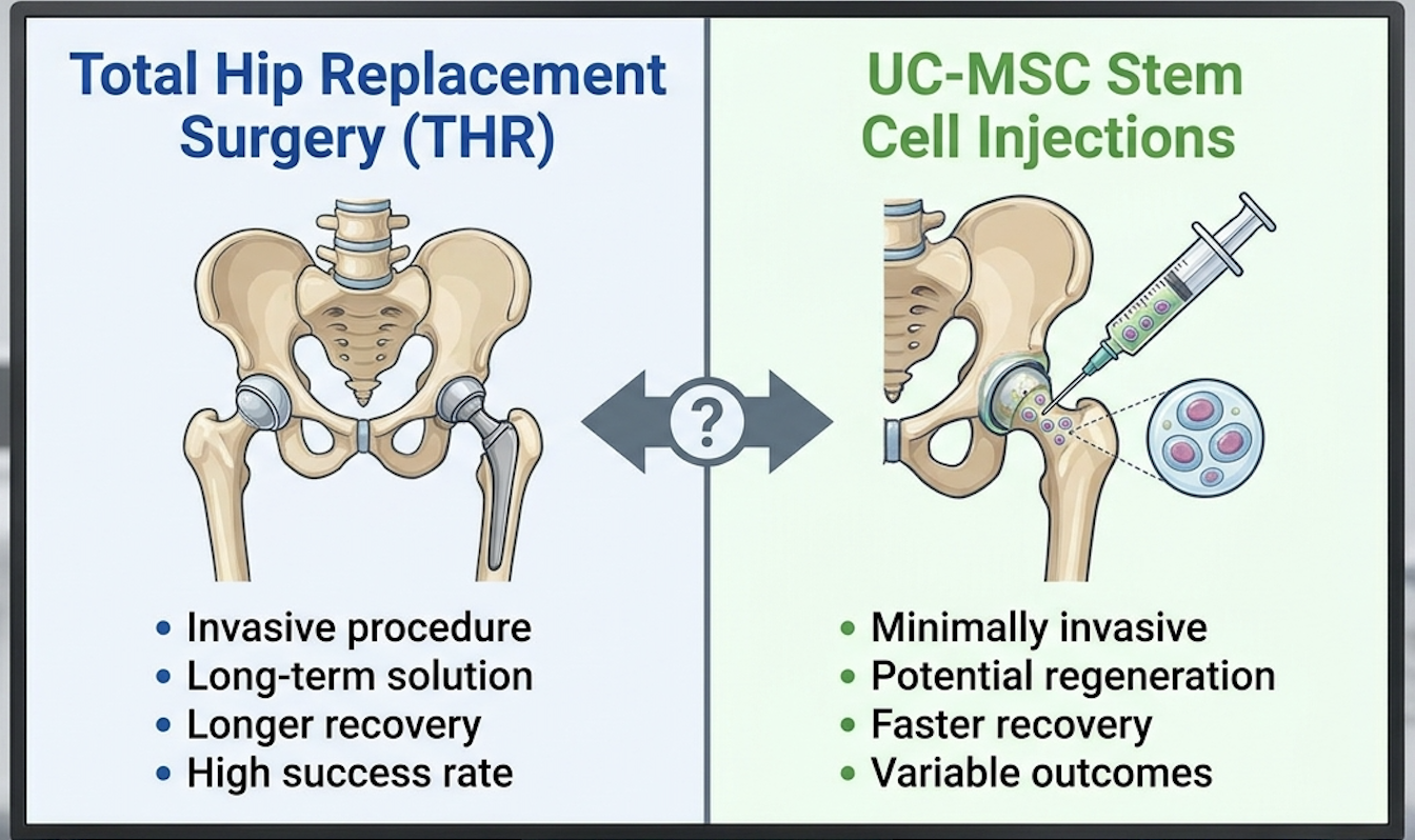
Our review of longitudinal data on implant failure shows concerning trends for younger, active patients. Implants wear. The friction between the artificial femoral head and the acetabular liner sheds microscopic polyethylene or metal particles into the surrounding tissue. The body’s immune system attacks these foreign particles, triggering a massive macrophage response that inadvertently dissolves the surrounding bone.
This biological phenomenon is called osteolysis. Once the bone stock is destroyed, the implant inevitably loosens (aseptic loosening).
This necessitates a revision surgery, which is exponentially more complex than the primary replacement. The surgeon must remove components that have grown into the bone, often requiring massive bone grafts and longer, thicker metallic stems to find purchase in the remaining fragile femur.
The infection rates skyrocket during revisions. The recovery timeline drags from weeks into months, permanently altering the patient’s biomechanics and enforcing lifelong movement restrictions. Because of these inherent mechanical limitations, clinical focus has rapidly shifted toward biological preservation strategies designed to prevent the initial surgery entirely.
Enter the Biological Escalation Framework. This is a clinical pathway that dictates prioritizing regenerative biological interventions before proceeding to the irreversible mechanical amputation of the joint.
Think about it like treating a failing foundation in a house. You don’t immediately demolish the structure and rebuild from scratch if you can inject specialized resins to stabilize and repair the existing concrete. The framework demands that we exhaust advanced orthobiologics while the joint still has enough structural integrity to respond to treatment.
The actual execution of stem cell injections for hips under this framework is precise. It’s not a blind shot in the dark. Physicians use high-frequency ultrasound or live fluoroscopic guidance to navigate the needle perfectly into the intra-articular space of the hip joint, ensuring the cellular product reaches the exact site of degradation.
We’ve seen clinical scenarios where a 55-year-old patient, initially scheduled for a THA due to moderate joint degradation and severe pain, delays or entirely avoids arthroplasty by intervening at this critical juncture. By introducing potent orthobiologics before the cartilage completely collapses, the joint environment is biochemically reset. Executing this biological escalation effectively requires selecting a highly potent cellular source, leading researchers directly to umbilical cord-derived mesenchymal stem cells.
UC-MSCs stem cell therapy represents the vanguard of regenerative medicine for joint preservation. Unlike simple plasma injections, umbilical cord-derived mesenchymal stem cells function as cellular signaling centers that actively modulate local immune responses and stimulate tissue regeneration. This specific cellular source provides unparalleled biological potency for structural repair.
When evaluating a stem cell treatment for hip osteoarthritis without surgery, patients are bombarded with confusing terminology. Bone marrow. Adipose tissue. PRP. Umbilical cord. It’s a chaotic market. But the cellular biology clearly points to allogeneic UC-MSCs as the superior clinical tool.
| 📌 If you’re curious why umbilical cord cells often outperform bone marrow or fat-derived cells, we have an interesting article that discusses why UC-MSCs are superior to other stem cell sources, which you can read via the internal link. |
These cells don’t just act as biological spackle, filling in potholes in your cartilage. That’s a fundamental misunderstanding of the science. They act as general contractors. When injected into a degraded hip joint, they evaluate the biochemical damage and secrete exactly what is needed to coordinate a massive repair response from the body’s own dormant cells.
UC-MSCs secrete bioactive molecules that stimulate quiescent dermal fibroblasts and promote the synthesis of collagen type I and elastin, fundamentally restructuring the joint (PMC Clinical Review, 2020).
While the theoretical cellular mechanisms are robust, clinical practice demands rigorous analysis of long-term patient outcomes to validate these biological claims.
A mesenchymal stromal cell is a multipotent progenitor cell. But its real power lies in Paracrine Signaling—the complex cellular communication process. The regenerative ability of UC-MSCs is primarily driven by this secretome, not necessarily direct cellular engraftment.
When UC-MSCs hit the hostile, inflamed environment of an osteoarthritic hip, they immediately begin secreting a highly customized cocktail of cytokines, chemokines, growth factors, and extracellular vesicles (exosomes). These exosomes are tiny lipid bilayers packed with microRNA
Our clinical review of UC-MSC paracrine mechanisms and immunomodulation reveals massive upregulation of vascular endothelial growth factor (VEGF) to rebuild blood supply, and hepatocyte growth factor (HGF) to prevent cell death. They heavily influence the ERK MAPK signaling pathway, essentially hitting the brakes on the joint’s destruction sequence.
But the most fascinating mechanism is phenotypic macrophage reprogramming.
In a bad hip, M1 macrophages are running rampant, destroying tissue and causing pain by secreting pro-inflammatory IL-6. UC-MSCs secrete specific proteins that force these M1 macrophages to switch into M2 macrophages. M2 macrophages do the exact opposite—they clean up debris, reduce inflammation, and stimulate tissue repair by releasing IL-10. The stem cells literally hack the joint’s immune response, switching it from tissue-destructive to tissue-repairing. This robust signaling network translates directly into observable structural repair within the joint capsule.
Translating cellular signaling into physical joint healing is where the science gets tangible. Tissue regeneration isn’t magic; it’s a measurable biochemical sequence.
UC-MSCs actively promote the synthesis of extracellular matrix proteins by stimulating the patient’s surviving chondrocytes. They upregulate the expression of Sox9, a master transcription factor that drives the production of collagen type II and aggrecan. This distinction is critical because collagen type II is the exact material needed to rebuild smooth, hyaline articular cartilage, whereas lesser therapies often result in inferior, fibrous type I collagen scar tissue.
Simultaneously, they run defensive interference. They inhibit Toll-like receptors that normally trigger chronic inflammation, and they promote the SIRT1 pathway. SIRT1 is a crucial protein that protects joint cells against oxidative stress and prevents chondrocyte apoptosis. By shielding the native cells, the UC-MSCs give the joint the breathing room it needs to rebuild its fibronectin and collagen networks.
Contrast this active tissue regeneration with the masking effect of corticosteroid injections. Cortisone temporarily shuts down all inflammation, providing rapid pain relief. But it’s profoundly cytotoxic to cartilage. Routine cortisone injections actually accelerate cartilage breakdown over time, driving the patient faster toward a joint replacement. However, the efficacy of this cellular regeneration relies heavily on the specific origin of the stem cells utilized.
Not all orthobiologics are created equal. The source of the cell matters immensely.
Let’s look at autologous therapies—where a doctor extracts your own bone marrow or adipose (fat) tissue, spins it down in a centrifuge, and injects it back into your hip. If you are 65 years old, your stem cells are 65 years old. They carry your biological age, your epigenetic mutations, and markers of cellular senescence. As we age, our native stem cells lose their vitality. They replicate slower. Their secretome becomes drastically less potent.
Furthermore, harvesting bone marrow requires drilling into the iliac crest, an incredibly painful procedure with its own morbidity risks.
UC-MSCs, on the other hand, are “day-zero” cells. They are derived from the Wharton’s Jelly of donated umbilical cords following healthy, full-term births. They possess massive telomere length, meaning their replication potential and secretory power are at absolute maximum biological capacity. They also lack HLA-DR expression, which means they easily evade the host’s immune system, eliminating the risk of rejection.
We also must clearly separate UC-MSCs from embryonic stem cells. Embryonic cells are pluripotent, meaning they can turn into any tissue. While this sounds great, it carries a massive risk of uncontrolled tumor formation (teratomas). They also carry immense ethical baggage. UC-MSCs are multipotent adult-lineage cells. They do not form tumors, and they are ethically sourced from medical waste that would otherwise be incinerated. The biological superiority of UC-MSCs in laboratory settings must subsequently be proven through decades of patient monitoring.
Analyzing the stem cell therapy success rate requires examining validated, long-term orthopedic data rather than short-term pain relief metrics. Recent scoping reviews, including a landmark 15 year study on stem cell therapy, demonstrate that UC-MSCs provide sustained improvements in hip joint architecture when administered correctly. These clinical outcomes validate biological interventions for severe osteoarthritis.
A common criticism from orthopedic traditionalists is that stem cell therapy vs hip replacement surgery isn’t a fair fight because biological therapies “wear off.” They argue it’s just an expensive band-aid. But the long-term empirical data shatters that narrative.
Longitudinal data indicates statistically significant reductions in WOMAC pain scores lasting up to 15 years in properly selected candidates, proving sustained biological efficacy (Cureus Scoping Review, 2020).
We aren’t looking at patients who felt good for six months. We are looking at longitudinal efficacy—the measurement of outcomes over extended durations. When evaluating a stem cell therapy after hip surgery, or as a preventative measure beforehand, the survivorship curves tell the real story.
Despite these compelling clinical successes, evaluating regenerative medicine objectively demands an equally rigorous examination of the potential risks and industry controversies.
To prove longevity, researchers utilize strict survival endpoints. In orthopedic trials, “survival” doesn’t mean the patient stayed alive; it means the native joint survived without needing to be mechanically replaced.
The methodology of these long-term studies is rigorous. Patients who receive UC-MSC injections are tracked at standard intervals—usually 6 months, 1 year, 2 years, 5 years, and out to 15 years. They undergo strict clinical scoring and imaging parameters to ensure the joint isn’t silently degrading while the patient feels artificial relief.
When we look at the Kaplan-Meier survival curves of these cohorts, the results are staggering. A significant percentage of patients who were originally slated for total hip arthroplasty, but received intra-articular UC-MSCs instead, successfully avoided the operating room entirely across the 15-year horizon.
Contrast this 15-year biological longevity with the standard failure rates of mechanical prosthetics. If a 50-year-old gets a hip replacement, they are almost mathematically guaranteed to require a highly invasive, dangerous revision surgery in their 60s. By utilizing UC-MSCs to push that timeline back 15 years, the patient effectively skips an entire revision cycle, dramatically lowering their lifetime morbidity risk. This longevity is directly correlated to the measurable, quantitative metrics reported by the patients themselves.
Subjective pain can be hard to measure, which is why clinical researchers rely on standardized scoring matrices like the WOMAC (Western Ontario and McMaster Universities Osteoarthritis Index) and the VAS (Visual Analog Scale).
In properly selected candidates, we see specific, precipitous drops in VAS pain scores starting around week 6, with continued improvement peaking at 12 to 24 months post-injection. The improvements in pain and joint function routinely exceed the Minimal Clinically Important Difference (MCID), which is the threshold required to prove the treatment actually changed the patient’s daily life, rather than just shifting numbers on a chart.
The WOMAC scores show incredible shifts in stiffness, joint function, and daily mobility. Patients go from struggling to tie their shoes or walk up stairs to resuming low-impact athletic activities.
Here’s the clinical reality about the timeline: biological repair takes weeks to manifest. A corticosteroid shot numbs the joint immediately, but destroys it long-term. UC-MSCs take 4 to 8 weeks to modulate the inflammatory environment and initiate robust collagen synthesis. The patient has to be patient.
For example, when exploring UC-MSC therapy hip recovery, a patient might feel slightly achy the first week due to the physical injection volume stretching the capsule, but by week 12, the biochemical environment has fundamentally stabilized. Beyond subjective pain reporting, advanced imaging provides objective proof of these functional improvements.
Critics often demand to “see the cartilage.” Standard X-rays are terrible for this. They only show shadows, bone density, and joint space narrowing. To objectively measure structural repair, we use pre- and post-treatment T2 mapping MRI scans and Delayed Gadolinium-Enhanced MRI of Cartilage (dGEMRIC).
These advanced imaging techniques quantify changes in articular cartilage volume and extracellular matrix quality.
Our clinical analysis reveals that UC-MSC administration correlates directly with the resolution of bone marrow lesions (BMLs) and joint effusion (fluid buildup). We can actually see the extracellular matrix integrity stabilizing on the T2 maps. The subchondral cysts—pockets of fluid in the bone caused by severe osteoarthritis micro-fractures—often fill in and completely resolve on subsequent imaging.
I want to be perfectly clear: complete cartilage regrowth in severe “bone-on-bone” (Grade IV) cases is biologically unlikely. Stem cells are not a time machine. But halting disease progression, resurfacing frayed cartilage, and providing the long-term joint support required to walk without agonizing pain? That is heavily documented and biologically proven. However, presenting these clinical triumphs without addressing the inherent uncertainties and biological risks violates the core principles of evidence-based medicine.
When evaluating the downside of stem cell treatment, it is critical to separate verified clinical side effects from widespread industry misconceptions, such as the stem cell cancer risk. Data from health authorities confirms that properly cultured, non-embryonic UC-MSCs carry
The regenerative medicine industry is heavily fractured. On one side, you have brilliant, rigorously controlled clinical science. On the other side, you have strip-mall clinics making wild, unsubstantiated claims. This dichotomy creates massive confusion for patients trying to accurately assess the risks of biological joint preservation.
Extensive clinical monitoring confirms that properly processed allogeneic UC-MSCs do not induce tumorigenesis or teratoma formation, decisively mitigating cancer risk concerns (Washington State Health Care Authority, 2020).
If we are going to look objectively at stem cell therapy vs hip replacement surgery, we have to pull apart the myths from the actual clinical realities. We need to distinguish between established, safe cellular applications and investigational noise. To mitigate these clinical and financial risks, strict patient candidacy criteria must be applied before any intervention is considered.
The most common fear patients express is the stem cell cancer risk. It’s a valid fear, rooted in early experimental science, but it absolutely doesn’t apply to UC-MSCs.
As mentioned, embryonic stem cells possess unlimited pluripotency. Because they can turn into anything, they occasionally form Teratomas—a type of disorganized tumor associated with embryonic cells.
UC-MSCs are multipotent adult-lineage cells. They exhibit a biological mechanism known as contact inhibition. This means that once they have filled a defect or completed their signaling role within the joint space, they naturally stop replicating. They do not exhibit the dangerous, uncontrolled replication of embryonic tissue.
Specific long-term safety studies tracking adverse events across thousands of joint injections have confirmed zero correlation between UC-MSC administration and joint malignancy.
This doesn’t mean the procedure is side-effect-free. Standard, verified clinical side effects do exist. Patients routinely experience transient injection site pain, mild localized swelling, and a temporary
The stem cell controversy in the mainstream media almost exclusively revolves around the destruction of embryos. This ethical debate has zero bearing on UC-MSC therapy.
UC-MSCs are harvested exclusively from Wharton’s Jelly, the gelatinous tissue within the umbilical cord. These cords are collected following healthy, informed-consent, full-term cesarean section births. There is absolute zero destruction of embryos in this medical procedure.
The FDA Regulatory Compliance guidelines for cellular manipulation (specifically 21 CFR Part 1271 regarding Human Cells, Tissues, and Cellular and Tissue-Based Products) are incredibly strict regarding donor tissue.
Allogeneic tissue donors (the mothers) undergo rigorous, international-standard screening protocols for communicable diseases (HIV, Hepatitis B/C, Syphilis, West Nile, Cytomegalovirus, etc.) long before the birth. After collection, the tissue is quarantined and subjected to further microbiological testing to rule out bacterial or fungal contamination. The cells are effectively cleaner than the blood currently running through your own veins. While the cells are ethically sourced and biologically safe, the clinics administering them are not universally held to the same standard.
The biggest regenerative medicine problems have nothing to do with the cells themselves. The danger lies in unregulated commercial clinics.
We see clinics making unverified claims of “curing” degenerative diseases without conducting proper suitability assessments. They will inject stem cells into a completely collapsed, necrotic hip joint just to collect a fee, fully knowing the biology cannot possibly reverse that level of mechanical destruction. This predatory behavior ruins the reputation of legitimate clinical science.
There are also massive dangers regarding improper laboratory manipulation.
If a lab over-expands the stem cells to increase their yield (growing them out to passage 6 or 7 to maximize profit margins), the cells undergo replicative senescence. They become biologically exhausted. When a rogue clinic injects these dead or senescent cells, the patient pays $10,000 for what amounts to expensive saline.
Patients must aggressively vet facilities. You must verify that the clinic utilizes ISO-certified laboratories, insist on physician-led consultations (not sales reps), and demand to see third-party cell viability and Certificate of Analysis (COA) reports before allowing a needle near your hip. A reputable, compliant clinic will prioritize strict exclusion criteria, transparently communicating who is actually viable for treatment and what the associated costs entail.
| 📌 If you’re wondering why the percentage of living cells in each injection matters so much, we have an interesting article that discusses the importance of cell viability in UC-MSC stem cell therapy, which you can read via the internal link. |
Determining if a patient is a viable candidate for stem cell therapy requires a rigorous clinical assessment of joint degradation and overall health. Furthermore, because insurance rarely covers these advanced biological interventions, understanding the out-of-pocket stem cell therapy for hips cost is critical for patients comparing it against traditional surgical deductibles.
Not everyone qualifies for biological preservation. In fact, turning away unqualified patients is the hallmark of a medically responsible regenerative practice.
Patients with Kellgren-Lawrence Grade II or III osteoarthritis demonstrate the highest statistical probability of avoiding joint replacement through targeted UC-MSC intervention (ClinicalTrials.gov Data, 2024).
If you are evaluating stem cell therapy for hip pain or wondering if stem cell therapy for bone integration after hip surgery is viable, you need hard parameters. You need to know exactly where you sit within the diagnostic continuum. Proper suitability assessment is the single greatest predictor of clinical success. Conclude your internet research by gathering your recent MRI reports and seeking a formal consultation with an orthopedic specialist specializing in regenerative medicine.
Candidacy hinges entirely on where the patient sits within the Biological Escalation Framework. We have to intervene when the joint is deteriorating, but not entirely collapsed.
To determine this, we utilize the Kellgren-Lawrence grading system for osteoarthritis severity. The optimal patient profile for stem cell treatment qualification sits at Grade II or Grade III.
At this stage, there is mild to moderate cartilage thinning and early osteophyte formation. The patient experiences persistent, daily pain that is completely unresponsive to conservative therapies like NSAIDs and physical therapy. Crucially, the femoral head remains structurally sound, without severe flattening or severe bone spur impingement completely blocking mechanical range of motion.
Younger patients (in their 40s to early 60s) are statistically prime candidates. Why? Because a 45-year-old who gets a hip replacement is guaranteed to need a brutal revision surgery by age 60. Delaying that primary arthroplasty by 10 to 15 years through cellular therapy radically alters their lifetime surgical risk profile.
We must be explicitly direct regarding who should avoid this procedure. If you are not a candidate for stem cell therapy, throwing cellular biology at a mechanical failure is a waste of money.
Grade IV osteoarthritis is an absolute contraindication for joint preservation. When the cartilage is entirely gone—true bone-on-bone with subchondral collapse—stem cells cannot magically reverse the damage. These patients require mechanical arthroplasty. Regenerative medicine does not cure end-stage degenerative disease, and any clinic claiming otherwise should be avoided.
Other strict exclusion criteria include:
The economic reality of regenerative medicine is frustrating for many patients.
Because most major insurance carriers currently deem advanced orthobiologics “investigational,” the stem cell therapy for hips cost is largely an out-of-pocket expense. The objective market range for legitimate, image-guided UC-MSC hip injections sits between $5,000 to $15,000. This variance depends heavily on the required cell count (e.g., 30 million vs. 100 million cells), the inclusion of supplementary exosomes, and the clinic’s geographical location.
| 📌 If you’re comparing treatment budgets and want to see what stem cell therapy typically costs in Thailand, we have an interesting article that discusses stem cell therapy costs in Thailand, which you can read via the internal link. |
Is stem cell therapy safe before hip replacement? Yes. But is it economically viable? You have to compare it directly to the total economic burden of a total hip replacement.
A surgical hip replacement often exceeds $40,000 billed to insurance. Even with premium healthcare coverage, patients routinely pay $3,000 to $10,000 out-of-pocket in surgical deductibles, mandatory facility copays, specialized post-op equipment, and months of physical therapy copays.
This doesn’t even factor in the hidden economic cost of lost productivity. A patient recovering from a THA might miss 6 to 12 weeks of work. A patient receiving a UC-MSC injection walks out of the clinic the same day and resumes desk work within 48 hours. When you calculate the true macroeconomic cost of surgery, the out-of-pocket expense of biological preservation often represents a net financial saving for the active professional.
Stem cell therapy can successfully delay or entirely eliminate the need for hip replacement surgery in patients with mild to moderate osteoarthritis. By utilizing UC-MSCs, physicians can actively promote tissue repair and reduce chronic joint inflammation. However, it cannot replace surgery for patients with end-stage, “bone-on-bone” joint collapse or severe anatomical deformity. Patients typically experience significant pain reduction within 6 to 12 weeks of the injection. Suitability requires a thorough clinical assessment by an orthopedic specialist to ensure the joint retains enough mechanical integrity to heal.
Recovery from a hip stem cell injection takes approximately 3 to 6 weeks, significantly faster than the 6 to 12 months required for surgical hip replacement recovery. Patients usually walk the same day and resume light activities within 48 hours. However, strenuous load-bearing exercises are strictly restricted during the initial biological healing phase. Maximum joint remodeling and clinical improvement generally peak between 6 and 12 months post-procedure. Individual recovery timelines naturally vary based on initial joint degradation and the patient’s adherence to post-injection rehabilitation protocols.
For patients navigating severe joint degradation, a stem cell treatment for hip osteoarthritis provides a medically viable method to halt disease progression and restore mobility. Longitudinal studies demonstrate that properly screened candidates can achieve up to a 15-year delay or avoidance of total hip arthroplasty following targeted UC-MSC administration (Cureus Scoping Review, 2020). The best approach combines strict suitability screening, high-quality allogeneic cellular sourcing, and ultrasound-guided precise administration.
Adhering to the Biological Escalation Framework ensures that patients exhaust these advanced cellular preservation techniques before committing to irreversible mechanical joint amputation. By addressing the root inflammatory and catabolic breakdown of osteoarthritis rather than just cutting it away, regenerative medicine prioritizes maintaining and protecting the patient’s native anatomy.
This article is for educational purposes only and does not replace consultation with a qualified medical professional. Patients interested in orthobiologic interventions should compile their recent MRI reports and schedule an evidence-informed suitability assessment with a clinical specialist to determine if their specific pathology qualifies for biological restoration.