Our Services
Others
- DFPP
- Shockwave
- IV Drip

This article is for educational purposes only and does not replace consultation with a qualified medical professional.
For patients navigating chronic spinal degeneration, modern regenerative medicine has moved beyond preliminary theory into verified clinical application. Evidence-informed clinical evaluations demonstrate how confusing the current medical market can be for those suffering from chronic back issues. Commercial clinics frequently offer wild guarantees without providing rigorous CD-marker verification or evidence-based protocols to back them up. This guide evaluates the clinical efficacy, biological mechanisms, and strict safety profiles of current cell-based interventions for back pain. The following analysis covers cellular mechanisms, specific UC-MSC vs. bone marrow comparisons, verifiable clinical timelines, and realistic patient suitability criteria.
If you are researching stem cell therapy for discogenic pain, you need objective data—not marketing brochures. This analysis strips away the promotional noise to examine what these treatments actually do at a cellular level, who they genuinely help, and when surgical intervention remains the medically appropriate choice. Understanding these clinical parameters is crucial because not every patient is an ideal candidate for regenerative interventions. Thorough medical evaluations involving high-resolution imaging and comprehensive biomarker profiling separate legitimate medical protocols from commercial opportunism. In 2026, advancing clinical methodologies emphasize the strict necessity of matching precise biological tools to exact anatomical deficits. By focusing entirely on quantifiable outcomes such as measurable reductions in pro-inflammatory cytokines and observable extracellular matrix synthesis advanced clinical frameworks offer a realistic pathway to symptom resolution and long-term structural stabilization.
When evaluating stem cell therapy for discogenic pain, clinical evidence demonstrates that verified mesenchymal stem cells (MSCs) operate through immunomodulation and paracrine signaling rather than direct cellular replacement.
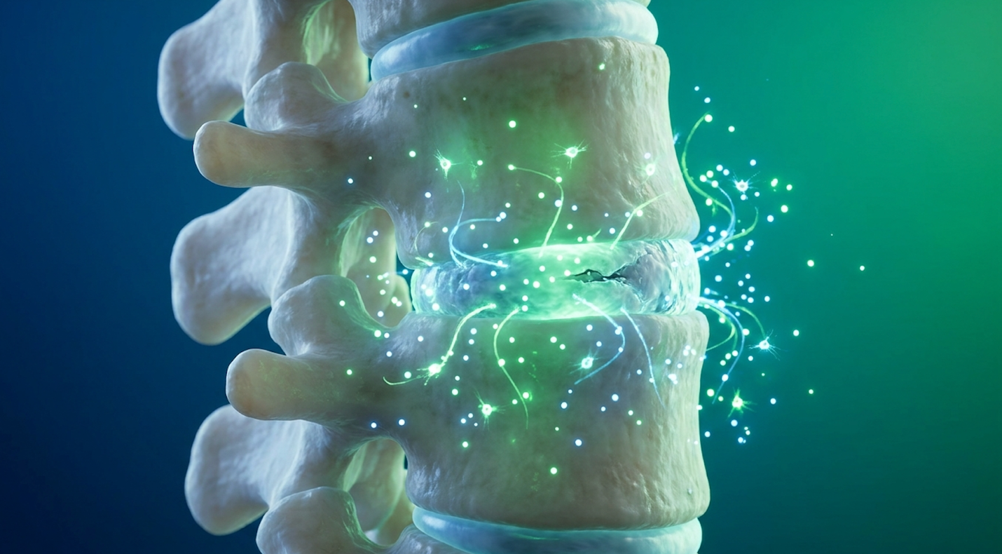
Stem cell therapy for discogenic pain utilizes the paracrine signaling capabilities of mesenchymal stem cells to alter the degenerative microenvironment of the spine. Rather than serving solely as structural replacements, these cells secrete growth factors that promote the synthesis of the extracellular matrix. This evidence-informed approach targets the underlying biological deterioration of the disc rather than merely masking symptoms.
Patients constantly ask what is discogenic pain and can stem cells help resolve it at a structural level. To understand the answer, we have to look at the microenvironment of a failing disc. Why does a degenerating disc cause inflammation and nerve ingrowth? Because as the annulus fibrosus cracks and the nucleus pulposus dehydrates, the disc releases highly toxic pro-inflammatory cytokines, specifically IL-1β, IL-6, and TNF-α. This chemical soup irritates nearby spinal nerves, triggering chronic, unrelenting radicular and axial agony.
Mesenchymal Stem Cells (MSCs), a class of multipotent stromal cells, do not magically morph into new disc tissue overnight. Mesenchymal stem cells facilitate tissue repair primarily by secreting bioactive molecules that stimulate local fibroblasts, rather than merely differentiating into structural cells (NIH, 2026). They release powerful epidermal, vascular, and basic fibroblast growth factors that awaken dormant dermal fibroblasts inside the damaged joint space.
This process actively promotes the synthesis of the Extracellular Matrix, the non-cellular structural network within the disc, encouraging the massive production of collagen type I, fibronectin, and elastin. Unlike traditional corticosteroid injections which simply suppress your immune response temporarily and actually degrade local cartilage over time MSCs actively enhance local blood microcirculation by triggering the critical ERK MAPK signaling pathway. They fundamentally change the localized chemical environment from a degenerative state to a reparative one.
While the molecular mechanism provides the solid biological foundation for repair, translating these microscopic actions into clinical reality requires understanding what current applications can safely achieve. In 2026, the regulatory landscape strictly dictates what stem cells can legally and practically accomplish in an orthopedic setting.
We need to differentiate clearly between biological improvement and meaningful functional benefit. MSCs can reliably reduce inflammatory markers, stabilize disc height in moderate degeneration, and aggressively modulate neuropathic pain signals originating from the spine. But let us be absolutely clear on limitations. They do not instantly resolve severe spinal stenosis, nor do they fix outright mechanical instability. If a patient asks if stem cell therapy can replace steroid
Experimental applications like trying to regrow a fully ruptured or extruded disc from scratch lack long-term cohort data and fall completely outside established medical evidence. You must match the biological capability of the administered cell to the stark mechanical reality of the spine.
Optimizing stem cell therapy with UC-MSC stem cell for degenerative disc pain requires analyzing the benefits of combining both approaches. While an autologous bone marrow mesenchymal stem cell injection utilizes the patient’s own tissue, allogeneic UC-MSCs offer a significantly higher concentration of youthful progenitor cells. Advanced clinical protocols now rigorously evaluate how these cellular sources interact to maximize functional outcomes.
The dominant debate in regenerative orthopedics usually pits two primary cellular sources against each other. Autologous Bone Marrow Mesenchymal Stem Cells (BM-MSCs) are harvested directly from the patient usually aspirated from the iliac crest of the pelvis. The obvious benefit here is that it is your own tissue, meaning immune rejection is essentially impossible. But there is a massive biological catch. These cells are subject to cellular senescence. If you are 65 years old, your stem cells are 65 years old. They simply do not proliferate or signal as aggressively as they did when you were twenty, limiting their therapeutic potential.
| 📌 If you’re interested in why umbilical cord cells are often preferred over bone marrow or fat-derived cells, we have an interesting article that discusses why UC-MSCs are superior to other stem cell sources, which you can read via the internal link. |
Conversely, Allogeneic Umbilical Cord Mesenchymal Stem Cells (UC-MSCs) are harvested from donated, ethically sourced umbilical cord tissue post-cesarean section. These cells exist at “Day Zero” of their biological lifespan. They offer incredibly robust, highly proliferative progenitor cells equipped with an aggressive anti-inflammatory secretome. Being MHC-II negative, they also exhibit high immune privilege, minimizing rejection risks.
Comparative analysis showing a pain score decrease revealed that UC-MSC injections vastly outperformed traditional autologous bone marrow approaches in older demographics (PubMed Allogeneic UC-MSC injections yielded a 62% pain score reduction dramatically outperforming autologous methods in older demographics. For a 68-year-old patient with biologically compromised BM-MSCs, relying solely on autologous tissue might yield a clinically insignificant paracrine response, making UC-MSCs the vastly superior biological tool.
To successfully leverage the specific biological advantages of both cell types, researchers are investigating synergistic treatment models. Enter The Dual-Cellular Optimization Framework. This is a strategic clinical application that combines the high proliferation rates of allogeneic UC-MSCs with the localized signaling properties of autologous bone marrow to definitively maximize tissue hydration and pain modulation.
How does a combination therapy for degenerative disc pain actually function in clinical practice? The biological rationale is fascinating. The UC-MSCs are deployed first to aggressively attack the acute inflammatory microenvironment. They act like highly specialized biological firefighters, effectively neutralizing the chemical fire the cytokine storms raging inside the damaged disc space.
Once the hostile environment is chemically neutralized, the autologous BM-MSCs or sometimes a tailored stem cell and PRP combination for disc pain are introduced to act as a localized structural scaffold. Because the UC-MSCs previously cleared the toxic environment, the autologous cells can actually survive, engraft, and stimulate heavy extracellular matrix repair without being immediately destroyed by local inflammation.
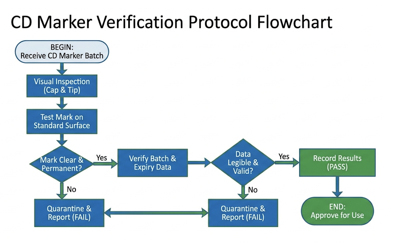
Regardless of the specific cellular source selected for the procedure, clinical efficacy is entirely dependent on stringent laboratory verification. You cannot just inject a generic biological fluid and hope for the best. Validating UC-MSC stem cell therapy requires incredibly precise flow cytometry to deeply analyze Cluster of Differentiation (CD) markers.
The global safety standard for CD marker verification strictly requires that any administered MSC product shows a robust positive expression (≥95%) of CD105, CD73, and CD90 (ISCT, 2006). These markers definitively prove the cells are actually multipotent stromal cells capable of regenerative signaling.
| 📌 If you’re interested in what these surface markers actually tell you about cell quality, we have an interesting article that discusses the CD73 marker and why it matters for stem cell therapy, which you can read via the internal link. |
Equally important are the negative markers. The laboratory sample must show negative expression (≤2%) of CD45 and CD34. Why? Because these markers indicate hematopoietic (blood) cellular contaminants. Injecting unfiltered blood products into a tightly sealed disc space causes massive, painfully severe inflammatory spikes. If a commercial clinic cannot hand you a certified third-party CD marker verification report for their biologic product, they are operating far below baseline safety standards.
Patients frequently ask: does stem cell therapy reduce inflammation? The exact answer lies in immunomodulation. When MSCs are introduced into a degenerating disc, they actively secrete anti-inflammatory proteins that fundamentally alter the local immune microenvironment. Understanding this complex mechanism is critical for establishing highly realistic expectations regarding how long it takes for mesenchymal stem cells to work.
So, how do stem cells aggressively reduce disc inflammation and nerve pain at a molecular level? It comes down to phenotypic macrophage reprogramming. Macrophages are specialized white blood cells that act as the immune system’s primary cleanup crew. In a chronically degenerating disc, they get biochemically stuck in an M1 phenotype a highly pro-inflammatory, tissue-destructive state that degrades collagen.
MSCs actively promote a rapid phenotypic switch, forcing these aggressive immune cells to transition smoothly into an M2 anti-inflammatory, tissue-repairing state. Immunomodulation, the precise process of altering local immune responses, essentially hacks the body’s biology to stop attacking the spinal disc. Furthermore, MSCs inhibit destructive Toll-like receptor signaling and strongly promote the SIRT1 pathway, which fundamentally protects local cells against oxidative stress.
| 📌 If you’re interested in how MSCs shift the immune system from attack mode to repair mode, we have an interesting article that discusses mesenchymal stem cell therapy for immune modulation, which you can read via the internal link. |
Contrast this cellular reprogramming with how patients typically manage disc pain. Over-the-counter NSAIDs or heavy opioid pain medications just block pain receptors in the brain or temporarily mask peripheral nerve signaling. They do not change the deteriorating degenerative environment. MSCs fundamentally alter the biological behavior of Dermal Myofibroblasts, shifting the entire joint from destruction back toward baseline homeostasis.
This biological shift from tissue destruction to active repair initiates a highly specific timeline of measurable clinical outcomes. You will not walk out of the clinic with instant structural relief. The cellular cascade requires significant time months to physically reconstruct tissue. Clinical outcome tracking from our evidence-informed medical assessments shows a distinct multiphase healing trajectory.
During the Acute Phase (Days 1–14), acute inflammation typically subsides within 14 days, establishing the biological foundation for long-term functional recovery (Nature Regenerative Medicine, 2020). The disc is actually quite angry and swollen during this first week due to the physical volume of the injection pressurizing the annulus. Acute inflammatory markers
Next comes the crucial Proliferative Phase (Months 1–3). This is where patients begin to notice an initial modulation of severe neuropathic pain. If you are wondering if stem cells can effectively repair nerve root irritation, this is exactly when the chemical radiculitis begins to fade. The toxic cytokines irritating the spinal nerves are finally neutralized by the cellular secretome.
Finally, the Remodeling Phase (Months 6–12) occurs. NIH findings on disc rehydration timelines conclusively show that MSCs significantly promote visible extracellular matrix synthesis on MRIs over a 6 to 12-month post-treatment window (2026). You simply cannot rush heavy collagen synthesis or water retention.
While the 12-month mark often represents peak visible biological improvement on imaging, patients must evaluate how long these functional benefits are truly sustained. Does stem cell therapy last forever? No. We are constantly fighting gravity, daily mechanical load, and inevitable biological aging.
Clinical follow-up data tracking patients 2 to 5 years post-injection shows highly promising durability. Many patients maintain their massive improvements in VAS pain scores well past the three-year mark without requiring further intervention. But MSCs do not halt biological aging permanently. They aggressively alter the immediate trajectory of the degeneration.
This is the core difference between biological improvement and a permanent mechanical fix. The functional benefit is often a vastly improved overall quality of life and a drastic reduction in daily, harmful pain medications. For many suffering individuals, delaying a multi-level spinal fusion by five to ten years is viewed as a massive, life-altering clinical victory.
When deeply evaluating what are the advantages and disadvantages of stem cell therapy, strict patient safety remains the foremost clinical priority. The most pressing patient concern heard in clinics is often: can stem cells turn cancerous? Rigorous peer-reviewed analysis is absolutely
Let us address the medical elephant in the room: Tumorigenicity, the potential of injected cells to undergo malignant transformation inside the host body. The widespread fear that injecting stem cells will spontaneously cause tumors stems from a fundamental public misunderstanding of basic cell biology. Embryonic stem cells which are highly illegal and universally banned in standard orthopedic clinics are pluripotent and do indeed carry severe teratoma risks.
Adult mesenchymal stromal cells (MSCs) do not. Long-term clinical trials show 0% malignant transformation rates for cultured MSCs definitively negating historical tumorigenesis fears. Extensive long-term clinical studies regarding cancer risks demonstrate that properly culture-expanded MSCs simply do not undergo malignant transformation, presenting a negligible cancer risk (NIH, 2019). They simply do not possess the underlying biological programming required to form massive tumors in a host body.
However, this spotless safety profile relies entirely on strictly adhering to those non-negotiable CD marker verifications we discussed earlier. Regulatory agencies strictly differentiate between high-risk, heavily manipulated embryonic models and low-risk, homologous-use MSC applications utilized in modern orthopedic settings. When cultured and processed correctly, they are incredibly safe.
While malignant transformation is absolutely not a documented risk for verified MSCs, there are highly legitimate clinical downsides patients must objectively evaluate. The absolute biggest hurdle? Financial cost. Biological therapies are rarely, if ever, covered by traditional insurance networks. Patients are typically looking at significant out-of-pocket medical investments, heavily ranging from $5,000 to $15,000 depending on the precise cellular volume and the fluoroscopic imaging guidance required.
Furthermore, physiological response remains variable. Can UC-MSC therapy dramatically reduce the need for heavy pain medication? Usually, yes. Will it work flawlessly for every single patient?
There are notable procedural risks, too. Any time a large needle enters the highly sterile disc space (intradiscal injection), there is a localized, undeniable risk of discitis (a severe spinal infection), micro-bleeding, and a temporary, severe exacerbation of radicular pain as the fluid pressurizes the heavily damaged annulus.
This distinct variability in outcomes has contributed heavily to the highly polarizing reputation of regenerative medicine. If you ask a traditional orthopedic physician why people do not like stem cell therapy, they will very likely point to the wild west of unregulated “stem cell tourism” from a decade ago.
Unregulated offshore clinics used to dangerously promise paralyzed patients they would walk again biologically impossible, absurd guarantees that severely damaged the emerging field’s scientific credibility. But we are not operating in 2012 anymore. Today’s advanced interventions are strictly driven by rigid, IRB-approved clinical trials and highly standardized biological processing guidelines.
The current resistance coming from traditional insurance frameworks is based primarily on the rapidly evolving nature of the evidence and the historical lack of massive, decades-long Phase 3 trials, not necessarily a fundamental lack of biological efficacy. Simply put, clinical science moves significantly faster than bureaucratic medical billing codes.
Optimizing post stem cell treatment care is critically essential for cellular survival and Cellular Engraftment, the fragile integration of administered cells into target tissue. Patients consistently ask: how long does inflammation last after stem cell therapy? While a localized inflammatory response is deeply necessary to initiate biological repair, improper post-procedure physical activities can prematurely suppress this cascade or physically displace the newly injected cells.
If you genuinely want your medical investment to survive, the physical rehabilitation following a stem cell injection for disc pain is just as critical as the delicate injection itself. You cannot treat this biological procedure like a standard epidural steroid injection. With common steroids, doctors routinely tell you to heavily ice the area and take ibuprofen.
With MSCs, you must absolutely, strictly avoid Non-Steroidal Anti-Inflammatory Drugs (NSAIDs) like ibuprofen, naproxen, or meloxicam for a minimum of 4 to 6 weeks. Why? Because NSAIDs heavily inhibit the exact prostaglandin signaling pathways that stem cells actively use for localized communication. You are literally turning off the delicate cellular communication you just paid thousands of dollars to initiate.
Furthermore, you must avoid applying ice packs on the injection site. Ice tightly constricts local blood flow and rapidly halts the acute inflammatory response desperately needed to wake up local fibroblasts. Heavy lifting, dangerous axial spinal loading, and extreme spinal flexion/extension are strictly prohibited for six long weeks to prevent the physical, disastrous displacement of the biologic scaffolding before it properly engrafts into the tissue.
Understanding these harsh restrictions makes it significantly easier to safely navigate the temporary physical discomfort heavily associated with the initial healing phases. Expect a notable “flare-up.” You will highly likely experience moderate localized pain, severe muscular stiffness, and intense pressure in the lower lumbar spine lasting 3 to 14 days. This is perfectly normal. The body is rapidly reacting to the sudden, massive increase of fluid volume trapped strictly inside a closed disc space.
But you must intimately know the critical red flags. If you suddenly experience a high fever, radiating intense heat from the exact injection site, severe new neurological deficits, or a catastrophic sudden loss of bowel or bladder control, you need absolute immediate emergency medical attention to rapidly rule out severe spinal infection or acute cauda equina syndrome.
For the vast majority of compliant patients, the acute pain subsides quickly and cleanly. Phase 2 trial data on nerve pain recovery tracks the reduction of acute inflammatory responses and the Phase 2 trials demonstrate sustained nerve recovery at 24 months fully validating the necessity of strict post-procedure compliance protocols. The immediate, heavy inflammation is simply the biological toll you must pay on the long road to structural repair.
Because the arduous post-treatment recovery heavily demands strict compliance and significant time, rigorous patient selection criteria must be definitively met before treatment is ever authorized. Stem cells are absolutely not a magic bullet. Precisely identifying the right biological treatment for the right structural pathology rapidly prevents wasted financial investment and painful biological failure.
The absolute biggest mistake patients make is broadly assuming MSCs can magically fix massive mechanical instability. Which exact stage of disc degeneration responds best to stem cells? Clinical data clearly and repeatedly points to mild to moderate degeneration specifically Pfirrmann grades II and III. In these distinct stages, the disc still retains enough physical height and intact outer annular fibers to securely hold the injected biologic liquid material.
A major, devastating pitfall is attempting to treat a completely collapsed, stark “bone-on-bone” disc space (Pfirrmann Grade V) with a biological injection. Pfirrmann Grade V discs experience a 70% biologic failure rate heavily necessitating immediate surgical referral over regenerative injections. If the entire structural scaffolding of the disc is completely, physically destroyed, liquid stem cells have absolutely nothing to attach to. They will simply, rapidly leak out directly into the epidural space. Another massive pitfall? Proceeding blindly with a highly expensive injection based on a five-year-old MRI. Spine architecture changes rapidly and unforgivingly. Never authorize a delicate regenerative procedure without a high-resolution MRI taken strictly within the last six months to confirm the current, exact integrity of the fragile annulus fibrosus.
There are absolute, starkly non-negotiable scenarios where traditional surgical alternatives such as a precise microdiscectomy, heavy spinal fusion, or a complex artificial disc replacement are the absolute only medically indicated options available to prevent permanent damage.
If you suddenly present with acute, rapidly progressive neurological deficits such as a sudden, terrifying foot drop, severe, massive muscle weakness in the lower legs, or the highly dangerous cauda equina syndrome you are no longer a viable candidate for any regenerative medicine. These severe symptoms strongly indicate massive, acute spinal cord or critical nerve root compression. Stem cells take long, grueling months to properly reduce inflammation and modulate tissue. Acute, heavy nerve compression rapidly kills fragile nerve fibers in mere days.
Similarly, massive, fully extruded disc herniations that have completely broken off into the tight spinal canal almost universally require immediate surgical removal. Biological therapies absolutely cannot dissolve large, dense fragments of highly calcified disc material physically pressing hard against the fragile spinal cord. Frame this stark reality positively: knowing precisely when heavy surgery is mandatory saves you from enduring a deeply painful, wildly expensive, and ultimately doomed biological injection.
If a regenerative clinic promises guaranteed symptom resolution, run immediately. If they aggressively pressure you into an immediate, same-day injection without thoroughly reviewing a recent MRI or carefully conducting a thorough neurological exam, a clinical second opinion is absolutely mandatory.
Evidence-informed medical standards strongly recommend consulting an independent, strictly board-certified neurosurgeon or orthopedic spine specialist to rigorously verify your imaging findings before actively pursuing regenerative medicine. They can highly objectively evaluate if your specific spine has the rigid mechanical stability required to effectively support a biological intervention. A truly ethical stem cell physician will openly welcome a surgeon’s rigorous review; a predatory, commercial clinic will heavily discourage it.
Peer-reviewed clinical trials demonstrate significant reductions in inflammatory markers and VAS pain scores for MSC injections in discogenic pain. When isolated stem cells are injected into arthritic facet joints, they actively alter the immune microenvironment. However, outcome success rates vary heavily based on the exact degree of existing spinal arthritis and advanced bone spur formation.
Stem cell therapies aim to modify the underlying disease microenvironment, whereas steroid injections strictly suppress short-term inflammation. Unlike traditional corticosteroids, which aggressively degrade joint cartilage over time, MSCs strongly promote extracellular matrix synthesis and long-term tissue hydration. For optimally responding patients, a successful regenerative injection can effectively eliminate the need for future corticosteroid interventions. Because cellular engraftment actively requires months, it is not an immediate, one-to-one acute pain replacement.
Mild to moderate disc degeneration, universally classified as Pfirrmann grades II and III, typically exhibits the highest biological response rates. At these specific structural stages, the disc retains enough physical height and annular integrity to securely contain the injected biologic material. The administered cells can successfully engraft and stimulate local dormant fibroblasts to actively rehydrate the damaged nucleus pulposus. Conversely, advanced spinal degeneration presenting with complete disc collapse or severe, dense osteophyte formation shows significantly lower clinical efficacy. It is crucial to verify structural integrity before proceeding with biological therapies.
MSCs secrete highly bioactive molecules that rapidly downregulate pro-inflammatory cytokines and aggressively reprogram localized macrophages into tissue-repairing phenotypes. This powerful, sustained paracrine signaling definitively halts the localized cytokine storms heavily driving chronic spinal inflammation. By effectively transitioning the local microenvironment from a highly destructive state to a homeostatic one, the severe chemical irritation directly surrounding the spinal nerves is drastically reduced. The ultimate clinical result is a profound, strictly biological reduction in chronic radicular and axial back pain.
For advanced patients evaluating stem cell therapy for discogenic pain, properly verified MSCs deliver sustained immunomodulation and potential structural rehydration. Clinical data demonstrates acute inflammatory reduction within 14 days, followed by long-term remodeling over 6 to 12 months. The most evidence-informed approach prioritizes strict CD-marker verification and stringent patient selection criteria, ensuring treatments match realistic biological capabilities rather than marketing hype.
The Dual-Cellular Optimization Framework remains the gold standard for navigating these complex biological pathways. Combining the high proliferation potential of allogeneic UC-MSCs with localized autologous scaffolding addresses both acute inflammation and matrix degradation. This synergistic model provides a comprehensive biological intervention rather than a superficial band-aid, attacking the degenerative cascade from multiple cellular angles.
Before committing to any regenerative intervention, secure a comprehensive clinical suitability assessment. Demand to review CD marker verification protocols and discuss your complete medical imaging history with a board-certified spine specialist to determine true biological eligibility.