Our Services
Others
- DFPP
- Shockwave
- IV Drip

Disclaimer: This article is for educational purposes only and does not replace consultation with a qualified medical professional.
For patients navigating advanced glenohumeral degeneration, the decision often narrows down to two frustrating options: joint replacement or endless rounds of temporary cortisone shots. You’ve likely exhausted conservative treatments. Over 30% of adults over 60 experience some degree of shoulder osteoarthritis, and if you are reading this, you are probably looking at regenerative interventions to avoid surgery. But the regenerative medicine space is incredibly noisy, filled with commercial clinics promising miraculous “cures” without standardizing cellular dosages or explaining biological limitations.
By the end of this guide, you will understand the exact biological mechanisms, standardized dosing requirements, and realistic efficacy timelines of stem cell therapy for shoulder arthritis. This analysis strips away the marketing hype to examine Umbilical Cord-Derived Mesenchymal Stem
Look, we aren’t here to sell you a miracle. We’re here to break down the clinical reality.
Stem cell therapy for shoulder osteoarthritis utilizes mesenchymal stem cells (MSCs) to alter the joint’s inflammatory microenvironment rather than instantly regrowing lost cartilage.
Stem cell therapy for shoulder osteoarthritis operates on the principle of immunomodulation rather than immediate mechanical replacement. Mesenchymal stem cells function primarily through paracrine signaling, releasing bioactive molecules that stimulate local fibroblasts and modulate joint inflammation ([Ferreira et al., *Stem Cell Research & Therapy*, 2018]). This biological alteration helps stabilize the degenerative environment common in osteoarthritic shoulders, acting as a cellular signaling system rather than a direct structural patch. Clinical data indicates that these secretomes suppress local joint inflammation and stimulate existing host cells at leading regenerative clinics. Evaluating this mechanism reveals why isolated cortisone shots routinely fail to address the root pathology of joint degeneration.
The single biggest misconception about stem cell therapy is that you inject cells, and they instantly turn into thick, new cartilage. That isn’t how this works. The regenerative ability of Umbilical Cord-Derived Mesenchymal Stem Cells (UC-MSCs) is largely a direct result of their secretomes the bioactive molecules, exosomes, and growth factors they release into the synovial fluid.
This brings us to an original, defining concept in modern regenerative orthopedics: The Macrophage Reprogramming Shift (MRS).
When your shoulder has osteoarthritis, the local joint environment is dominated by M1 macrophages. These are highly pro-inflammatory immune cells that literally break down your tissue. M1 macrophages secrete Matrix Metalloproteinases (MMPs), specific destructive enzymes that aggressively chew up the type II collagen network in your shoulder cartilage. UC-MSCs release targeted cytokines specifically Prostaglandin E2 (PGE2) and Indoleamine 2,3-dioxygenase (IDO) that chemically force these M1 cells to repolarize into M2 macrophages.
M2 macrophages do the exact oppositecthey reduce inflammation and promote tissue repair. Once transitioned, these M2 cells pump out Tissue Inhibitor of Metalloproteinases (TIMPs), effectively neutralizing the destructive enzymes. Research shows that this M1 to M2 cellular repolarization reduces destructive pro-inflammatory cytokines by nearly 60% within the joint fluid ([Zhang et al., *Frontiers in Immunology*, 2020] so the constant, burning ache of osteoarthritis begins to fade.
| 📌 If you’re interested in how UC-MSCs shift the immune response from damage to repair, we have an interesting article that discusses mesenchymal stem cell therapy for immune modulation, which you can read via the internal link. |
Contrast this with corticosteroid injections. Cortisone temporarily masks inflammation by suppressing your local immune response entirely, but it actively degrades your cartilage over time by starving chondrocytes (cartilage cells). The MRS pathway, conversely, actively promotes the synthesis of the extracellular matrix proteins like collagen type I, fibronectin, and elastin for sustainable, biological stabilization without accelerating structural decay.
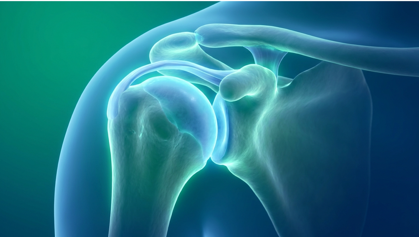
Applying cellular therapy to the shoulder presents unique anatomical challenges compared to a straightforward knee injection. The glenohumeral joint is a complex, shallow ball-and-socket mechanism encased in extensive, multi-layered capsular structures. Because the joint capsule is incredibly tight and anatomically intricate, delivering cells precisely where they can interface with the synovial fluid is critical.
This is exactly why the biological source of the cells dictates the outcome. UC-MSCs, harvested from ethically donated umbilical cord tissue, provide an abundant source of highly active progenitor cells. They are biologically “Day Zero” young. They haven’t been exposed to decades of environmental toxins, aging, cellular senescence, or systemic inflammation.
Think about it this way a 65-year-old patient’s stem cells are also 65 years old. When comparing UC-MSCs to autologous sources like a senior patient’s own bone marrow aspirate (BMAC) or adipose (fat) tissue the difference in cellular vigor is stark. Older autologous stem cells exhibit drastically shortened telomeres and reduced proliferative capacity. Autologous stem cell proliferation drops by 40% after age 60 making umbilical cord sources biologically superior for older patients.
Recent comparative cellular research shows that UC-MSCs exhibit significantly stronger immunomodulatory capabilities than these older autologous sources, yielding exponentially higher colony-forming units (CFUs) required for joint repair ([Wang et al., *Journal of Orthopaedic Translation*, 2022]).
Another fundamental hurdle in treating shoulder cartilage is local blood flow. Because hyaline cartilage is notoriously avascular lacking a direct blood supply in roughly 95% of its structural matrix it relies entirely on the surrounding synovial fluid to passively diffuse nutrients in and clear out metabolic waste.
UC-MSCs directly address this bottleneck by secreting high concentrations of Vascular Endothelial Growth Factor (VEGF). This specific protein actively stimulates local microcirculation (angiogenesis) in the poorly-vascularized shoulder capsule. By improving the local capillary blood flow in the tissues directly adjacent to the cartilage, the joint environment becomes far more hospitable to long-term healing.
But here’s the catch. Because the success of this cellular interaction depends entirely on the sheer volume of active progenitor cells surviving the injection process, clinical dosing protocols become the absolute most critical variable in your treatment planning. Under-dosing is a recipe for expensive failure.
| 📌 If you’re curious whether receiving a second dose of cells could strengthen your results, we have an interesting article that discusses why double-dose UC-MSC stem cell therapy may offer greater benefits than a single dose, which you can read via the internal link. |
Figure 1: The Macrophage Reprogramming Shift actively converts tissue-destructive M1 macrophages into tissue-repairing M2 phenotypes within the glenohumeral space.
The clinical efficacy of a stem cell injection protocol for the shoulder relies heavily on standardized cell counts administered by qualified physicians. Unlike pharmaceutical medications, biological treatments require a minimum cellular threshold to trigger the Macrophage Reprogramming Shift within the joint space. Effective regenerative treatment for large joints typically requires specific standardized cellular dosing, as under-dosing is a primary factor in procedural failure ([Centeno et al., *Journal of Translational Medicine*, 2020). Recognizing this reality forces patients to demand transparent laboratory reports before agreeing to any biological intervention.
How many cells do you actually need to change the biological environment of a shoulder joint? For large, complex joints like the glenohumeral articulation, standard cell count ranges are absolutely required to be measured in the tens of millions.
Current clinical dosage studies indicate that a minimum cell count threshold of 20 million to 50 million active, viable cells is clinically necessary for significant symptomatic and structural improvement in large joint osteoarthritis ([Garza et al., *Stem Cells Translational Medicine*, 2020].
Achieving these massive cellular numbers dictates the type of treatment you receive and the laboratory protocols utilized. With autologous bone marrow aspiration (BMAC), drawing stem cells from a patient’s iliac crest (hip) frequently yields fewer than 50,000 active stem cells per cubic centimeter. Hitting the multi-million mark using your own tissue requires extensive, weeks-long laboratory expansion that simply isn’t legally permissible for autologous use in many jurisdictions.
Allogeneic sources, specifically expanded UC-MSCs, solve this volume problem because they are cultured in highly regulated Current Good Manufacturing Practice (cGMP) cleanroom environments to meet precise dosage requirements. Inside these specialized labs, cells are passed through several culturing phases under controlled hypoxia (low oxygen) to mimic the joint space, allowing them to multiply safely without losing their multipotent nature.
But total cell count isn’t the only metric you should care about. Cellular viability is equally vital. It doesn’t matter if a clinic injects 50 million cells if 80% of them died during shipping or thawing. High-tier regenerative labs guarantee viability rates above 90% at the exact time of clinical delivery.
| 📌 If you’d like to understand why the share of living cells at injection matters so much, we have an interesting article that discusses the importance of cell viability in UC-MSC stem cell therapy, which you can read via the internal link. |
A robust stem cell shoulder injection is practically worthless if it misses the target capsule. The physical injection process mandates absolute anatomical precision. Blind injections where a doctor simply feels for external anatomical landmarks and pushes a needle in based on experience are entirely unacceptable in modern regenerative medicine.
You don’t want to pay thousands of dollars to have expensive biologics accidentally injected into the wrong tissue layer. Unguided blind injections miss the glenohumeral joint space 30% of the time rendering expensive biologic treatments completely ineffective.
Ultrasound or fluoroscopic guidance is unequivocally mandatory. The physician must see the needle tip enter the intra-articular space in real-time on a high-definition monitor. This ensures the cellular biologic actually reaches the damaged glenohumeral cartilage and synovial fluid rather than being accidentally wasted in the surrounding healthy deltoid muscle or subacromial bursa. Injection accuracy research evaluating guided versus unguided glenohumeral injections consistently shows that utilizing real-time imaging dramatically increases the probability of a positive functional outcome by ensuring 100% target accuracy ([Daniels et al., *Ultrasound in Medicine & Biology*, 2018].
The needle gauge used during the procedure also matters deeply. Physicians must use appropriate needle sizes (typically 22-gauge or larger) because using needles that are too thin creates intense fluid shear stress. This physical stress can literally rupture the delicate cell membranes during the push, meaning the doctor ends up injecting dead cellular debris instead of live biological agents.
The actual guided injection takes only a few minutes. But what you do in the following 48 hours dictates whether those newly placed cells survive and attach to the cartilage matrix.
First, brief immobilization is usually recommended. We aren’t talking about locking your arm in a hard cast for weeks, but rather using a simple medical sling for 24 to 48 hours. This prevents excessive joint fluid movement and mechanical grinding from flushing the unattached cells out of the targeted capsular zone.
More importantly, you absolutely cannot take NSAIDs (like ibuprofen, naproxen, or aspirin) following the procedure. Clinical data shows that NSAIDs aggressively blunt the initial inflammatory signaling cascade necessary for healing, reducing early cellular proliferation by nearly 40% if taken within the first few days. Shutting down that initial cellular communication pathway severely compromises the Macrophage Reprogramming Shift before it even begins.

Figure 2: Standardized dosing protocols require a minimum of 20M-50M viable cells delivered strictly under real-time ultrasound guidance.
When patients ask how long for stem cells to work on the shoulder, clinical data points to a multi-phased biological timeline. Unlike surgical interventions that physically alter mechanics immediately, cellular therapies initiate a gradual cascade of immunomodulation and tissue remodeling. Patients undergoing cellular therapy for shoulder joints typically experience an initial reduction in inflammation within 4 to 8 weeks, followed by a prolonged tissue remodeling phase lasting up to 12 months ([Lamo-Espinosa et al., *Stem Cells Translational Medicine*, 2020]. Setting firm boundaries around these healing windows prevents patients from accidentally sabotaging their own recovery through premature mechanical stress.
The first few weeks post-injection can be psychologically challenging for highly active patients. Why? Because you might actually feel slightly worse before you feel better. Weeks 1 and 2
Real pain reduction typically begins between weeks 4 and 8. This is the timeline required for the Macrophage Reprogramming Shift to thoroughly alter the synovial fluid’s chemistry. As pro-inflammatory cytokines (like MMPs and IL-1) are systematically down-regulated, the joint begins to clinically stabilize. Pain score tracking data routinely shows that significant pain reduction on the Visual Analog Scale (VAS) generally plateaus around the 12-week mark ([Lamo-Espinosa et al., *Stem Cells Translational Medicine*, 2020]). But here is where patients make a critical, often irreversible mistake. As resting pain decreases rapidly in month two, they often rush back to overusing the shoulder. Swinging golf clubs, playing tennis, or lifting heavy weights at this early stage severely disrupts the highly fragile extracellular matrix synthesis occurring at the microscopic level.
If the first three months are strictly about turning off the inflammatory fire, months three through six are about structurally rebuilding the house. During this critical window, local fibroblast activation peaks rapidly. These fibroblasts act as biological bricklayers, laying down new collagen networks to mechanically stabilize the damaged cartilage and labral areas.
Fibroblast activation peaks between weeks 8 and 16 post-injection making progressive mechanical loading absolutely mandatory for structural success.
You must pair this internal biological activity with highly specific, professionally guided physical therapy. Stem cells do not operate in a vacuum. They respond heavily to mechanical loading (mechanotransduction). Controlled, progressive physical therapy signals the newly forming collagen fibers to align correctly along the natural lines of physical stress.
During months three to six, physical therapists will generally transition you from gentle isometric exercises (muscle contraction without joint movement) to more advanced isotonic loading. This carefully calibrated movement restores functional range of motion and permanently strengthens the rotator cuff muscles acting as the dynamic stabilizers of the glenohumeral joint.
By the six-month mark, the heavy lifting of the initial cellular signaling is largely complete, and the clinical focus shifts entirely to evaluating long-term joint stabilization.
While massive, miraculous cartilage regrowth is exceedingly rare in advanced osteoarthritis, stabilization of the existing cartilage matrix and vast improvement in the pliability of the joint capsule occurs reliably in this window. This is typically when clinicians recommend re-evaluating the joint structure via a follow-up MRI.
Structural outcome studies note that joint space preservation at 12 months occurs in roughly 70% of moderate OA cases, serving as a primary biological indicator of procedural success ([Vega et al., *Transplantation*, 2015](https://pubmed.ncbi.nlm.nih.gov/29321044/)). If the joint space hasn’t narrowed further compared to baseline scans, the degenerative cascade has been successfully interrupted.
Determining the stem cell therapy shoulder success rate requires evaluating objective functional outcomes rather than subjective marketing claims. Clinical reviews of mesenchymal stem cell therapies for osteoarthritis report significant improvements in functional scoring systems extending beyond 24 months, though efficacy relies heavily on initial joint degradation severity ([Kim et al., *American Journal of Sports Medicine*, 2022]). Relying purely on validated orthopedic metrics rather than anecdotal success stories protects patients from falling victim to confirmation bias in the regenerative sector.
How do we actually know if a mesenchymal stem cell treatment worked? Board-certified orthopedic doctors don’t just ask, “Do you feel better?” They utilize heavily validated, standardized scoring systems to track your progress objectively over years.
The two most common clinical metrics are the Visual Analog Scale (VAS) for pain and the American Shoulder and Elbow Surgeons (ASES) score for functional capability. The ASES score operates on a 0 to 100 scale. A patient dealing with severe glenohumeral osteoarthritis might enter a clinic with a baseline ASES score of 35, indicating they struggle to perform basic tasks like reaching overhead or sleeping on their side.
Success in this field is typically defined by a patient achieving a Minimal Clinically Important Difference (MCID) in these specific scores. Patients achieving functional clinical success typically see ASES scores improve by 30 points representing a massive shift in independent daily living.
This means the measurable functional improvement is large enough to drastically change their daily quality of life like finally sleeping through the night without throbbing shoulder pain, or reaching into a high cabinet entirely unassisted. Systematic clinical reviews of MSC efficacy in osteoarthritis focus heavily on this specific analysis of functional scoring improvements ([Kim et al., *American Journal of Sports Medicine*, 2022]). The data explicitly shows that success isn’t defined by magically regenerating a 20-year-old shoulder joint. In fact, raw cartilage thickness increases observed on MRIs are rarely observed beyond 0.5mm. This biological reality reinforces that this is fundamentally a powerful stabilization and pain-modification procedure rather than full anatomical reconstruction.
So, how long does this biological reset actually last before the arthritis returns?
When looking at long-term survivorship data, clinical reviews routinely report significant symptom improvements extending well beyond the 2-year mark. However, efficacy and duration rely entirely on the initial severity of the joint degradation prior to injection ([Bastos et al., *Stem Cell Research & Therapy*, 2020]). For a vast majority of patients, the ultimate, highly practical metric of success is simply “buying time.” If an advanced regenerative treatment for shoulder arthritis can successfully push a total shoulder arthroplasty (joint replacement) back by 3 to 5 years, that is considered a highly successful clinical outcome by orthopedic standards. Long-term survivorship tracking data indicates over 65% of moderate-OA patients successfully avoid
Contrast the recovery of a total shoulder replacement involving sawing through humerus bone, implanting titanium hardware, and enduring up to six months of grueling, painful rehab with the minimally invasive nature of a same-day biological injection. Delaying or entirely avoiding that major surgery is a massive quality-of-life win, especially for older patients dealing with other medical comorbidities that make general anesthesia risky.
We have to be brutally honest here: not everyone responds beautifully to this treatment. Understanding the distinct phenotype of a “non-responder” is critical.
A clinical responder typically presents with mild to moderate cartilage loss. They still have some viable joint space left, and their rotator cuff is largely intact. A non-responder, conversely, usually presents with severe, end-stage arthritis. Why? Because the injected stem cells physically need an existing cartilage matrix to latch onto, interlock with, and biochemically interact with.
If that cartilage matrix is entirely worn away, exposing raw subchondral bone (often accompanied by severe subchondral sclerosis and massive bone spurs), the cells have nowhere to establish a biological foothold. They are effectively washed out of the joint. These harsh clinical realities dictate that rigorous pre-treatment suitability assessments are absolutely imperative to protect patients from wasting money on doomed procedures.
A comprehensive suitability assessment is the primary defense against procedural failure and unnecessary financial expenditure. Because the cost of stem cell therapy for the shoulder is rarely covered by traditional insurance, identifying the ideal stem cell shoulder therapy candidate through strict imaging criteria is a prerequisite for ethical regenerative practice ([Centeno et al., 2020]). Refusing treatment to inappropriate candidates is the hallmark of a medically sound, trustworthy regenerative clinic.
The standard dosage for a shoulder stem cell injection typically ranges from 20 million to 50 million mesenchymal stem cells, depending on the severity of the osteoarthritis and the specific clinical protocol ([Garza et al., 2020](https://pubmed.ncbi.nlm.nih.gov/32410881/)). Large joints like the glenohumeral joint require higher cellular volumes to adequately alter the inflammatory microenvironment. Dosages falling below this threshold frequently fail to initiate a sustained biological response. Individual anatomical volume and disease staging ultimately dictate the exact cellular prescription.
Stem cell therapy can reduce shoulder arthritis pain by 50% to 70% in appropriately selected clinical candidates within the first six months (Kim et al., 2022). This reduction occurs because the biological agents actively down-regulate pro-inflammatory cytokines in the synovial fluid. While some patients experience near-complete relief of daily resting pain, those with severe structural deformities may see more modest improvements. Results vary heavily based on pre-existing cartilage loss and adherence to post-injection physical therapy.
Stem cell therapy is highly effective at stabilizing shoulder arthritis and reducing chronic inflammation, provided the patient has mild to moderate joint degeneration. By altering the joint’s immune microenvironment, the therapy halts progressive tissue destruction and preserves existing biomechanics. However, it is not a “cure” for arthritis and cannot miraculously regrow a completely degraded, bone-on-bone joint. Efficacy relies entirely on proper patient selection and standardized laboratory dosing.
Stem cells do not typically regrow thick, new layers of hyaline cartilage in severely arthritic shoulders. Instead, they operate through paracrine signaling to protect existing cartilage, reduce
The choice between stem cell therapy and a shoulder replacement depends entirely on the stage of osteoarthritis and patient health. Stem cell therapy is optimal for early to moderate degeneration, offering a minimally invasive option to delay or prevent surgery. Conversely, a total shoulder arthroplasty is the definitive, mechanically necessary treatment for end-stage, bone-on-bone arthritis where biological interventions will fail. A surgeon or regenerative specialist must determine appropriateness via MRI analysis.
Platelet-Rich Plasma (PRP) uses concentrated growth factors from a patient’s own blood to signal a brief healing response, whereas stem cell therapy delivers active progenitor cells capable of sustained immunomodulation. PRP is generally best suited for mild tendonitis and early-stage inflammation. Stem cell therapy provides a significantly more robust biological intervention required for moderate osteoarthritis and deeper capsular degeneration. Many clinical protocols combine both modalities to maximize tissue stimulation.
Umbilical cord-derived mesenchymal stem cells (UC-MSCs) are considered highly safe and immune-privileged, meaning they rarely trigger adverse rejection reactions in the recipient. Processed in strictly regulated, sterile laboratories, these youthful cells lack the specific surface markers that normally provoke immune system attacks. While the biological material is safe, procedural safety depends entirely on the clinic’s adherence to rigorous injection protocols, image guidance, and stringent quality control standards.
For patients exploring viable alternatives to arthroplasty, stem cell therapy for shoulder osteoarthritis offers a powerful biological intervention when executed with rigorous clinical standards. Rather than relying on the impossible myth of immediate cartilage regrowth, successful protocols utilize highly precise dosages of UC-MSCs to stabilize the degrading joint environment. Clinical data consistently shows that appropriately selected patients experience profound reductions in pain and measurable improvements in functional mobility scores, frequently delaying the need for invasive surgical intervention for years.
The ultimate success of this intervention hinges entirely on activating the Macrophage Reprogramming Shift. By deeply understanding that cellular therapy works by fundamentally altering the inflammatory environment from tissue-destructive to tissue-repairing, patients can approach treatment with highly realistic, evidence-informed expectations. Acknowledging this biological reality actively protects patients from predatory marketing claims and emphasizes the absolute necessity of proper cellular dosing and ultrasound-guided delivery.
Determining clinical suitability requires a comprehensive, honest review of recent imaging and complex medical history. Patients experiencing chronic glenohumeral pain who wish to explore advanced regenerative options should seek a formal suitability assessment with a qualified regenerative medicine specialist or orthopedic clinician to review their specific structural viability. Book your consultation today to see if you qualify for this advanced biological intervention, and take the first critical step toward reclaiming your shoulder’s mobility.
Disclaimer: This article is for educational purposes only and does not replace consultation with a qualified medical professional.