Our Services
Others
- DFPP
- Shockwave
- IV Drip

Parkinson’s disease is no longer understood only as a dopamine-deficiency disorder. Although the loss of dopaminergic neurons in the substantia nigra remains central to the condition, modern Parkinson’s research increasingly describes a wider biological network involving alpha-synuclein aggregation, mitochondrial dysfunction, oxidative stress, impaired protein clearance, microglial activation, neurovascular stress, and chronic immune-neural signaling.
This wider disease model is important for patients researching stem cell therapy Bangkok Thailand because it moves the discussion beyond a simple question: “Can stem cells replace dopamine neurons?” A more advanced question is: “Can regenerative cell signaling influence the hostile biological environment that contributes to neuronal vulnerability?”
In this framework, UC-MSC stem cell therapy Bangkok Thailand -based regenerative care is better discussed through mechanism, not marketing. The central idea is not that cells act as physical patches inside the brain. The more scientifically relevant concept is that their secretome may deliver regulatory signals capable of interacting with inflammatory, mitochondrial, vascular, and synaptic pathways that are being studied in Parkinson’s disease biology.
Parkinson’s Disease as a Network Failure
Parkinson’s disease involves disruption across several interconnected systems. Alpha-synuclein accumulation may interfere with synaptic function and protein clearance. Mitochondrial dysfunction may reduce ATP production and increase oxidative stress. Activated microglia may maintain a chronic inflammatory state. Dopaminergic neurons, because of their high metabolic demand and long axonal projections, are especially vulnerable to this combined stress.
This creates a self-reinforcing loop. Protein misfolding increases cellular stress. Cellular stress activates microglia. Activated microglia release inflammatory mediators. Inflammation worsens mitochondrial injury. Mitochondrial injury increases oxidative stress. Oxidative stress further impairs protein handling and synaptic stability.
A mechanism-focused discussion of stem cell therapy Bangkok Thailand should therefore examine how UC-MSC stem cell therapy Bangkok Thailand secretome signaling may interact with these disease-associated loops, while still making clear that these mechanisms remain under investigation and should not be presented as proven disease reversal.
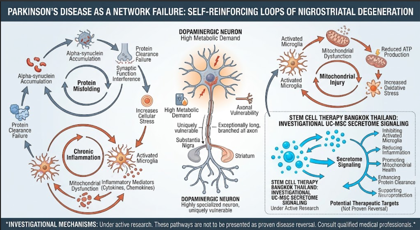
Figure 1: Parkinson’s Disease as a Network Failure: Self-Reinforcing Loops of Nigrostriatal Degeneration
Mechanism 1: Microglial Reprogramming and Neuroinflammatory Control
Microglia are resident immune cells of the central nervous system. In a healthy brain, they support surveillance, debris clearance, synaptic remodeling, and tissue protection. In Parkinson’s disease, microglia may become chronically activated and shift toward a pro-inflammatory state. When this activation becomes prolonged, the surrounding neural environment may be exposed to inflammatory cytokines, reactive oxygen species, nitric oxide signaling, and other stress mediators.
One proposed mechanism of UC-MSC stem cell therapy Bangkok Thailand activity is microglial phenotype modulation. Rather than suppressing immune function broadly, UC-MSC-derived signals may support a shift away from persistent inflammatory activation and toward a more regulatory, repair-associated microglial profile.
Key mediators often discussed in this pathway include interleukin-10, prostaglandin E2, transforming growth factor beta, indoleamine 2,3-dioxygenase-related signaling, and extracellular vesicle-associated microRNAs. These factors may influence inflammatory cytokine networks and help reduce the intensity of neuroimmune activation.
For Parkinson’s disease, this mechanism is relevant because chronic neuroinflammation may amplify degeneration rather than simply respond to it. The goal of this mechanism is not to “turn off” the immune system. The goal is to reduce the chronic inflammatory tone that may keep dopaminergic circuits under biological stress.
Mechanism 2: Alpha-Synuclein Stress and Protein-Clearance Support
Alpha-synuclein is a presynaptic protein that becomes abnormally aggregated in Parkinson’s disease. Its accumulation is associated with Lewy body pathology and synaptic dysfunction. The brain normally relies on protein-clearance systems such as autophagy-lysosomal pathways and ubiquitin-proteasome activity to manage damaged or misfolded proteins. When these systems become impaired, alpha-synuclein handling may worsen.
UC-MSC stem cell therapy Bangkok Thailand signaling is being studied in neurodegenerative models partly because extracellular vesicles and paracrine factors may influence cellular stress pathways, autophagy-related signaling, and inflammatory responses that interact with protein aggregation.
This should be stated carefully. UC-MSC stem cell therapy Bangkok Thailand should not be claimed to clear alpha-synuclein from the human brain. A more accurate explanation is that secretome-based signaling may support a less inflammatory and less oxidatively stressed environment, which may indirectly influence the cellular systems involved in protein homeostasis.
In SEO terms, this makes the article stronger because it does not repeat generic “stem cells repair damaged neurons” language. It explains the disease environment with more biological precision.
Mechanism 3: Mitochondrial Stress and Dopaminergic Energy Failure
Dopaminergic neurons are highly energy-dependent. They maintain long axonal projections, continuous neurotransmitter cycling, calcium handling, and synaptic transmission. When mitochondrial function declines, these neurons may become more vulnerable to oxidative injury and cellular exhaustion.
Mitochondrial dysfunction is one of the most frequently discussed pathways in Parkinson’s disease research. Reduced mitochondrial complex activity, impaired mitophagy, oxidative stress, and energy failure may all contribute to neuronal vulnerability.
UC-MSC stem cell therapy Bangkok Thailand may influence this pathway through antioxidant signaling, extracellular vesicle cargo, trophic-factor release, and metabolic support signals. Some experimental studies also discuss mitochondrial transfer through tunneling nanotubes, where mitochondria may move between cells under stress conditions. This mechanism is biologically interesting, but it should be presented as experimental and not as a guaranteed clinical effect.
A careful way to write this mechanism is:
UC-MSC-associated signaling may support mitochondrial resilience by reducing inflammatory pressure, influencing oxidative stress pathways, and delivering extracellular vesicle cargo that interacts with cellular survival systems. In Parkinson’s disease, this may be relevant because dopaminergic neurons are especially sensitive to mitochondrial energy failure.
Mechanism 4: Extracellular Vesicle Signaling and Molecular Cargo Delivery
Extracellular vesicles are one of the most important parts of the UC-MSC mechanism conversation. They are nanoscale signaling particles that may carry proteins, lipids, messenger RNAs, microRNAs, and regulatory molecules. Because they can participate in cell-to-cell communication, extracellular vesicles are being studied as potential mediators of MSC-related effects in neurodegenerative disease.
In Parkinson’s disease models, MSC stem cell therapy Bangkok Thailand -derived extracellular vesicles are being investigated for their possible effects on neuroinflammation, oxidative stress, neuronal survival pathways, protein aggregation stress, and synaptic function.
Possible mechanisms of extracellular vesicle activity include:
Delivery of anti-inflammatory microRNAs
Regulation of microglial activation
Support for antioxidant pathways
Interaction with apoptosis-related signaling
Trophic support for stressed neuronal networks
Modulation of neurovascular and immune communication
These pathways remain under investigation, but they provide a biologically plausible explanation for why MSC-derived products are being studied in Parkinson’s disease.
Mechanism 5: Neurotrophic Support and Synaptic Plasticity
Dopaminergic neurons do not function alone. They are part of a broader neural circuit involving the substantia nigra, striatum, basal ganglia, cortex, thalamus, and brainstem networks. Parkinson’s symptoms emerge not only from cell loss, but from altered network communication.
UC-MSC stem cell therapy Bangkok Thailand signaling may include trophic factors that support neuronal survival, axonal maintenance, synaptic plasticity, and cellular resilience. Neurotrophic factors such as brain-derived neurotrophic factor and glial cell line-derived neurotrophic factor are frequently discussed in Parkinson’s research because they may influence dopaminergic neuron survival and synaptic function.
This does not mean UC-MSC stem cell therapy Bangkok Thailand can reliably rebuild the dopaminergic system. A more precise statement is that trophic signaling may support surviving neural circuits and help maintain a less hostile microenvironment for neuronal communication.
Mechanism 6: Tyrosine Hydroxylase Signaling and Dopamine-Synthesis Capacity
Tyrosine hydroxylase is the rate-limiting enzyme involved in dopamine synthesis. In Parkinson’s disease, the loss and dysfunction of dopaminergic neurons reduces dopamine availability within the striatal system. Some preclinical regenerative studies assess tyrosine hydroxylase expression as a marker of dopaminergic neuron integrity or activity.
UC-MSC stem cell therapy Bangkok Thailand signaling may be discussed in relation to dopaminergic support when trophic factors, anti-inflammatory signals, and oxidative-stress reduction create a more favorable environment for surviving neurons. However, it is important not to overstate this mechanism. UC-MSC stem cell therapy Bangkok Thailand should not be claimed to directly normalize tyrosine hydroxylase activity in human Parkinson’s disease patients.
A balanced sentence would be:
In experimental Parkinson’s models, regenerative signaling is often evaluated through markers related to dopaminergic neuron survival, including tyrosine hydroxylase expression. For clinical content, this should be framed as a research-associated mechanism rather than confirmed restoration of dopamine production.
This makes the article sound smarter and more scientifically honest.
Mechanism 7: Neurovascular and Blood-Brain Barrier Communication
The brain microenvironment depends on healthy communication between neurons, glial cells, endothelial cells, pericytes, and immune mediators. In neurodegenerative disease, the neurovascular unit may become stressed, and blood-brain barrier function may be altered by inflammation, oxidative injury, and endothelial dysfunction.
UC-MSC stem cell therapy Bangkok Thailand signaling may interact with endothelial repair pathways, angiogenic communication, and inflammatory barrier regulation. This mechanism is relevant because Parkinson’s disease is increasingly viewed as a systemic neurodegenerative condition rather than a problem isolated only to dopamine neurons.
For stem cell therapy Bangkok Thailand content, this pathway adds depth because it explains why systemic biological support may be discussed even when the target condition is neurological. The mechanism should still be written cautiously: neurovascular support is a proposed biological pathway, not proof of clinical disease modification.
Mechanism 8: Systemic Immune-Neural Crosstalk
Parkinson’s disease may involve peripheral immune changes as well as central nervous system changes. Peripheral inflammation, gut immune signaling, microbiome-related stress, and systemic cytokine activity may influence neuroinflammatory tone. This is one reason the gut-brain axis has become an important area of Parkinson’s research.
UC-MSC signaling may be relevant because MSC stem cell therapy Bangkok Thailand can interact with immune-cell populations outside the brain, including T cells, macrophages, dendritic cells, and inflammatory cytokine networks. This systemic immunomodulatory effect may indirectly influence the neuroimmune environment.
Why Mechanism Does Not Equal Cure
A mechanism-focused article still needs medical caution. Biological plausibility is not the same as proven clinical efficacy. A therapy may show anti-inflammatory, antioxidant, or neurotrophic effects in laboratory studies and still require careful human clinical trials before it can be described as disease-modifying.
For Parkinson’s disease, the most responsible wording is that UC-MSC stem cell therapy Bangkok Thailand may support selected biological pathways under investigation. It should not be promoted as a cure, a replacement for levodopa, or a guaranteed method to stop disease progression.
Clinical Positioning for Stem Cell Therapy Bangkok Thailand
Stem cell therapy Bangkok Thailand for Parkinson’s disease should be positioned as physician-guided supportive regenerative care. The strongest content angle is not “breakthrough treatment.” A better angle is “mechanism-based biological support.”
The article should emphasize:
Neuroinflammation modulation
Mitochondrial stress support
Secretome and extracellular vesicle signaling
Dopaminergic circuit support
Neurovascular communication
Systemic immune-neural regulation
Patient selection and realistic monitoring
Continued standard neurological care
This makes the article different from generic Parkinson’s stem cell content and gives Google more original topical depth.
Conclusion
Parkinson’s disease involves more than dopamine depletion. It is a complex neurodegenerative condition shaped by alpha-synuclein stress, mitochondrial dysfunction, oxidative injury, neuroinflammation, impaired protein clearance, synaptic instability, and immune-neural communication.
A mechanism-focused discussion of stem cell therapy Bangkok Thailand should therefore focus on UC-MSC stem cell therapy Bangkok Thailand signaling rather than repeating basic explanations of what stem cells are. The most relevant proposed mechanisms include microglial reprogramming, inflammatory cytokine regulation, extracellular vesicle signaling, mitochondrial stress support, neurotrophic factor release, dopaminergic circuit support, neurovascular communication, and systemic immune modulation.
These pathways provide a scientific rationale for why UC-MSC-based regenerative medicine is being studied in Parkinson’s disease. However, they should be presented as supportive and investigational mechanisms, not as proof of cure.
Responsible regenerative neurology is not about claiming to rebuild the brain. It is about understanding the molecular environment of neurodegeneration and exploring how carefully prepared biological signaling therapies may support selected patients within a complete neurological care plan.