Our Services
Others
- DFPP
- Shockwave
- IV Drip

The clinical resolution of systemic bone mineral density reduction, pathologically categorized as Osteoporosis, stands as an increasingly complex challenge within modern metabolic medicine and degenerative orthopedics. Characterized by the progressive thinning of cortical walls and the structural fragmentation of trabecular networks, this skeletal decay drastically lowers bone tensile strength. Affected individuals face a silent but aggressive vulnerability to low-trauma fragility fractures, particularly within the femoral neck, distal radius, and vertebral bodies.
For generations, conventional therapeutic frameworks have relied on systemic pharmacological management: antiresorptive agents such as bisphosphonates or RANK-ligand inhibitors, alternated with seasonal injections of anabolic parathyroid hormone analogs. While these standard chemical pathways alter bone turnover markers, their long-term efficacy is limited by biological counter-reactions.
Bisphosphonates artificially shut down osteoclasts, but this prolonged suppression halts the natural cycle of micro-damage removal, leading to brittle bone matrices and rare structural failures like atypical femoral fractures or jaw osteonecrosis. Anabolic hormone options are limited by strict continuous usage thresholds due to cell fatigue.
Translational molecular biology introduces an alternative strategy that moves past short-term endocrine suppression to target the microenvironmental environment directly at the source. By delivering high-potency cellular matrices into the systemic circulation, advanced clinical frameworks utilizing stem cell therapy bangkok thailand address the true biological imbalances driving the disease.
Instead of placing passive mineral coatings onto a weakening skeleton, these targeted interventions deploy active paracrine signaling vectors. They navigate into the marrow niche, reset faulty gene networks, clear out chronic senescent signaling, and reactivate the body’s natural bone-building systems to reverse the structural decline associated with advanced Osteoporosis.
1. The Skeletal Equilibrium Crisis: Accelerated Osteoclastogenesis and Marrow Adipogenesis
To see why traditional pharmaceutical options eventually plateau and how advanced cell-based therapies change long-term clinical trajectories, the biological lens must look past superficial bone density T-scores and analyze the deep cellular environment inside the trabecular marrow niche.
Healthy skeletal structure relies on a continuous, tightly coupled cycle of bone remodeling governed by two opposing cell groups: bone-resorbing osteoclasts and bone-building osteoblasts.
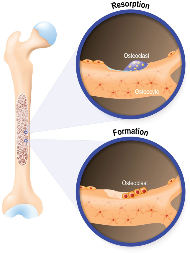
Figure 1: The physiological mechanisms of bone resorption vs. bone formation within the skeletal matrix.
As shown in the bone remodeling overview above, a healthy skeletal environment relies on a balanced synchronization between cell lines. In the development of advanced Osteoporosis, this homeostatic coupling breaks down completely. The primary driver of this structural failure is a major shift in cell differentiation within the bone marrow niche.
Healthy bone marrow contains native multipotent progenitor cells that face a constant biological choice: differentiate into bone-building osteoblasts or fat-storing adipocytes.
As the body ages or experiences chronic low-grade inflammation, the genetic wiring behind this selection path fails. The body upregulates the transcription factor PPAR-, which commands local progenitor cells to stop transforming into osteoblasts and instead shift into fat cells. This process, known as marrow adipogenesis, floods the bone cavity with fat tissue while starving the skeleton of fresh, bone-building cells.
Concurrently, the remaining marrow cells display an un-balanced overproduction of RANKL (Receptor Activator of Nuclear Factor B Ligand) while downregulating its natural protector, Osteoprotegerin (OPG).
This major collapse in the RANKL/OPG ratio triggers massive osteoclastogenesis an over-activation of multinucleated osteoclasts that aggressively tunnel through trabecular bridges faster than the remaining osteoblasts can repair them. The resulting structural decay creates the porous, micro-fractured skeletal networks that define advanced Osteoporosis.
Traditional medications cannot alter this fat-versus-bone lineage mistake; they merely freeze the existing broken matrix. Suppressing this decline requires a targeted biological intervention that can step in to completely rewrite the cell differentiation pathway.
2. Molecular Signaling Transduction: Re-Activating the Wnt/-Catenin Pathway
Advanced applications of stem cell therapy bangkok thailand intervene within this broken skeletal niche by using a highly specialized biological strategy: paracrine transduction. When high-potency allogeneic cell lines are introduced, they utilize the body’s natural circulatory highways to migrate directly toward the high concentrations of SDF-1 chemokines released by damaged, thinning bone tissues.
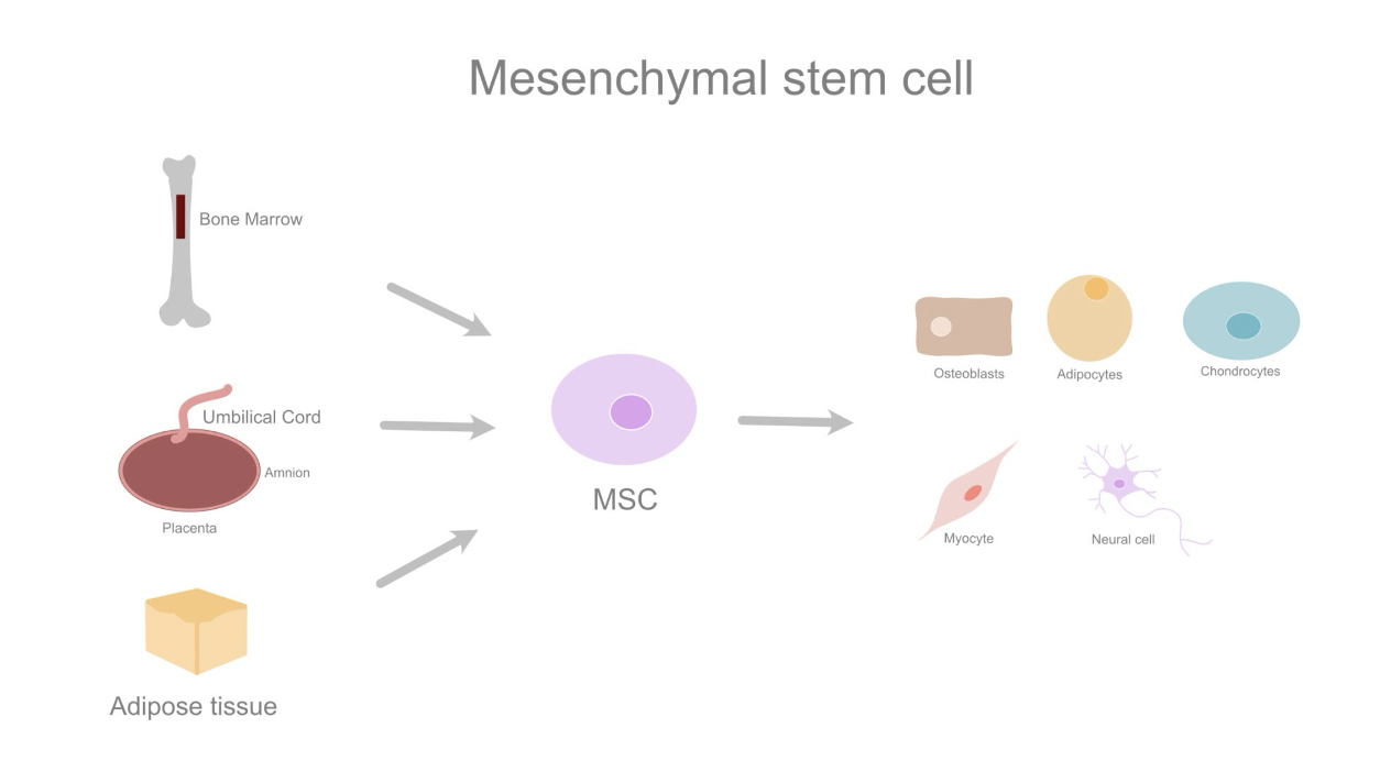
Figure 2: The multipotent differentiation paths and secretome mechanics of mesenchymal lineages.
As outlined in the lineage framework map above, these youth-derived cellular lines possess the distinct biological plasticity required to alter the marrow microenvironment. Rather than operating as passive building blocks that manually turn into bone cell-for-cell, their primary therapeutic mechanism is the continuous manufacture and targeted deployment of a powerful anti-inflammatory and anabolic secretome payload.
Once settled within the porous marrow niche, the cell lines execute a multi-layered molecular override to reverse the progression of Osteoporosis:
Reactivation of the Canonical Wnt/-Catenin Pathway
In an osteoporotic skeleton, bone formation is permanently blocked because senescent cells continuously produce inhibitory proteins like Sclerostin and Dickkopf-1 (DKK-1), which shut down the cellular engines of bone growth. The transplanted cell lines counter this restriction by releasing a dense cloud of canonical Wnt ligands (such as Wnt-3a and Wnt-10b).
These signaling molecules bind directly to the LRP5/6 receptors on sluggish host progenitor cells, completely blocking the destructive GSK-3$beta$ enzyme complex. This molecular interaction allows -catenin to accumulate in the cytoplasm and move directly into the cell nucleus.
Once inside, -catenin activates the essential Runx2 and Osterix genes, commanding host progenitors to stop turning into fat cells, shut down PPAR- production, and immediately restart the mass synthesis of fresh, active osteoblasts.
Restoring the RANKL/OPG Ratio via Exosomal MicroRNA Transfer
To stop unchecked bone tunneling, the transplanted cell lines discharge millions of microscopic, membrane-bound extracellular vesicles called exosomes into the marrow fluid. These vesicles fuse with host stromal cells, dropping off highly concentrated payloads of regulatory microRNAs most notably miR-21, miR-29a, and miR-133.
This microRNA payload acts as an intracellular brake, systematically downregulating the production of destructive RANKL while actively increasing the secretion of protective Osteoprotegerin (OPG). OPG acts like a biological shield, binding to RANKL before it can touch osteoclasts, completely freezing their hyper-activated tunneling and stopping progressive bone loss.
Upregulating Type I Collagen Synthesis and Extracellular Mineralization
As the newly activated osteoblasts multiply, paracrine growth factors primarily Transforming Growth Factor-beta 3 and Bone Morphogenetic Protein 2 (BMP-2) command them to ramp up the manufacture of Type I Collagen strands. These strands are woven into an organized osteoid matrix, which serves as a tight structural net that captures circulating calcium and phosphorus. This targeted deposition forms fresh hydroxyapatite crystals along the thinning trabecular arches, progressively restoring structural bone density.
3. Microenvironmental Target Mapping Across the Marrow Niche
To see how a targeted cell protocol maps across a degraded skeletal axis to alter structural outcomes, it is useful to analyze the specific biological targets and their corresponding regenerative interventions within Osteoporosis:
| Target Bone Niche Layer | Pathological Malfunction in Osteoporosis | Cellular Regenerative Intervention | Key Semantic Entity |
| Marrow Progenitors | Overexpress PPAR-γ; switch from bone-building to fat cell production. | Secretes Wnt-10b to activate β-catenin, shutting down PPAR-γ and driving osteoblastogenesis. | Marrow adipogenesis, Osteoblastogenesis, β-catenin accumulation |
| Osteoclast Matrix | Overstimulated by excessive RANKL levels; aggressively tunnels through bone arches. | Delivers exosomal microRNAs to restore the RANKL/OPG ratio, freezing unchecked bone resorption. | Osteoclastogenesis, RANKL/OPG ratio, Trabecular fragmentation |
| Extracellular Osteoid | Suffers from severe Type I collagen depletion and matrix fragmentation. | Produces BMP-2 and TGF-β3 to stimulate fresh collagen weaving and capture mineral deposits. | Extracellular matrix (ECM), Type I Collagen, Extracellular mineralization |
| Bone Marrow Capillaries | Experience progressive narrowing and tissue ischemia, starving local cell networks. | Releases high concentrations of VEGF to activate micro-capillary sprouting and restore marrow circulation. | Angiogenesis, VEGF signaling, Skeletal microvascular ischemia |
4. The Viability Imperative: Protecting Cellular Integrity in Bangkok Cultivation Centers
The clinical success of advanced stem cell therapy bangkok thailand for systemic disorders like Osteoporosis relies entirely on a single technical metric that is frequently overlooked in mainstream discussions: cellular viability at the exact point of care. The human secretome is an incredibly complex, active mix of signaling proteins that can only be produced and released by living, metabolically active cells. If a formulation contains high percentages of dead or dying cells, it cannot perform targeted chemotaxis, cannot manufacture exosomes, and will be quickly cleared away by the recipient’s immune system as biological waste.
Many international clinics source their cellular products from distant manufacturing facilities, requiring the cells to be deeply frozen and thawed right at the patient’s bedside. This cryopreservation process utilizing chemical cryoprotectants introduces profound thermodynamic stress to delicate plasma membranes, frequently causing cell lysis and destroying the vital surface receptors required for targeted bone homing.
Advanced biomedical facilities delivering stem cell therapy bangkok thailand bypass this logistical bottleneck by leveraging a continuous, closed-system cultivation framework operating locally. By executing aseptic processing within state-of-the-art laboratory environments close to the clinical suites, technicians expand neonatal lineages without the need for deep freezing.
The formulated allogeneic grafts remain suspended in a temperature-regulated, nutrient-dense transport matrix right up to the exact minute of clinical delivery. Automated cytometry and fluorescence-based live/dead assays confirm verified viability scores exceeding 95%. This logistical integration ensures that the recipient receives an uncompromised secretome payload, maximizing paracrine signaling efficiency and optimizing structural tissue remodeling within heavily compromised bone environments.
5. Real-World Expectations: Tracking Skeletal Turning Points
When discussing advanced cell-based applications for bone density restoration, maintaining absolute transparency and an honest, grounded perspective is essential. Stem cell therapy bangkok thailand is not a magical overnight treatment that will instantly eliminate bone porosity or wipe away years of physical degradation in a single day. Instead, it serves as a powerful biological accelerant that works from the inside out to slow the progression of tissue decay and create conditions where lost skeletal density can naturally recover.
Patients responding well to advanced, fresh cell protocols typically observe gradual, steady skeletal improvements over a window of three to nine months:
Stabilization of Bone Turnover Markers: A measurable reduction in circulating serum cross-laps ( -CTX) paired with a steady increase in bone-specific alkaline phosphatase (BSAP), providing clear evidence of shifted cell dynamics.
Arrest of Microstructural Height Loss: A noticeable stabilization of vertebral body height, accompanied by a systematic reduction in chronic, deep dull bone pain across the axial skeleton.
Progressive Trabecular Stabilization: High-resolution follow-up dual-energy X-ray absorptiometry (DEXA) scans or peripheral quantitative CT panels typically reveal a visible stabilization or progressive recovery of trabecular micro-architecture.
Reduction in Fragility Incidents: A significant increase in the mechanical loading threshold of weight-bearing structures, helping to lower overall fracture risks during daily physical activities.
Conclusion: Securing Your Skeletal Future
Marrow adipogenesis, osteoclast overactivation, and progressive trabecular fragmentation involve complex, aggressive biological processes, but patients do not have to remain locked in a purely reactive cycle of managing bone loss with temporary chemical blocks while their underlying skeletal health undergoes permanent degradation. Treating a deep cellular and microenvironmental failure with simple surface-level mineral supplements masks the physical decline without addressing the true biological crisis.
By choosing advanced, fresh cellular protocols through stem cell therapy bangkok thailand , you give your body the highly potent, youth-derived resources it needs to cool chronic marrow inflammation, reactivate the canonical Wnt pathway, and rebuild a resilient parallel collagen scaffold from the inside out. Embracing the cutting edge of regenerative medicine under strict quality control standards represents a powerful, proactive choice to avoid the constraints of progressive disease, protect your long-term mobility, and build a resilient foundation for your skeletal future.