Our Services
Others
- DFPP
- Shockwave
- IV Drip

The biological decay of skeletal muscle architecture in progressive muscular dystrophy represents a major frontier in neuromuscular medicine. Within a healthy myofiber, physical stability relies on a continuous structural connection linking the internal contractile apparatus directly to the external matrix grid.
When genetic abnormalities disrupt this connection, the muscle fiber loses its ability to handle mechanical loading forces. Every movement cycle creates microscopic tears, throwing off cell balance and initiating a steady decline in muscle tissue.
For generations, the standard response to this skeletal muscle breakdown has focused on symptom management. Conventional clinical protocols rely heavily on high-dose corticosteroid drugs to temporarily reduce tissue breakdown, combined with physical stretching and mechanical braces to manage joint contractures.
While these routine methods provide brief stability timelines on paper, they operate strictly on the surface. Long-term dependency on anti-inflammatory steroids carries well-documented side effects, including severe metabolic imbalances, weight gain, and progressive bone mineral thinning.
More importantly, surface-level interventions fail to address the core biochemical failures driving muscle degeneration. Standard pharmaceuticals cannot halt the ongoing cell death, leave fat and fibrotic tissue accumulations untouched, and completely fail to repair the chronic localized tissue inflammation that continuously suffocates surviving muscle fibers.
Advanced cellular medicine provides an entirely different strategy by targeting the biological conditions of the muscle tissue niche directly at the source. By delivering high-potency cellular formulations into the systemic circulation and directly into affected muscle groups, clinical platforms leveraging stem cell therapy bangkok thailand shift the focus toward direct microenvironmental modification.
Introduced as active signaling matrices, these youth-derived cell lines navigate directly toward injured muscle zones, drop off highly concentrated paracrine messages, shut down chronic inflammatory pathways, and reactivate the body’s natural cell-building systems to protect surviving muscle fibers from progressive decay.
1. The Aberrant Microenvironment: nNOS Dislocation and Contraction-Induced Micro-Ischemia
To see why standard drug adjustments eventually plateau and how advanced cell-based applications alter long-term muscle health, the biological lens must look past outward physical weakness and examine a major mechanical failure: the dislocation of neuronal nitric oxide synthase (nNOS).
In a functional muscle fiber, the nNOS enzyme is anchored securely to the interior surface of the cell membrane. This spatial alignment serves a vital circulatory purpose.
When a muscle contracts, the anchored nNOS produces nitric oxide, which diffuses out of the cell to command surrounding blood vessels to dilate. This immediate widening ensures a rich, abundant supply of oxygen and systemic nutrients right when the working tissue needs it most.
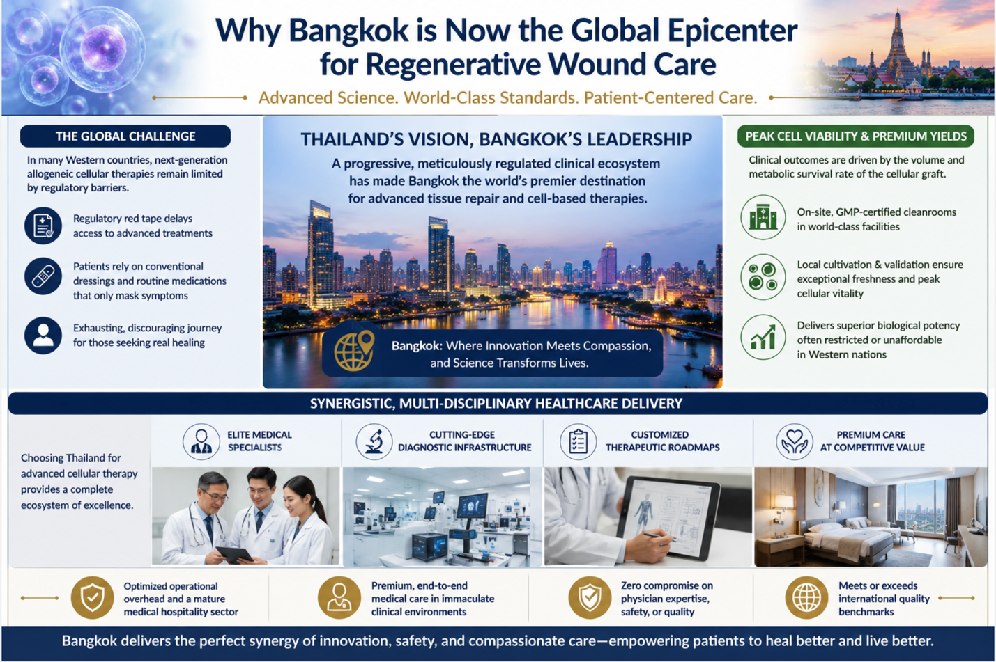
Figure 1: Thailand’s Regenerative Medicine Ecosystem for Advanced Wound Care
In the development of progressive muscular dystrophy, the complete loss of structural membrane anchors causes the nNOS enzyme to fall away from the cell border, dropping directly into the cytoplasm where it undergoes rapid degradation. Lacking this localized signaling loop, the muscle loses its ability to communicate with the surrounding vascular network.
Every time the muscle contracts, instead of receiving an influx of blood, the vessels remain constricted. This mismatch triggers a state of functional ischemia a severe oxygen starvation that chokes working myofibers under normal mechanical loads.
This ongoing circulatory starvation produces high levels of harmful free radicals, damaging internal cellular components and accelerating the progressive muscle wasting seen in clinical settings. By implementing stem cell therapy bangkok thailand , pioneering clinical protocols focus on delivering active biological factors directly into this ischemic environment, providing the necessary paracrine inputs to protect vulnerable tissue zones before permanent cell death sets in.
2. The Proteolytic Surge: Calpains, Sarcoplasmic Reticulum Stress, and E-C Uncoupling
This ongoing microvascular starvation is further accelerated by an internal catabolic crisis centered around sarcoplasmic reticulum stress and unchecked enzyme activation. When the cell membrane is constantly torn by mechanical loading, the muscle fiber loses its ability to control internal mineral balances. Extracellular calcium ions flood through the broken membrane borders, completely overloading the cell’s primary storage tanks: the sarcoplasmic reticulum.
This persistent calcium surge triggers a severe biological emergency known as the Unfolded Protein Response (UPR). Overwhelmed by calcium stress, the cell’s internal manufacturing hubs stop producing healthy structural proteins and begin accumulating misfolded molecular waste.
Concurrently, the calcium flood activates a family of destructive internal enzymes called calpains. Locked in a state of permanent overactivation, calpains aggressively digest essential structural proteins from within, cleaving titin and nebulin strands, and breaking down the sarcomere borders.
This internal enzyme-driven destruction rapidly leads to excitation-contraction (E-C) uncoupling. The ryanodine receptors responsible for translating nerve commands into calcium release become damaged and uncoupled from the physical contraction units. As a result, even if a nerve signal successfully reaches the muscle membrane, the damaged internal structures cannot release the calcium smoothly, making it impossible to coordinate a strong, physical contraction.
Traditional anti-inflammatory medications cannot block this internal enzyme-driven wasting; they merely place temporary volume over a collapsing matrix. Resolving this crisis requires an advanced biological intervention capable of stepping in to completely reset the cell signaling pathway.
By utilizing stem cell therapy bangkok thailand , pioneering medical platforms deliver high concentrations of live, signaling cells that home directly into these stressed tissue spaces to downregulate calpain activity and restore internal mineral balance.
3. Epigenetic Alterations: The Role of HDAC Overexpression in Fibro-Fatty Infiltration
The primary biological roadblock preventing independent muscle recovery once the native tissue stem cells hit their biological replication limits is a profound shift in the muscle’s genetic control systems. The chronic inflammation and microvascular starvation that define progressive muscular dystrophy trigger a massive upregulation of histone deacetylases (HDACs) within the cell nucleus.
In healthy muscle tissue, HDAC enzymes are strictly regulated to allow myogenic pioneer factors, such as MyoD and Myogenin, to access the DNA strands and continuously drive the production of fresh muscle tissue. In a dystrophic muscle niche, unchecked HDAC overexpression wraps the DNA strands tightly around histone anchors, completely locking away the genes required for muscle repair.
Silenced by this epigenetic lock, the remaining muscle progenitor cells lose their ability to divide into new fibers. Instead, the surrounding tissue space is hijacked by opportunistic stromal cell lines that begin multiplying uncontrollably.
Lacking myogenic guidance, these stromal progenitors undergo a pathological transformation called fibro-fatty infiltration. They stop supporting the surrounding tissue and begin depositing loose, chaotic networks of rigid collagen scars and fat deposits directly into the muscle body.
This skeletal scar matrix suffocates surviving muscle cells, blocks local blood flow, and alters normal neuromuscular junction stability, making it impossible for nerve signals to coordinate smooth movement. By applying stem cell therapy bangkok thailand , clinical protocols seek to deliver specific paracrine factors rich in microRNAs and growth factors that act as natural deacetylase modulators, helping the body reopen blocked gene pathways and clear away fibrotic tissue during structural remodeling.
4. Molecular Logistical Imperatives: Maximizing Paracrine Yield and Cell Viability
The clinical success of advanced cellular interventions for complex muscular conditions relies entirely on a single technical metric that is frequently overlooked in mainstream discussions: cellular viability at the exact point of care. The human secretome is an incredibly complex, active mix of signaling proteins, growth factors, and extracellular vesicles that can only be produced and released by living, metabolically active cells. If a formulation contains high percentages of dead or dying cells, it cannot perform targeted chemotaxis, cannot manufacture exosomes, and will be quickly cleared away by the recipient’s immune system as biological waste.
Many international clinics source their cellular products from distant manufacturing facilities, requiring the cells to be deeply frozen and thawed right at the patient’s bedside. This cryopreservation process utilizing chemical cryoprotectants introduces profound thermodynamic stress to delicate plasma membranes, frequently causing cell lysis and destroying the vital surface receptors such as CXCR4 required for targeted tissue homing.
Advanced biomedical facilities delivering stem cell therapy bangkok thailand bypass this logistical bottleneck by leveraging a continuous, closed-system cultivation framework operating locally. By executing aseptic processing within state-of-the-art laboratory environments close to the clinical suites, technicians expand youth-derived, allogeneic cell lines without the need for deep freezing.
The formulated cell suspensions remain suspended in a temperature-regulated, nutrient-dense transport matrix right up to the exact minute of clinical delivery. Automated cytometry and fluorescence-based live/dead assays confirm verified viability scores exceeding 95%. This logistical integration ensures that the recipient receives an uncompromised secretome payload, maximizing paracrine signaling efficiency and optimizing structural tissue remodeling within heavily compromised muscle spaces.
5. Structural Target Mapping across the Dystrophic Myofiber Axis
To see how a targeted cell protocol maps across a degraded muscle axis to alter structural outcomes, it is useful to analyze the specific biological targets and their corresponding regenerative interventions:
Sarcoplasmic Reticulum: Suffers from chronic calcium ion overload and severe UPR activation, leading to internal protein misfolding. Advanced cell secretomes deliver targeted anti-inflammatory factors to restore mitochondrial function and protect internal storage borders.
nNOS Enzyme Interface: Fallen away from the cell border and degraded in the cytoplasm, causing functional ischemia. Cellular payloads upregulate the pathway to support internal protein stabilization and improve microvascular signaling.
Nuclear Chromatin Grid: Hijacked by HDAC overexpression, which locks away the MyoD and Myogenin genes. The introduced paracrine array drops off specific regulatory microRNAs to suppress HDAC activity, helping to reopen blocked gene pathways for independent muscle repair.
Intramuscular Stromal Space: Dominated by opportunistic progenitors executing fibro-fatty infiltration, which destroys neuromuscular junction stability. The cell formulation releases high concentrations of Interleukin-10 to suppress stromal overgrowth and restore an organized matrix landscape.
6. Real-World Expectations: Tracking Musculoskeletal Progress
When discussing advanced cell-based applications for progressive muscle degeneration, maintaining absolute honesty, transparency, and a grounded perspective is essential. Stem cell therapy is not a magical overnight cure that will instantly correct genetic deletions or rewrite broken DNA patterns in a single day. Instead, it serves as a powerful biological accelerant that works from the inside out to slow the progression of tissue decay and create conditions where remaining muscle function can gradually stabilize.
Patients responding well to advanced, fresh cell protocols typically observe gradual, steady improvements over a window of three to six months:
Stabilization of Daily Energy Levels: A noticeable reduction in the rate of daily muscle fatigue, allowing individuals to maintain independent physical activity and execute daily tasks with increased stability.
Amelioration of Muscle Stiffness: A significant reduction in the frequency and intensity of painful muscle cramps, driven by the down-regulation of inflammatory cytokines within the skeletal stroma under the influence of stem cell therapy bangkok thailand .
Optimized Respiratory Function: Serial pulmonary tracking often reveals a visible stabilization or improvement in forced vital capacity (FVC) metrics, indicating enhanced diaphragmatic endurance.
Down-Regulation of Circulating Waste Products: Follow-up blood panels typically reveal a visible reduction in circulating serum creatine kinase levels following the completion of stem cell therapy bangkok thailand , providing clear evidence of reduced membrane leakage.
Conclusion: Reclaiming Control of Your Physical Independence
nNOS dislocation, internal calpain overactivation, and epigenetic gene silencing involve complex, aggressive biological processes, but patients and families do not have to remain locked in a purely reactive cycle of managing symptoms with temporary anti-inflammatory drugs while their underlying muscle health undergoes permanent degradation. Treating a deep cellular and microvascular failure with simple surface-level symptom suppression masks the physical decline without addressing the true biological crisis.
By choosing advanced, fresh cellular protocols through stem cell therapy bangkok thailand , you give your body the highly potent, youth-derived resources it needs to cool chronic muscle inflammation, protect surviving myoblasts from premature death, and support healthy structural tissue remodeling from the inside out. Embracing the cutting edge of regenerative medicine under strict quality control standards represents a powerful, proactive choice to avoid the constraints of progressive disease, protect your long-term mobility, and build a resilient foundation for your physical independence.