Our Services
Others
- DFPP
- Shockwave
- IV Drip

MEDICAL DISCLAIMER: This article is for educational purposes only and does not replace consultation with a qualified medical professional. Emerging biological therapies require individualized clinical assessment. The FDA has not approved any stem cell treatments for hearing restoration.
For patients with permanent sensorineural impairment, conventional auditory devices manage symptoms without addressing the underlying cellular deterioration. Stem cell therapy for hearing loss represents a shift from mechanical amplification to biological repair, targeting the regeneration of cochlear structures.
Look, the clinical reality is far more complex than the headlines suggest. The confusion surrounding regenerative medicine is massive, with unproven commercial clinics often blurring the lines between legitimate, highly regulated clinical trials and expensive, experimental procedures. Many patients fly overseas expecting a guaranteed cure, only to return with lighter wallets and unchanged audiograms.
By the end of this guide, you will understand the exact biological mechanisms of regenerative audiology so you can evaluate clinical trial viability and prepare for specialized consultations. We’re going to examine cellular feasibility, intratympanic delivery protocols, and current regulatory statuses for emerging auditory treatments. Our research team evaluated the most recent clinical data to separate molecular facts from medical tourism fiction.
Stem cell therapy for hearing loss utilizes biological mechanisms to repair damaged cochlear tissue, though treatments remain strictly investigational.
Stem cell therapy for hearing loss focuses on repairing auditory nerve pathways and cochlear structures using advanced biological models. Current investigational approaches utilize targeted cells to secrete proteins that stabilize local tissue environments, rather than attempting to construct entirely new anatomical structures. This precise biological intervention is currently restricted to highly regulated clinical trials. sensorineural hearing loss accounts for 90% of cases (ASHA) making biological regeneration the ultimate frontier in modern audiology.
Can stem cells restore hearing loss? To answer that, we have to isolate exactly what is broken inside the ear. The clinical reality is that while stem cells offer a fascinating biological scaffolding, they cannot reverse decades of established cochlear fibrosis.
First, we draw an absolute line between conductive hearing loss and sensorineural hearing loss. Conductive loss is purely mechanical. It involves the physical obstruction of sound waves whether that is fluid trapped in the middle ear, a ruptured tympanic membrane, or ossicular chain fixation where the tiny bones fuse together (like in advanced otosclerosis). Stem cells do absolutely nothing for this. Surgery and prosthetics fix mechanical problems.
Sensorineural hearing loss, however, targets cellular and nerve decay. Sensorineural damage occurs when the delicate stereocilia (hair cells) within the cochlea or the spiral ganglion neurons (SGNs) of the auditory nerve die off. These structures do not naturally regenerate in mammals. In a healthy human ear, sound waves vibrate the organ of Corti, causing the stereocilia to bend. This physical bending stretches microscopic protein threads called tip links, which open mechanotransduction ion channels and allow a massive influx of potassium from the endolymph. That rapid chemical change fires the electrical signal to your brain.
When you are exposed to extreme acoustic trauma or ototoxic drugs (like high-dose aminoglycosides or cisplatin chemotherapeutics), reactive oxygen species (ROS) flood the inner ear. These free radicals brutally destroy the mitochondria inside the hair cells, causing them to undergo apoptosis (programmed cell death). Once a human hair cell is destroyed, it is gone permanently. Worse, once the hair cell dies, the underlying spiral ganglion neuron loses its primary source of electrical stimulation and neurotrophic support, triggering a secondary wave of nerve death known as Wallerian-like retrograde degeneration.
This is where Mesenchymal Stem Cells (MSCs) enter the clinical picture. MSCs don’t just transform into new ear parts like Lego blocks. Instead, research on neurotrophic factors secreted by MSCs demonstrates that MSCs act as biological factories (PubMed). They secrete specialized neurotrophic proteins specifically Brain-Derived Neurotrophic Factor (BDNF), Glial Cell Line-Derived Neurotrophic Factor (GDNF), and Neurotrophin-3 (NT-3). These specific proteins promote the survival of spiral ganglion neurons and protect the remaining, undamaged cochlear hair cells from further deterioration.
To make sense of this, our research team utilizes The Regenerative Audiology Matrix. This is a three-step evaluation framework to determine if a patient is even a viable candidate for emerging clinical trials.
Contrast replacing a faulty cochlear implant electrode with the biological nightmare of regenerating microscopic auditory hair cells. One is hardware engineering; the other is directing complex cellular differentiation in a fluid-filled, highly pressurized, and deeply encased organ.
The mammalian cochlea is incredibly hostile to foreign cellular integration. The endolymph fluid that bathes the hair cells maintains a highly specific, toxic potassium concentration (+80mV endocochlear potential). Any biological therapy introduced to this environment must survive this extreme chemical gradient while simultaneously making exact synaptic connections with the auditory nerve. This immense biological hurdle explains exactly why functional restoration in human trials remains deeply challenging and heavily delayed.
UC-MSC stem cells do not directly turn into new cochlear hair cells; rather, they secrete powerful neurotrophic factors that stabilize the inner ear environment. When discussing sensorineural hearing loss stem cell therapy, we are specifically looking at Umbilical Cord Mesenchymal Stem Cells.
Why the umbilical cord? Because UC-MSCs extracted primarily from Wharton’s Jelly possess unique regenerative, paracrine, and immunomodulatory properties that far exceed adult stem cells harvested from bone marrow or adipose (fat) tissue. Bone marrow extraction requires a painful iliac crest aspiration, yields fewer viable cells, and those specific cells often carry the epigenetic baggage and chromosomal wear-and-tear of the aging donor. Furthermore, adult stem cells trigger higher immune responses.
UC-MSCs, conversely, are immunologically privileged. They lack Major Histocompatibility Complex (MHC) Class II antigens and crucial co-stimulatory molecules like CD40, CD80, and CD86. This means they don’t trigger aggressive graft-versus-host immune rejections when introduced into a patient’s inner ear. They essentially fly under the radar of the host’s immune system.
Critically, they produce a far more robust “secretome.” The secretome is the highly concentrated cocktail of proteins, growth factors, and exosomes that the cell secretes into its local environment. In the inner ear, this secretome mitigates excessive tissue fibrosis. When the cochlea is damaged, it doesn’t just sit there quietly. It often fills with fibrous scar tissue through a process called fibro-osseous degeneration. This dense scar tissue effectively blocks any chance of natural or artificial signal transmission, rendering future therapies (even cochlear implants) useless.
UC-MSCs create a highly protective environment, halting that fibrotic cascade by actively downregulating pro-inflammatory cytokines like TNF-alpha, Interleukin-1 beta, and Interleukin-6 (IL-6). These secreted proteins modulate local inflammation and encourage the survival of existing, damaged spiral ganglion neurons. In laboratory organoid models, this biological scaffolding prevents further cellular degradation. Researchers are currently attempting to use these secreted factors to trigger surrounding supporting cells (like Deiters’ cells or pillar cells) to transdifferentiate into functional sensory cells. But this precise biological mechanism remains strictly in the investigational phase.
But we need to set realistic, hard boundaries here. UC-MSCs can promote local healing, stabilize a dying auditory nerve, and prevent further degradation of the cochlear microenvironment. They cannot magically rebuild a completely destroyed, heavily ossified cochlea from scratch. Once the fluid spaces turn to bone, cellular therapy is permanently off the table.
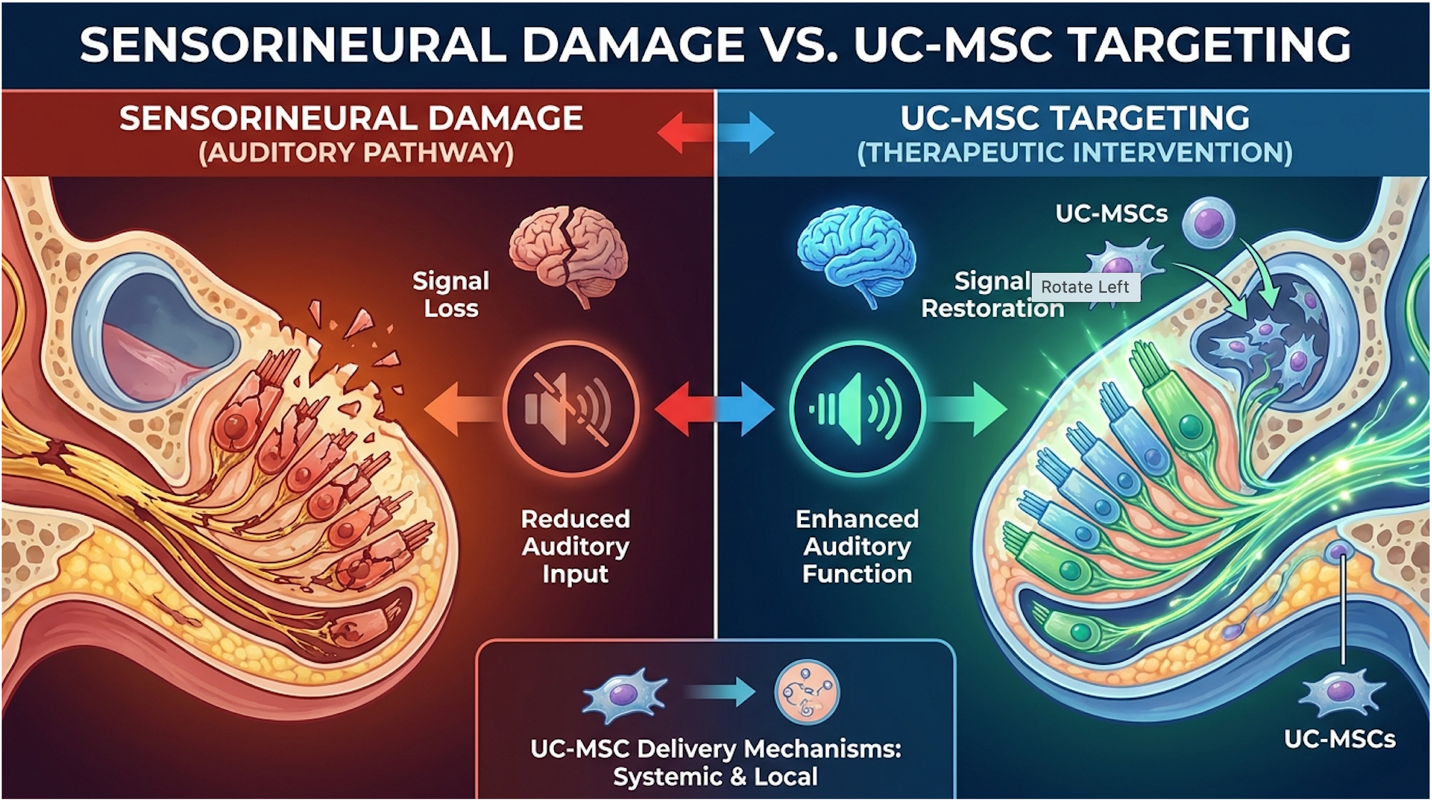
Figure 1: UC-MSCs target cochlear fibrosis by secreting localized neurotrophic factors.*
This isn’t purely theoretical. In an active FDA-approved clinical trial evaluating umbilical cord blood, autologous umbilical cord blood is being administered to children with acquired sensorineural hearing loss to evaluate its safety and preliminary efficacy (ClinicalTrials.gov). These trials rigorously monitor auditory brainstem responses (ABR) and otoacoustic emissions (OAEs) to see if these paracrine effects translate to measurable, real-world decibel gains in pediatric patients.
The most recent treatment for hearing loss extends beyond traditional amplification to include molecular pharmacology and regenerative biology. Researchers are actively developing pharmaceutical agents and advanced biological models designed to halt cellular degeneration in the cochlea. These therapies target the molecular pathways responsible for auditory nerve survival, aiming for biological preservation rather than mechanical compensation. using stem cell-derived organoids to test new drugs allows scientists to bypass animal models safely (Nature) massively accelerating the research pipeline.
There is currently no commercially available drug that restores chronic sensorineural hearing loss. Everyone asks about a new drug that restores hearing. However, clinical trials are actively testing novel molecular compounds designed to trigger the regeneration of supporting cells within the cochlea. The pharmaceutical pursuit of auditory restoration splits into two distinct categories: otoprotective drugs and regenerative drugs. Overpromising pharmaceutical timelines only hurts patients; the biological reality of penetrating the cochlea remains the industry’s biggest hurdle.
Otoprotective drugs aim to prevent damage before or immediately after it occurs. Think of military personnel taking a pill prior to artillery training to shield their stereocilia from acoustic trauma. Researchers are exploring compounds like D-methionine, sodium thiosulfate, and ebselen, which act as powerful free-radical scavengers. When loud noise traumatizes the ear, it creates reactive oxygen species (ROS) that poison the hair cells. Otoprotective drugs neutralize these toxins before permanent mitochondrial collapse and cell death happens. They must be administered within a very tight timeframe usually hours to be effective.
Regenerative drugs, conversely, attempt to trigger hair cell regrowth after the damage is already permanent. This is infinitely harder. Researchers are investigating complex genetic triggers, such as ATOH1 gene therapy. During embryonic development, the ATOH1 (or Math1) gene acts as the master switch that tells embryonic cells to become auditory hair cells. Scientists are trying to use Adeno-Associated Viral (AAV) vectors to deliver ATOH1 directly into the supporting cells of a deaf adult, attempting to reboot this developmental program.
Simultaneously, researchers are exploring small-molecule inhibitors targeting the Notch signaling pathway. During normal fetal development, Notch signaling uses a process called lateral inhibition to prevent too many cells from turning into sensory hairs (forcing them to become structural supporting cells instead). By delivering highly targeted gamma-secretase inhibitors, researchers block this Notch signal, effectively lifting the biological brake and encouraging those existing supporting cells to transdifferentiate into new hair cells.
The primary barrier to all of this is anatomical delivery. The inner ear is encased in the temporal bone the hardest bone in the human body. To get a drug into the cochlea, it typically must cross the round window membrane. The molecular weight and hydrophilicity of the drug dictate its success. Large molecule drugs and heavy viral vectors simply fail to pass through the membrane’s microscopic pores into the perilymph fluid.
| Drug Goal | Mechanism of Action | Clinical Challenge | Current Status |
| Otoprotective | Scavenges reactive oxygen species (ROS) | Timing; must be given near exposure | Phase II/III Trials |
| Regenerative | Triggers ATOH1 gene expression | Molecule size; penetrating round window | Phase I/II Trials |
| Anti-inflammatory | Suppresses immune response via steroids | Systemic side effects if absorbed | Standard of Care |
Despite some compelling early data showing mild speech recognition improvements in quiet environments, no regenerative drug has achieved total auditory restoration. The official FDA warnings regarding unapproved therapies explicitly state that no stem cell-based products or regenerative pharmaceuticals are currently approved for hearing loss (FDA). Patients must be highly cautious of medical practitioners claiming they have early, exclusive access to miracle pharmaceuticals.
To accelerate the testing of these molecular compounds without risking human hearing, scientists have turned to inner ear organoids for hearing loss.
Inner ear organoids are complex 3D biological tissue models developed entirely from stem cells. Instead of growing a flat, two-dimensional layer of cells in a plastic petri dish, researchers cultivate a three-dimensional, miniaturized version of actual cochlear tissue. They start with induced pluripotent stem cells (iPSCs) often derived from a patient’s own skin or blood cells and carefully guide them through highly specific developmental stages using precise Wnt, BMP4, and FGF signaling pathways.
They push the iPSCs to form an otic placode, which then folds inward into an otic vesicle, flawlessly mimicking exact human embryonic development. These models actually develop the distinct layers, supporting cells, and sometimes even primitive hair cell structures found in the human ear. The architectural complexity of an organoid behaves much more like a real human cochlea than a flat cell culture ever could. They feature the necessary spatial arrangement and transcriptomic profiles of the organ of Corti.
Why does this matter to a patient waiting for a cure? Because it speeds up the clinical testing pipeline exponentially. If a pharmaceutical company develops a new compound they believe will trigger Notch pathway transdifferentiation, they no longer have to rely solely on guinea pigs or wait years for human Phase I safety data.
By using stem cell-derived organoids to test new drugs, scientists can rapidly execute high-throughput screening on thousands of compounds for ototoxicity (ear-damaging side effects) and regenerative potential simultaneously. The Regenerative Audiology Matrix relies heavily on this organoid response data to validate which biological agents actually warrant the immense cost and risk of human trials. It provides a crucial biological bridge between raw stem cell theory and human pharmacological application.
While clinical solutions undergo rigorous organoid testing, patients often want to know how to improve hearing naturally today.
Let’s be direct. No natural supplement, dietary change, or holistic exercise will resurrect dead hair cells. However, natural strategies are absolutely critical for preserving existing vascular health which is strictly necessary to keep the surviving auditory nerve alive while awaiting future therapeutics. A dead, withered nerve cannot be revived by future drugs.
An injection for hearing loss utilizes an intratympanic delivery method, bypassing the eardrum to administer therapeutics directly into the middle ear. Currently, this 15-to-30 minute in-office procedure is the standard medical protocol for delivering high-dose steroids to treat sudden sensorineural hearing loss. In the future, this same precise anatomical pathway will be required to deliver experimental regenerative biologics. Success rates of intratympanic steroid injections hit 60% when administered rapidly highlighting the critical importance of swift medical intervention.
If you’re researching treatments, you’ve likely seen ads for intravenous stem cell therapy for hearing loss. Biologically speaking? It’s highly inefficient and clinically flawed. Systemic IV infusions scatter cells throughout the lungs and liver, rarely crossing the blood-labyrinth barrier into the ear. Intravenous delivery is a marketing tactic, not an otologic protocol.
Targeted therapies require an intratympanic injection. Here’s exactly how intratympanic steroid injections administered in a clinical setting work. The patient lies on an exam table with their affected ear facing the ceiling and their head turned about 45 degrees. The otolaryngologist (ENT) applies a topical anesthetic usually a phenol solution or viscous lidocaine drops to heavily numb the tympanic membrane (eardrum). They wait 10 to 15 minutes for the anesthesia to take full effect.
Using an operating microscope or an endoscope for precise, magnified visualization, the physician inserts a fine, 25-gauge or 27-gauge spinal needle directly through the inferior-posterior quadrant of the eardrum. They specifically target this quadrant to avoid damaging the delicate ossicular chain (the malleus, incus, and stapes bones) located higher up.
They slowly inject the liquid therapeutic (typically dexamethasone or methylprednisolone) into the middle ear space, specifically aiming to pool the liquid over the round window niche.
The round window membrane (RWM) is not just an empty opening; it is a highly complex, semi-permeable biological barrier separating the air-filled middle ear from the fluid-filled cochlea. The RWM is roughly 70 micrometers thick and consists of three distinct microscopic layers: the outer squamous epithelium, the middle connective tissue core containing active fibroblasts and collagen, and the inner mesothelium that bathes directly in the perilymph fluid of the scala tympani.
The injected medication must sit flush against the outer epithelium and slowly diffuse across all three cellular layers into the cochlear fluid. This diffusion is governed strictly by molecular weight and hydrophilicity. Generally, molecules smaller than 1000 Daltons pass easily. Large stem cells, heavy viral vectors, or massive neurotrophic proteins struggle massively.
Sometimes, micro-adhesions, mucosal folds, or a false membrane physically block the round window niche, requiring the surgeon to adjust patient positioning or surgically clear the obstruction to ensure the fluid correctly pools over the target area. Because standard liquid medications drain quickly down the Eustachian tube, pharmaceutical researchers are actively developing hyaluronic acid hydrogels sticky, viscous nanocarriers designed to hold the drug against the RWM for days instead of minutes.
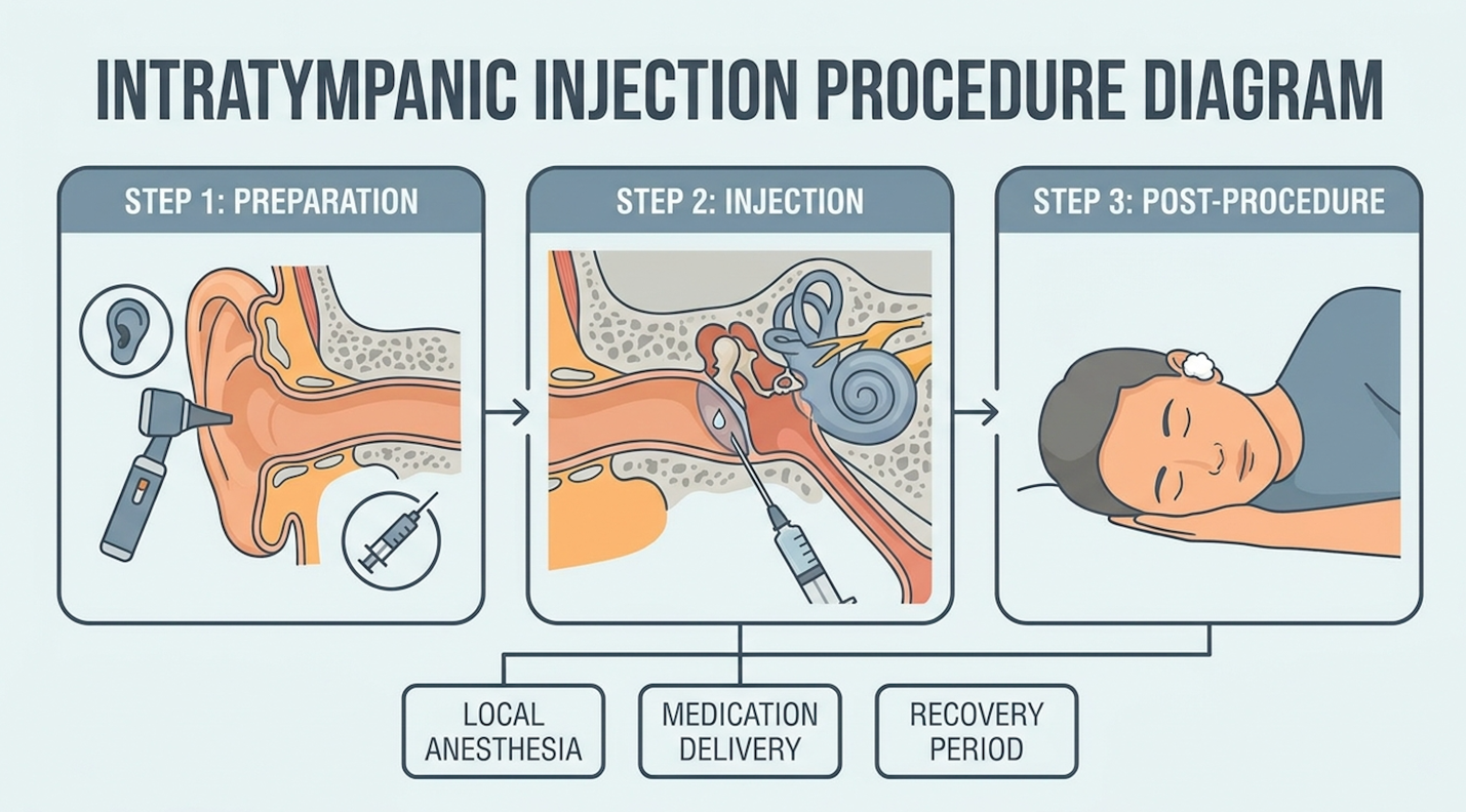
Figure 2: The needle bypasses the tympanic membrane to pool medication over the round window.
This exact physical mechanism currently used routinely for steroids will be the exact delivery system used for future UC-MSC therapies. The intratympanic injection procedure and associated risks carry minor complications, such as temporary dizziness, localized pain, or in rare cases, a persistent perforation of the eardrum that requires a minor surgical paper patch (UTHealth).
Intratympanic steroid injections are typically administered in a series of three to four injections spaced out over a two-to-three-week period. This rapid, aggressive protocol is the standard clinical response for Idiopathic Sudden Sensorineural Hearing Loss (ISSNHL). It aims to deliver concentrated anti-inflammatory medication before permanent nerve death occurs. Administration frequency relies heavily on the patient’s immediate audiometric response following the initial dose.
When evaluating the success rate of intratympanic injections, we have to meticulously isolate the medication from the mechanism. For sudden sensorineural hearing loss—where a patient wakes up completely deaf in one ear due to a suspected viral infection or autoimmune attack—time is absolutely critical. Clinical observations show that intratympanic steroid injections can improve auditory function (defined as a >10dB recovery across two contiguous frequencies) in up to 60% of patients dealing with ISSNHL, provided they are administered within the “golden window” of 14 days from symptom onset.
Patients typically notice auditory changes within one to two weeks, as the localized medication requires several days to adequately permeate the round window membrane and heavily reduce cochlear inflammation. If the injection is administered 30 to 60 days after the sudden loss, the efficacy rate plummets dramatically.
However, applying this 60% success rate to chronic, long-term, age-related presbycusis or noise-induced hearing loss using experimental biologics is scientifically fraudulent. Steroids reduce acute, active inflammation; they do not resurrect hair cells that died a decade ago. If no functional improvement is measured within four weeks, the steroid therapy is generally considered unsuccessful.
Let’s break down the intratympanic injection cost. Standard steroid injections in a US clinic typically range from $300 to $800 per session. Because this is a standard-of-care medical procedure (often billed under CPT code 69801), it is usually fully covered by commercial health insurance and Medicare after deductibles are met.
Conversely, experimental biologic injections are entirely out-of-pocket. Domestic investigational clinics often charge $5,000 to $10,000 per ear. This exorbitant price combines steep facility fees with expensive, proprietary cellular processing fees. Because of stringent FDA regulations strictly limiting the expansion and manipulation of stem cells in the US, many desperate patients explore stem cell therapy for hearing loss in countries like Thailand, Panama, or Mexico.
These international medical tourism packages routinely scale from $15,000 to $25,000. This massive cost factors in international travel, multi-day luxury hospital stays, and unproven multi-day IV/intratympanic combination treatments. The Regenerative Audiology Matrix strongly cautions against this financial outlay without independent, third-party audiometric validation of the clinic’s past results. You are paying for a highly experimental trial, not a guaranteed medical service.
Regardless of what liquid is pushed through the eardrum, the physical recovery process dictates stringent post-procedure behavior. Proper rest after a steroid injection isn’t merely a suggestion; it’s an absolute structural necessity. If you sit up too fast, the medication drains straight down the Eustachian tube into the back of your throat before it can absorb into the cochlea.
Patients must remain completely recumbent (lying on their side or back with the injected ear up) in the clinic for 20 to 30 minutes immediately following the injection. Do not talk. Do not yawn. Do not swallow heavily. The biophysics of swallowing naturally opens the Eustachian tube, creating a sudden vacuum effect that physically pulls the liquid away from the round window niche and empties it into the nasopharynx.
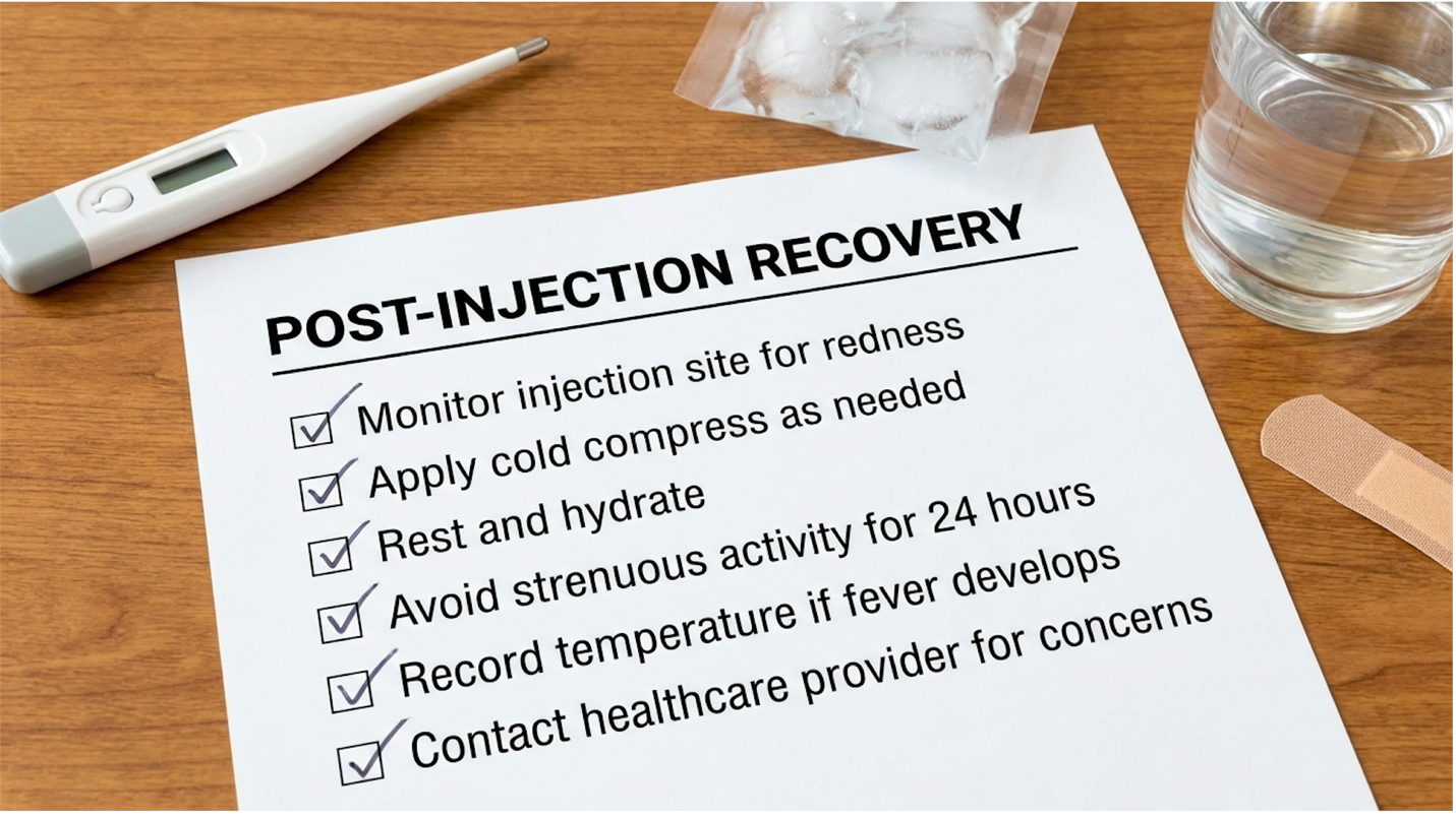
Figure 3: Avoid pressure changes and water exposure for 72 hours post-injection.
For the next 48 to 72 hours at home, strict otologic rules apply. You must avoid any Valsalva maneuvers. This means absolutely no forceful nose blowing, no bearing down during bowel movements, and no heavy lifting at the gym. Popping your ears to clear pressure can force the medication right out of the round window niche and can easily re-rupture the healing eardrum puncture site.
Keep the ear completely dry for 7 to 10 days to prevent middle ear infections (otitis media). When showering, do not let the showerhead spray directly onto the ear. Use a cotton ball heavily coated in petroleum jelly to block the outer ear canal, as the surface tension of the jelly actively repels water.
Flying in an airplane or SCUBA diving is strictly prohibited for several weeks until your ENT visually confirms the tympanic membrane has fully sealed. The cabin pressure changes during a flight will wreak havoc on an unhealed eardrum. If you experience severe, room-spinning vertigo that lasts more than an hour, or notice persistent purulent (pus-like) drainage on your pillow, immediate clinical reassessment is required.
While biological therapies hold immense future potential, patients must navigate current treatment options with intense skepticism regarding unverified commercial claims. The gap between peer-reviewed scientific trials and for-profit medical tourism creates significant safety hazards for desperate patients.
Patients desperately seeking auditory recovery often fall into predictable, highly dangerous clinical traps.
The first major pitfall is assuming that the phrase “stem cell” equates to a guaranteed cure for chronic dead hair cells. This misunderstanding leads patients to confuse the temporary reduction of inner-ear inflammation (which might slightly clear up tinnitus) with permanent cellular regeneration. They are not the same biologically.
The second pitfall is financial exploitation via medical tourism. Paying thousands of dollars out-of-pocket for non-FDA approved offshore treatments exposes you to massive risk. The official FDA warnings regarding unapproved cellular therapies explicitly state that no stem cell-based products are approved for hearing loss (FDA). These offshore clinics operate without Phase III efficacy data, often injecting uncharacterized cell suspensions that could theoretically trigger unwanted cellular proliferation (tumors or teratomas) deep within the temporal bone.
The third pitfall is abandoning standard-of-care rehabilitation while waiting for a miracle. Delaying conventional treatment leads to auditory deprivation, where the brain’s auditory processing centers literally atrophy from lack of stimulation. The longer the auditory nerve goes without electrical or acoustic input, the less effective any future biological therapy will be.
Biological therapies are not appropriate for everyone. If a patient presents with profound, total deafness accompanied by severe cochlear ossification (where the fluid spaces of the inner ear have turned to solid bone due to severe meningitis or advanced otosclerosis), stem cells have absolutely no fluid environment in which to survive.
In these specific scenarios, standard Cochlear Implants remain the proven, medical gold standard for functional recovery. CIs bypass the damaged hair cells entirely, directly stimulating the auditory nerve with precise electrical impulses. For detailed protocols on proven surgical interventions, patients should review the American Speech-Language-Hearing Association’s guide on cochlear implants.
Before making any financial decisions, patients must step away from wellness clinic sales pitches and seek expert help. Mandate a consultation with a board-certified neurotologist a subspecialist in inner ear neurology to review your high-resolution MRI and audiogram. Accessing specialized neurotology resources provides the objective data required to determine if you are a candidate for future clinical trials or if established surgical alternatives are your only viable path forward.
Stem cells cannot currently restore profound, chronic hearing loss in human patients outside of highly experimental trials. While preclinical research demonstrates that Mesenchymal Stem Cells can secrete proteins to protect existing neural structures, regenerating functional cochlear hair cells remains biologically complex. Current human trials focus strictly on safety and minor functional improvements. Patients should rely on established auditory devices while biological therapies undergo mandatory Phase III efficacy testing.
The primary treatment options for hearing loss remain mechanical amplification through hearing aids and surgical cochlear implants. For sudden onset sensorineural loss, clinicians immediately utilize intratympanic steroid injections to preserve nerve function. Experimental regenerative therapies, including cellular injections and targeted molecular pharmacology, are actively being researched but are not yet commercially approved. Over 90% of patients currently manage their condition through advanced digital amplification devices. Patients must consult a board-certified audiologist to determine their appropriate clinical pathway.
There is no definitive biological cure for chronic sensorineural hearing loss slated for commercial release. The medical consensus indicates that while regenerative trials and inner-ear organoid research are advancing rapidly, FDA-approved cellular restoration is still several years away. Current pharmacological developments focus primarily on otoprotective drugs that prevent future damage rather than reversing existing profound deafness. Unverified clinics offering immediate “cures” lack peer-reviewed efficacy data.
UC-MSC stem cells do not directly turn into new cochlear hair cells; rather, they secrete powerful neurotrophic factors that stabilize the inner ear environment. These secreted proteins modulate local inflammation and encourage the survival of existing, damaged spiral ganglion neurons. In laboratory organoid models, this biological scaffolding prevents further cellular degradation. Researchers are currently attempting to use these secreted factors to trigger surrounding supporting cells to transdifferentiate into functional sensory cells. This precise biological mechanism remains strictly in the investigational phase.
Intratympanic steroid injections are typically administered in a series of three to four injections spaced out over a two-to-three-week period. This rapid protocol is the standard clinical response for sudden sensorineural hearing loss, aiming to deliver concentrated anti-inflammatory medication before permanent nerve death occurs. Administration frequency relies heavily on the patient’s immediate audiometric response following the initial dose. The procedure is performed in-office by an ENT specialist.
There is currently no commercially available drug that restores chronic sensorineural hearing loss. However, clinical trials are actively testing novel molecular compounds designed to trigger the regeneration of supporting cells within the cochlea. These experimental pharmaceuticals utilize biological pathways discovered through stem cell and organoid research. While some early-phase trials show mild improvements in speech recognition in quiet environments, no drug has achieved total auditory restoration. Regulatory approval for these regenerative agents requires extensive long-term safety data.
Patients receiving an intratympanic steroid injection for sudden hearing loss typically notice auditory changes within one to two weeks. The localized medication requires several days to adequately permeate the round window membrane and reduce cochlear inflammation. While some individuals experience rapid improvement following the first dose, optimal recovery usually manifests after the full series of injections is completed. If no functional improvement is measured within four weeks, the therapy is generally considered unsuccessful.
For patients managing sensorineural impairment, stem cell therapy for hearing loss represents an investigational frontier aiming to repair inner ear structures rather than merely amplifying sound. While intratympanic steroid injections successfully improve sudden hearing loss in up to 60% of acute cases, experimental cellular regeneration remains in Phase I and II clinical trials. The safest approach combines rigorous clinical evaluation with continued utilization of advanced digital amplification.
Navigating this evolving medical landscape requires applying The Regenerative Audiology Matrix to separate commercial hype from clinical reality. By evaluating your specific diagnosis against delivery feasibility and exact regulatory statuses, you can avoid unproven cellular interventions. This framework ensures that your pursuit of biological restoration is grounded in verified, peer-reviewed molecular science.
Before pursuing any experimental biologic or regenerative treatment, compile your complete audiometric history, MRI imaging, and previous treatment records. Schedule a comprehensive evaluation with a board-certified neurotologist at a regulated academic medical center. A specialized assessment is the only reliable method to determine if you are a viable candidate for emerging clinical trials.