Our Services
Others
- DFPP
- Shockwave
- IV Drip

Type 2 diabetes gets described, most of the time, as a story about insulin resistance tissues that stop responding properly to insulin, forcing the pancreas to work harder until it eventually can’t keep up. That’s accurate, but it skips the more specific question: what is actually happening inside a beta cell that causes it to fail, years before a diagnosis shows up on a blood test? The answer involves protein folding, energy production, and a slow accumulation of misfolded material inside the pancreatic islets three mechanisms rarely discussed outside specialist endocrinology literature, and three of the more active areas current research into stem cell therapy Bangkok Thailand programs is examining.
The Beta Cell’s Impossible Job
A single beta cell produces and packages enormous quantities of proinsulin protein continuously, folding each molecule correctly before it can be converted to functional insulin. This is a lot of manufacturing work for one cell type to sustain for decades. In someone with rising insulin resistance, the pancreas compensates by demanding even more insulin output per cell, pushing beta cells into a chronic state of high manufacturing load long before blood glucose numbers look abnormal.
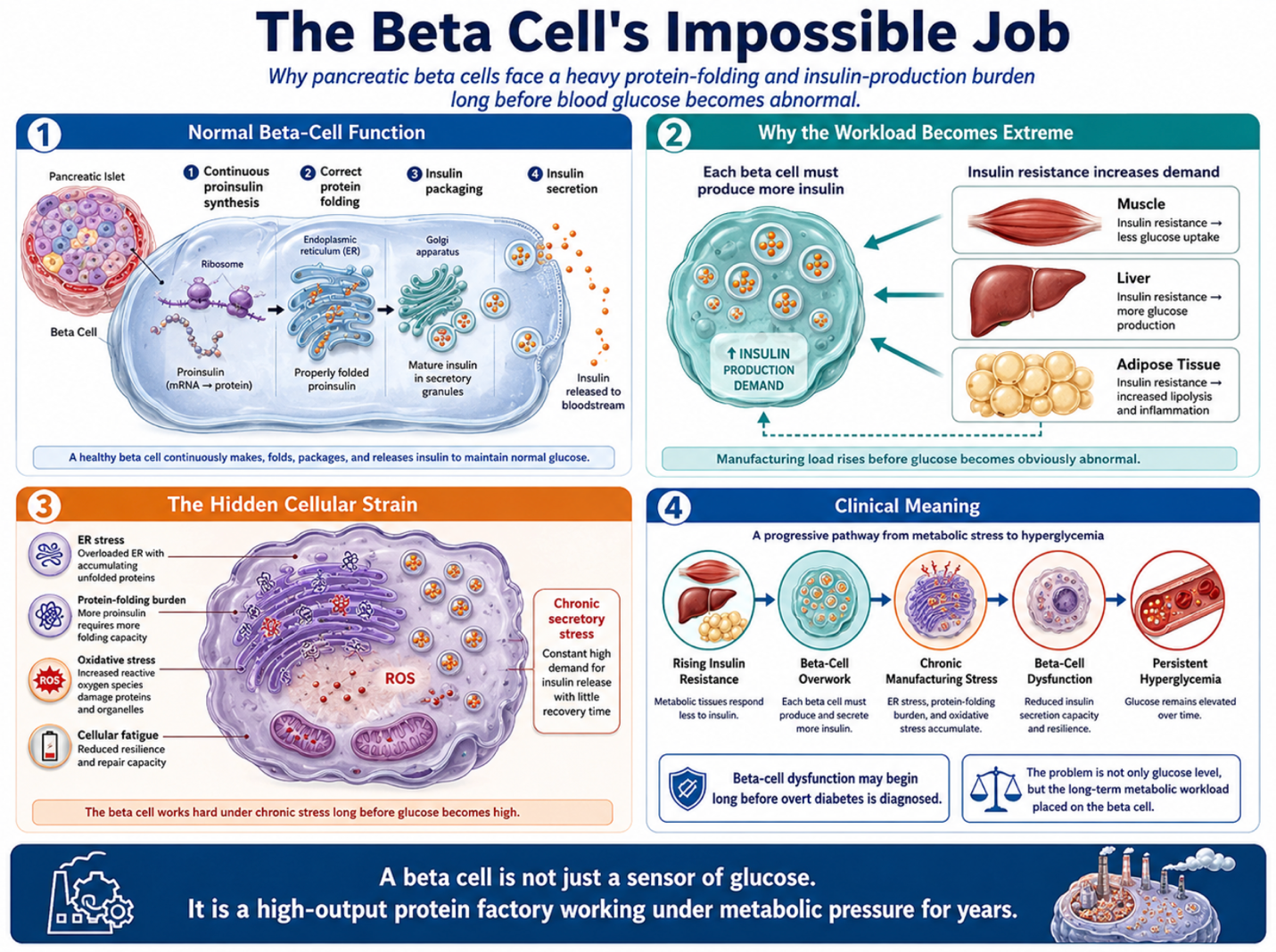
Figure 1: How Beta Cells Carry the Burden of Continuous Insulin Manufacturing
Endoplasmic Reticulum Stress: When Protein Folding Falls Behind
The endoplasmic reticulum is the cellular structure responsible for folding proteins like proinsulin correctly. Under sustained high demand, misfolded and unfolded proteins begin accumulating inside it faster than the cell can process them a state called ER stress. The cell has a built-in response to this, the unfolded protein response, which initially tries to reduce protein production and increase folding capacity to cope. But when the overload is prolonged, as it typically is in the years leading up to a type 2 diabetes diagnosis, this same protective response flips into a trigger for programmed cell death. In other words, the beta cell’s own quality-control system becomes the mechanism that kills it. Research into stem cell therapy secretions suggests some paracrine factors may help ease this ER stress load, though the evidence here is early and mostly confined to laboratory models rather than confirmed clinical outcomes.
Islet Amyloid: A Second, Separate Problem
Alongside insulin, beta cells co-secrete a smaller protein called islet amyloid polypeptide, or IAPP. In a healthy pancreas this isn’t an issue. In a chronically overworked beta cell, IAPP molecules can misfold and aggregate into amyloid deposits inside and around the islets a process with real similarities to amyloid buildup seen in some neurodegenerative conditions. These deposits are physically disruptive to islet architecture and are toxic to neighboring beta cells, and autopsy studies of long-standing type 2 diabetes consistently show significant islet amyloid accumulation. This is a distinct mechanism from insulin resistance or general inflammation, and it’s one reason beta cell loss in type 2 diabetes can continue even when a patient’s other metabolic markers are reasonably controlled.
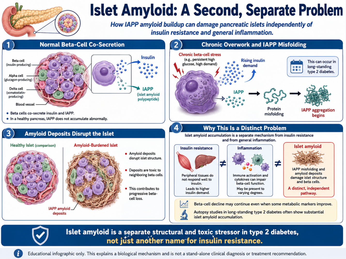
Figure 2: IAPP Misfolding, Amyloid Deposits, and Progressive Beta-Cell Dysfunction
Mitochondrial Bioenergetics: The Glucose-Sensing Failure
Beta cells rely on a tight coupling between glucose metabolism and mitochondrial ATP production to know when to release insulin rising glucose leads to more ATP, which is the actual trigger for insulin secretion at the cellular level. Chronic oxidative and ER stress damages mitochondrial function over time, weakening this coupling. The practical result is a beta cell that may still be alive but has lost the fine-tuned ability to sense glucose and secrete insulin appropriately, a state functionally different from beta cell death but still contributing meaningfully to poor glycemic control. Some laboratory data on mesenchymal cell paracrine signaling points to a possible supportive effect on mitochondrial function in stressed cells, a mechanism distinct from and additional to general antioxidant activity.
Exosomes and microRNA Cargo: A More Targeted Signal Than Growth Factors Alone
Beyond the broader paracrine factors typically discussed, stem cell therapy Bangkok Thailand release exosomes small membrane-bound vesicles carrying a specific cargo of microRNAs, small non-coding RNA molecules capable of directly influencing gene expression in recipient cells. Certain microRNAs found in these exosomes have been studied for their potential role in reducing beta cell apoptotic signaling and modulating the unfolded protein response described above. This is mechanistically different from a growth factor binding a receptor; it’s a more direct form of cell-to-cell instruction, and it’s an area receiving increasing research attention as exosome isolation and characterization techniques improve.
Why Mechanism Detail Matters for Evaluating a Program
Type 2 diabetes management increasingly recognizes that beta cell failure isn’t one single process it’s ER stress, amyloid burden, and mitochondrial decline layered on top of insulin resistance, often progressing at different rates in different patients. A stem cell therapy Bangkok Thailand protocol that only describes itself in terms of “reducing inflammation and improving insulin sensitivity” is describing a small part of the picture. Patients evaluating a clinic have reasonable grounds to ask more specific questions: what cell dosing and delivery protocol is used, whether pre-treatment C-peptide and beta cell function testing is part of the workup, and how the clinic monitors for meaningful change versus general symptom improvement over time.
What the Evidence Currently Supports
The cellular mechanisms described above ER stress, islet amyloid, mitochondrial dysfunction, and microRNA-mediated signaling are well documented in diabetes research generally. The extent to which mesenchymal stromal cell therapy meaningfully reverses these specific processes in human beta cells, at a scale that changes long-term insulin independence or HbA1c outcomes, remains an area of ongoing clinical study rather than settled consensus. This therapy is best considered alongside standard diabetes management under a physician’s supervision, not as a replacement for it.
Closing Thought
The insulin-resistance narrative around type 2 diabetes is true but incomplete. Underneath it, individual beta cells are dealing with a protein-folding crisis, an amyloid burden, and a slow mitochondrial decline three separate and compounding mechanisms. Understanding that layered reality is useful groundwork for anyone trying to evaluate what a stem cell therapy Bangkok Thailand program is actually targeting, and at what level of specificity.
This article is for informational purposes and does not constitute medical advice. Anyone considering cellular therapy for diabetes should consult both an endocrinologist and the treating clinic directly regarding candidacy, risks, and expected outcomes.