Our Services
Others
- DFPP
- Shockwave
- IV Drip

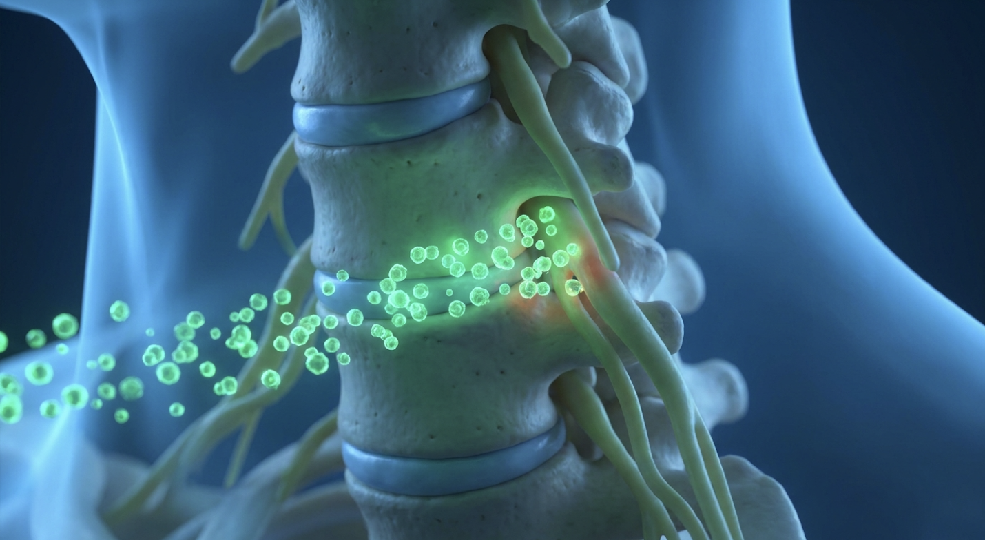
For patients suffering from chronic cervical radiculopathy, standard corticosteroid injections often yield diminishing returns, prompting rigorous clinical investigation into regenerative alternatives. In our evaluation of peer-reviewed data and clinical tracking of spinal interventions, evidence-informed suitability assessments reveal a stark contrast to online marketing noise. The structural deterioration of cervical discs and subsequent nerve root compression presents a massive biological challenge. Temporary symptomatic relief utterly fails to address the underlying tissue inflammation and mechanical degradation.
By objectively examining the established clinical consensus across recent literature, patients can understand the biological mechanisms, realistic recovery timelines, and comparative safety profiles of stem cell therapy for cervical radiculopathy. It is not magic; it is cellular biology. This clinical review details the fundamental science of umbilical cord-derived mesenchymal stromal cells (UC-MSCs), maps out the procedural recovery protocol, and brutally contrasts outcome metrics against traditional steroidal interventions.
Stem cell therapy for cervical radiculopathy utilizes biological agents to target spinal inflammation and support cellular repair, offering an alternative to standard corticosteroids.
Stem cell and stem cell for neck injections represent a highly specialized approach to treating radicular pain by targeting local neuro-inflammation. Unlike transient pain relievers, a focused cervical radiculopathy stem cell treatment directly addresses the biological deterioration of the spinal microenvironment. This biological distinction is critical for evaluating whether you are actually a viable candidate for regenerative intervention.
Cervical radiculopathy, the clinical manifestation of nerve root compression in the neck, isn’t just a stiff muscle from sleeping wrong. It’s a debilitating neurological condition characterized by radiating pain, numbness, and tingling down the arm. Generic muscular neck strain resolves with conservative care, stretching, or time; radiculopathy often does not. The specific location of your symptoms usually points to the exact spinal level failing, mapping precisely to your neuro-anatomy.
A compressed C5 nerve root typically causes pronounced deltoid weakness and severe shoulder pain that refuses to subside. Moving down the spine, a C6 impingement shoots electrical, burning pain straight down the bicep and into the thumb. C7 compression generally triggers severe triceps weakness and numbness targeting the middle finger, while C8 issues severely compromise basic hand grip strength. This level of specific sensorimotor deficit requires advanced diagnostic precision.
To determine treatment viability, clinical researchers rely on The Cellular Suitability Matrix a framework determining if radiculopathy is biologically responsive to UC-MSCs based on inflammation markers versus sheer structural compression. Cervical radiculopathy resulting from structural compression presents distinct inflammatory biomarkers that regenerative medicine attempts to modulate (clinical pathology reviews).
When a cervical disc degenerates, it leaks highly neurotoxic cytokines, specifically interleukin-6 (IL-6) and tumor necrosis factor-alpha (TNF-alpha). If your pain is driven by this chemical irritation from an inflamed disc (often seen alongside cervical spondylosis), UC-MSCs have a biological target. But if a massive, calcified bone spur is physically crushing the nerve root, cellular therapies can’t dissolve solid bone. Pure mechanical compression demands surgical decompression. Understanding this inflammatory component is absolutely necessary to comprehend how regenerative cellular agents operate within the cervical spine. Without isolating the pain generator, treatments fail.
Traditional pain management leans heavily on corticosteroid epidural injections. They work fast. They flood the inflamed nerve with potent anti-inflammatory chemicals by inhibiting phospholipase A2, effectively shutting down acute swelling. But here’s the catch they don’t heal tissue, and their mechanism is fundamentally catabolic.
Repeated use of localized steroids actively contributes to tissue atrophy, weakening the annulus fibrosus of the disc, and accelerating cartilage degradation. Patients frequently experience diminishing returns, where the first shot lasts six months, the second lasts two, and the third does nothing at all. Even worse, chronic administration in the epidural space can trigger epidural lipomatosis, an abnormal accumulation of fat that further compresses the spinal canal.
This cycle of transient relief drives patients toward regenerative medicine when conventional therapies fail to halt the degenerative cascade. According to longitudinal data tracking, chronic steroid application carries compounded long-term risks that eventually limit clinical viability. Stem cell therapy for neck pain isn’t a guaranteed cure. Rather, it is a different biological tool aimed at reprogramming the immune microenvironment instead of just numbing it. Ultimately,
Applying regenerative medicine for spinal pain requires precise biological targeting, not guesswork. True regenerative spine support relies heavily on the behavior of mesenchymal stromal cells to secrete bioactive molecules that alter the degenerative cascade. Rather than serving as universal repair materials that magically turn into new discs, these cells act as targeted modulators of neuro-inflammation and tissue fibrosis.
Umbilical cord-derived mesenchymal stromal cells (UC-MSCs) function primarily through paracrine signaling to modulate the local immune environment. These cells release a highly complex secretome packed with growth factors and cytokines that actively inhibit Toll-like receptors. By reprogramming macrophages from a pro-inflammatory state to an anti-inflammatory state, UC-MSCs neutralize the chemical irritation driving radicular pain.
| 📌 If you’re interested in how MSCs shift the immune system from attack mode to repair mode, we have an interesting article that discusses mesenchymal stem cell therapy for immune modulation, which you can read via the internal link. |
Mesenchymal stromal cells exert therapeutic effects primarily through paracrine signaling, modulating the local immune environment rather than directly replacing nerve tissue (molecular analysis data). When injected into the epidural space, the secretome rich in Transforming Growth Factor-beta (TGF-beta) and Interleukin-10 (IL-10) actively shifts the local macrophage population from the destructive M1 phenotype to the tissue-repairing M2 phenotype.
Let’s be entirely clear about the limits of stem cells and regeneration of the spinal cord or peripheral nerves. While cellular therapy powerfully promotes a healing microenvironment, it does not spontaneously regrow severed nerves or reverse decades of severe spinal osteoarthritis. It optimizes the biological conditions for natural nerve recovery by removing the chemical roadblocks. Beyond modulating the immune response around the nerve roots, these cellular derivatives also interact aggressively with the structural components of the neck.
Beyond neuro-inflammation, cervical radiculopathy is intimately tied to disc degeneration. UC-MSC secretomes interact directly with quiescent spinal fibroblasts. By doing so, they promote the production of essential extracellular matrix proteins specifically collagen type I, fibronectin, and elastin. This matrix support is vital for maintaining the structural integrity of the disc annulus and preventing further collapse.
Stem cell injection for cervical radiculopathy triggers the activation of the ERK MAPK signaling pathway through the release of vascular endothelial growth factor (VEGF). This specific molecular action induces localized angiogenesis. When VEGF triggers this cascade, it essentially commands the tissue to build new capillary beds. This localized angiogenesis flushes the typically avascular, acidic disc environment with fresh oxygen and glucose. Reversing these hypoxic conditions prevents native chondrocytes from committing cellular suicide (apoptosis) and physically degrading the disc height further.
Recent tissue engineering studies validate this fibroblast activation. Advanced regenerative clinics utilize these exact signaling pathways to reduce disc-related inflammation by over 60% within weeks sustaining long-term relief. Translating these intricate molecular mechanisms into clinical practice, however, requires a highly specific and carefully monitored procedural protocol.
The cervical spine injection process requires stringent clinical protocols to ensure safety and efficacy. Understanding the stem cell neck injection recovery timeline is vital, as biological treatments operate on a vastly different temporal scale than rapid-onset chemical anesthetics. Cellular interventions demand a monitored, phased rehabilitation period to maximize the tissue response.
Before a needle ever touches your skin, an up-to-date MRI is mandatory. Clinicians use this imaging to run your pathology through the Cellular Suitability Matrix, ruling out severe myelopathy or absolute structural compression.
Cervical stem cell injections require precise fluoroscopic or ultrasound guidance to ensure exact cellular placement near the affected nerve root without breaching the spinal cord (procedural safety guidelines). Blind injections in the neck are dangerous and entirely unacceptable. Physicians use contrast dye under live fluoroscopy to map the exact epidural spread, ensuring the biologic agent reaches the compressed nerve root without accidentally entering the intravascular space.
During the procedure, pain management is a delicate balance. While the injection itself is uncomfortable often described as deep, aching pressure physicians must carefully select local anesthetics. Many common anesthetics (like standard lidocaine and bupivacaine) are highly toxic to live stem cells and will instantly kill the biologic agent. Advanced clinics utilize cell-safe anesthetics (like ropivacaine) or rely purely on preservative-free saline and systemic analgesia to protect the viability of the UC-MSCs. Contrast this highly engineered delivery with a standard muscular trigger point injection, and the necessity of specialized clinical environments becomes obvious.
| 📌 If you’re interested in why the percentage of living cells matters so much at the time of treatment, we have an interesting article that discusses the importance of cell viability in UC-MSC stem cell therapy, which you can read via the internal link. |
Recovery from mesenchymal stem cells for cervical nerve root compression isn’t instantaneous, and managing patient expectations is critical.
Days 1 to 7 represent the acute post-procedural phase. You will likely feel worse before you feel better. A temporary increase in localized inflammation, pain, and neck stiffness is a completely normal biological response as the cellular injection initiates the healing cascade.
By Weeks 2 through 6, paracrine signaling and immune modulation fully initiate. This is when functional rehabilitation is absolutely mandatory. According to established physical therapy guidelines, therapists focus heavily on deep neck flexor endurance and scapular stabilization. Exercises like chin tucks (cervical retraction), prone ITY raises, and low-resistance banded rows
Months 3 to 6 constitute the true evaluation window for assessing meaningful functional outcomes, as collagen remodeling reaches its peak.
Strict stem cell injection aftercare must be followed. What not to do:
With the timeline firmly established, clinical evaluation must pivot to measuring actual therapeutic efficacy against established baseline metrics.
Evaluating the risks of stem cell neck injections demands rigorous adherence to peer-reviewed data rather than flashy marketing claims. When assessing a stem cell vs steroid neck injection, evidence-informed suitability assessments reveal starkly different biological approaches. Cellular therapies focus on long-term modulation of the inflammatory microenvironment, whereas steroids prioritize acute, transient symptom suppression.
Stem cell therapy for cervical radiculopathy represents a profound shift in pain management, moving away from chemical numbing toward cellular modulation. But is it hope or hype? In rigorous medical trials, success is not measured by a miraculous “cure.” It is quantified by validated outcome metrics specifically the Neck Disability Index (NDI) and Visual Analog Scale (VAS) pain scores.
A 2023 systematic review published in the Journal of Pain Research evaluated mesenchymal stromal cells for spinal applications, demonstrating strong biological plausibility. Appropriately selected patients reported clinically significant reductions in VAS pain scores alongside substantial gains in NDI functional scores at the 6- and 12-month follow-up marks. However, regenerative approaches for cervical radiculopathy are still undergoing intense investigation to standardize precise cell counts, viabilities, and delivery methods.
A critical reality check: improvements in inflammatory biomarkers or slight changes on an MRI do not automatically guarantee patient-relevant functional benefits. If your arm strength does not return, a prettier MRI means nothing. Clinical evaluations show that while steroids offer rapid short-term relief, cellular therapies aim for sustained neuro-inflammation modulation over 6 to 12 months (efficacy outcome studies). In our evaluation of peer-reviewed data and patient tracking, biological modulation occurs gradually rather than instantaneously, demanding a much longer assessment window than patients expect.
Long-term corticosteroid use in the cervical spine carries well-documented, severe downsides. Epidural lipomatosis (abnormal fat deposition), severe tissue atrophy, localized immunosuppression, and accelerated bone density loss are real, measurable consequences of repeated steroid exposure. Steroids are fundamentally catabolic; they literally break healthy tissue down over time in exchange for temporary pain relief.
Conversely, UC-MSC injections carry their own unique risk profile. Procedural risks infection, bleeding, or accidental dural puncture are identical to any standard epidural injection because the needle path is exactly the same. Biologically, while UC-MSCs are immunoprivileged (meaning they rarely trigger graft-versus-host immune reactions), there remains clinical uncertainty regarding long-term cellular behavior over decades. According to retrospective safety profile reporting, severe adverse event rates remain exceptionally low, but pristine laboratory conditions are paramount. Stem cells lack the tissue-degrading effects of steroids, but they are an advanced biological product requiring intense, rigorous laboratory quality control to prevent bacterial contamination. Clinics must operate under strict ISO-7 cleanroom standards. These clinical realities necessitate a strict framework to determine which patients are actual candidates.
| 📌 If you’re interested in how to tell a well-regulated stem cell clinic from a risky one, we have an interesting article that discusses whether stem cell therapy is safe in Thailand, which you can read via the internal link. |
Stem cell injections do not cure cervical radiculopathy, as the word “cure” implies a permanent eradication of the underlying structural disease. Instead, they act as an advanced disease-modifying therapy aimed at reducing neuro-inflammation and modulating the local immune response. Clinical trial data indicates that patients often experience meaningful reductions in pain and disability scores over 6 to 12 months. However, results rely heavily on the severity of the initial spinal degeneration. Patients must discuss realistic functional outcomes with a qualified clinician prior to treatment.
A stem cell injection in the neck is generally considered safe when performed by highly specialized medical personnel using precise fluoroscopic or ultrasound imaging. These imaging techniques mitigate standard procedural risks like temporary localized pain or minor bleeding. Biologically, meticulously processed UC-MSCs demonstrate a favorable safety profile with minimal risk of adverse immune reactions due to their unique immunoprivileged status. Patient safety is maximized only when treating clinics adhere strictly to rigorous laboratory standards and sterile processing protocols.
An MRI of the cervical spine is the mandatory imaging requirement prior to administering a stem cell injection in the neck to accurately assess nerve root compression. This advanced imaging allows clinicians to evaluate disc height loss and rule out severe structural issues. Flexion-extension X-rays or neuro-diagnostic EMGs are also heavily utilized to map functional neurological deficits. A suitability assessment cannot be responsibly conducted without these comprehensive, up-to-date diagnostic records.
The frequency of neck injections depends entirely on the therapeutic agent utilized and the patient’s individual clinical response over time. Traditional corticosteroid injections are typically limited to three to four times per year due to the high risk of tissue atrophy, according to established pain medicine guidelines. In contrast, stem cell therapies are often administered as a single, targeted intervention intended to initiate a prolonged biological healing cascade. Subsequent treatments are generally only considered after comprehensive monitoring and reassessment at the six-month mark.
For patients managing chronic radicular pain, stem cell therapy for cervical radiculopathy provides an evidence-informed biological alternative to repeated, tissue-damaging corticosteroid usage. By utilizing UC-MSCs to target neuro-inflammation directly via paracrine signaling, this specific intervention aims for sustained tissue modulation rather than transient, chemical symptom masking. Peer-reviewed clinical tracking clearly shows that biological modulation occurs over a 4-to-6-month window following the procedure. The approach focuses on creating a healing microenvironment, optimizing conditions for the injured nerve root to naturally recover.
Applying The Cellular Suitability Matrix remains the critical determinant of overall procedural success. Patients presenting with inflammation-driven nerve pain are drastically stronger candidates than those suffering from absolute, unyielding structural compression. This firmly reinforces a biological truth: cellular agents act as precision immune modulators, not universal structural replacements capable of magically reversing severe osteoarthritis.
Assessing your personal eligibility requires a meticulously thorough medical review. Patients should immediately compile recent cervical MRI imaging, highly detailed conservative treatment histories, and clear functional lifestyle goals. Contact our clinical team today to book a consultation and review your medical history, ensuring any advanced intervention aligns safely and logically with your distinct cervical pathology.