Our Services
Others
- DFPP
- Shockwave
- IV Drip

REQUIRED DISCLAIMER: This article is for educational purposes only and does not replace consultation with a qualified medical professional.
For patients navigating chronic joint and spine conditions, the decision often narrows down to mechanical intervention versus biological restoration. Evaluating stem cell therapy vs hip replacement requires isolating clinical facts from commercial marketing. Chronic lower back and hip pain frequently suffer from misdiagnosis, leading patients toward irreversible mechanical surgeries that fail to address the underlying cellular senescence driving the degeneration.
By the end of this guide, you will understand the biomechanical connection between spinal and hip pain and how to critically compare conventional surgical options against advanced UC-MSC regenerative protocols. This analysis covers anatomical load-sharing, surgical limitations, the molecular science of UC-MSCs, and comparative clinical recovery timelines.
Deciding between stem cell therapy vs hip replacement requires understanding The Mechanical-Biological Disconnect surgery repairs mechanics, while UC-MSCs target the biological microenvironment.
Degenerative disc disease and hip pain are intrinsically linked through biomechanical load sharing rather than isolated joint failure. When lumbar discs lose hydration and structural integrity, the resulting spinal instability forces the pelvic girdle to absorb compensatory stress. This mechanical overload accelerates hip joint deterioration and produces overlapping clinical symptoms.
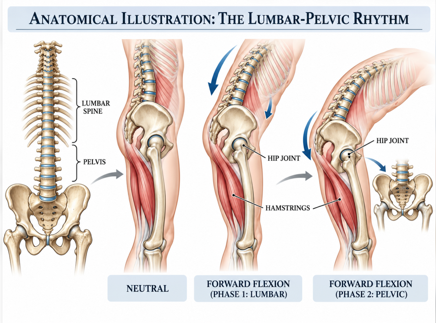
The spine and hip connection operates through a highly coordinated mechanical system known as the lumbar-pelvic rhythm. When you bend forward, your lumbar spine and pelvis rotate together in a precise sequence to distribute weight. But here’s the thing. When L4-L5 or L5-S1 discs desiccate and collapse, that rhythm shatters. The spine entirely loses its hydrostatic shock-absorbing capacity, fundamentally altering the kinematic chain of your entire lower body.
Consequently, the pelvis is forced into an anterior tilt to compensate for the lost lumbar lordosis. This altered tilt wreaks havoc on your lower extremities. It drastically increases friction within the acetabulofemoral joint (your hip socket) and places constant, exhaustive tension on the iliopsoas and gluteal musculature. The psoas major muscle—which originates on the lumbar
Furthermore, the quadratus lumborum and multifidus muscles are forced to act as primary stabilizers rather than dynamic movers. This chronic overactivation leads to rapid muscle fatigue, ischemic pain, and eventual fatty infiltration of the muscle bellies.
A recent biomechanical analysis in the Journal of Biomechanics established a clear consequence for this dysfunction. Lumbar disc degeneration shifts up to 30% of standard mechanical load down to the pelvic girdle accelerating rapid hip joint deterioration.
Orthopedic specialists refer to this interconnected breakdown as Hip-Spine Syndrome. When a patient presents with severe lateral hip pain, they frequently undergo hip-focused imaging,
This introduces what clinical researchers call The Mechanical-Biological Disconnect. While the mechanics shift the structural load, the resulting tissue micro-trauma triggers a cascade of inflammatory cytokines. Your biology essentially turns against itself. The environment inside the joint space becomes acidic and hostile, breaking down cartilage faster than your body can repair it. Treating the hip surgically when the root cause is actually lumbar disc degeneration leads to highly suboptimal outcomes because the underlying mechanical tether remains broken.
Before deciding on a treatment path, diagnosing the exact pain generator is absolutely mandatory. You have to know whether the pain in your hip actually originates in your hip. Referred pain from spine to hip is notoriously deceptive. Lumbar disc degeneration symptoms typically present as radicular pain a sharp, electrical sensation shooting down specific nerve pathways (dermatomes).
If a herniated lumbar disc compresses the L4 nerve root, you will feel intense pain radiating across the anterior thigh and into the hip, even if the hip joint itself is completely pristine. L5 radiculopathy typically shoots down the lateral leg. Conversely, true intra-articular hip pain caused by osteoarthritis typically presents as a deep, dull ache in the groin that worsens specifically with internal rotation of the leg.
Differentiating these two conditions requires highly specific physical provocation testing. A physician must perform the FABER (Flexion, Abduction, and External Rotation) test to isolate hip joint pathology, contrasting it with straight-leg raise tests that isolate lumbar radiculopathy. Additional testing, such as the Stinchfield test, can further differentiate intra-articular hip pathology from extra-articular or spinal sources. MRIs are incredibly sensitive but not overly specific. You might have an asymptomatic disc bulge, or you might have mild hip osteoarthritis that isn’t actually causing your primary pain. High false-positive rates for disc herniations in asymptomatic adults make physical provocation testing the definitive tie-breaker.
We can’t discuss anatomy without addressing the biochemical reality of joint decay. Chronic pain isn’t just a mechanical failure; it’s a localized chemical fire. When a lumbar disc herniates or hip cartilage wears down under sheer stress, the damaged tissues immediately release damage-associated molecular patterns (DAMPs).
Your immune system recognizes these DAMPs and deploys pro-inflammatory M1 macrophages directly to the site. These cells secrete aggressive cytokines like Tumor Necrosis Factor-alpha (TNF-a), Interleukin-1 beta (IL-1β), and Interleukin-6 (IL-6). In acute, temporary injuries, this inflammation is helpful for clearing cellular debris. In chronic degenerative disc disease, however, it is entirely disastrous.
The sustained presence of TNF-a and IL-1β upregulates matrix metalloproteinases (specifically MMP-1 and MMP-13) enzymes that literally eat your remaining healthy Type II collagen and aggrecan, the primary structural proteins of your cartilage. This biochemical breakdown is accompanied by neurochemical sensitization. The tissues release Substance P and Calcitonin Gene-Related Peptide (CGRP), massively lowering the firing threshold of your peripheral pain nerves. So, a mechanical problem in your back creates a runaway chemical problem that spills over into your systemic biology. Because these biomechanical faults manifest as severe pain, patients frequently evaluate invasive corrective measures to restore joint function without realizing their internal environment is actively hostile to tissue repair.
Conventional treatment for disc degeneration historically relies on mechanical stabilization and joint excision. While these interventions correct gross anatomical instability, patients increasingly seek hip replacement alternatives due to the invasive nature of orthopedic surgery. Mechanical interventions fundamentally alter physiological load distribution, initiating a cascade of adjacent structural failures over time.
To truly weigh stem cell therapy vs hip replacement, you must understand exactly what conventional surgery entails biologically. It is brutal, invasive, and permanent. In a standard lumbar spinal fusion, a surgeon performs a laminectomy to decompress nerves. This requires stripping the paraspinal muscles (like the multifidus) directly off the bone, denervating them and often causing them to atrophy into fat a process called fatty infiltration.
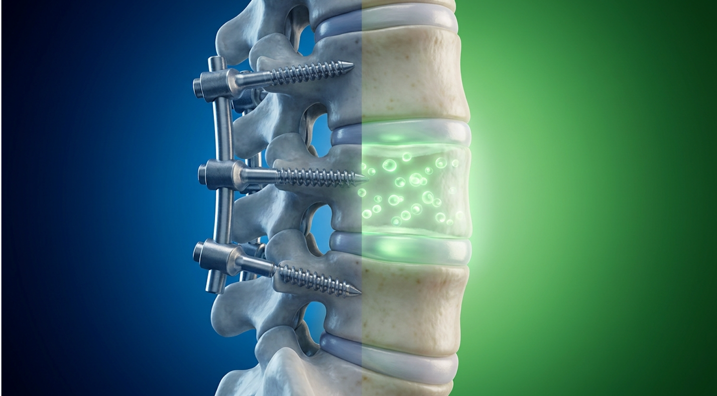
After removing the remnants of your degenerating disc, the surgeon inserts a synthetic spacer (cage) packed with bone graft. Finally, they drill titanium pedicle screws deep into your vertebrae, locking them together with heavy rods to eliminate absolutely all motion at that spinal segment. The biological environment is ignored; only the mechanical structure is addressed.
The disc surgery recovery time is grueling. You aren’t just healing an external incision; you are waiting for bone to literally grow across a synthetic bridge while your back muscles attempt to recover from severe surgical trauma.
Total hip arthroplasty (THA) is equally dramatic in its mechanical approach. The surgeon physically amputates the diseased femoral head with an oscillating saw. They then use a high-speed hemispherical drill to ream out the acetabulum (the hip socket), stripping away all subchondral bone until they reach a bleeding bone bed.
Into this hollowed-out space, they impact a titanium cup, secure it with bone screws, and hammer a titanium stem down the hollow canal of your femur. Hip replacement rehabilitation protocols demand prolonged physical therapy and strict movement restrictions to prevent prosthetic dislocation. Furthermore, the synthetic implant introduces the long-term risk of aseptic loosening where the bone eventually retreats from the metal, requiring a massive, highly complex revision surgery. The goal of these surgeries is to permanently eliminate movement to stop pain. The biological goal should be to restore tissue integrity to preserve movement.
Adjacent Segment Disease (ASD) is the dirty secret of mechanical spinal stabilization. When you lock one joint with titanium, that kinetic motion doesn’t just disappear. Physics dictates it
A comprehensive longitudinal review published in Spine revealed a stark clinical reality. Nearly 25% of patients require a secondary surgery within ten years to address Adjacent Segment Disease highlighting the systemic failure of isolated mechanical fixation.
Their adjacent discs herniate, degenerate, and collapse under the unnatural mechanical load transferred by the titanium rods. This perfectly illustrates The Mechanical-Biological Disconnect. The surgeon successfully fixed the mechanical instability at one level, but completely ignored the biological health of the surrounding tissues. The disease simply moves one floor up.
Perhaps the most devastating limitation of mechanical spine surgery is Failed Back Surgery Syndrome (FBSS). The definition is blunt and tragic: persistent or new chronic pain following an anatomically “successful” spinal surgery. Post-operative X-rays look perfect. The titanium screws are flawlessly placed. The bone fusion is solid. Yet, the patient is in agonizing, unrelenting pain.
Clinical epidemiology published in Translational Medicine Communications confirms the severity of this issue. Up to 40% of patients undergoing lumbar spinal fusion experience Failed Back Surgery Syndrome within five years requiring ongoing and intensive pain management.
Why does this happen? The etiology points directly back to biology. Post-surgical epidural fibrosis (massive scar tissue formation) chokes the sensitive spinal nerves. Altered biomechanics strain the paraspinal muscles constantly. The inflammatory cytokine presence never resolved. FBSS leaves patients trapped in a chronic pain loop, heavily dependent on pharmacology, opioid management, or surgically implanted neurostimulators. Recognizing these severe surgical limitations, modern clinical research has aggressively shifted focus toward interventions that alter the disease environment at the cellular level.
Advanced stem cell therapy for disc degeneration targets the root pathology of joint decay rather than merely masking it with titanium and screws. By utilizing highly potent umbilical cord mesenchymal stem cells, regenerative medicine addresses the localized biological deterioration, attempting to reverse cellular senescence and halt chronic inflammatory cascades.
Not all stem cells are created equal. This is a critical distinction that commercial clinics frequently fail to adequately explain to patients. Umbilical cord mesenchymal stem cells (UC-MSCs) are multipotent stromal cells derived exclusively from the Wharton’s Jelly matrix of an umbilical cord. This matrix is uniquely rich in hyaluronic acid and naturally shields the cells from immune detection. They are ethically sourced from healthy, full-term, scheduled cesarean births with full, stringent maternal consent.
Clinical-grade laboratory expansion involves rigorous, exhaustive quality control inside cGMP (Current Good Manufacturing Practice) bioreactors. The cells undergo extensive viability testing, pathogen screening, and flow cytometry. Flow cytometry ensures the cells express the exact correct surface markers they must be CD73, CD90, and CD105 positive, while remaining completely negative for hematopoietic markers like CD34 and CD45. Furthermore, UC-MSCs are immune-privileged; they lack MHC Class II molecules (HLA-DR), meaning your body will not reject them.
| 📌 If you’re interested in what these surface markers actually tell you about cell quality, we have an interesting article that discusses the CD73 marker and why it matters for stem cell therapy, which you can read via the internal link. |
If you want to learn about the safety of stem cell treatments in Thailand, you’ll find that top-tier international labs maintain these elite processing standards to ensure maximum cell viability, avoiding the contamination risks seen in lower-tier facilities.
To truly grasp the power of UC-MSC therapy as an alternative to surgery, we have to look closely at cellular senescence. Think of senescence as cellular retirement. As tissues in your hip and spine age and endure mechanical stress, their resident cells (chondrocytes in the hip, nucleus pulposus cells in the disc) hit the Hayflick limit. They stop dividing. They become senescent.
Worse, these retired cells don’t just sit there quietly. They secrete a toxic blend of proteins known as the Senescence-Associated Secretory Phenotype (SASP). SASP effectively poisons the surrounding healthy cells, accelerating the degeneration of the entire joint matrix.
When evaluating MSCs for joint repair, you must compare UC-MSCs against autologous (your own) bone marrow aspirate concentrate (BMAC) or adipose-derived stem cells (ADSCs). This is where the biological math heavily favors umbilical tissues. If you are 65 years old, your autologous stem cells are also 65 years old. Research through 2026 continues to demonstrate that BMAC from patients over 60 shows a significant reduction in colony-forming unit-fibroblast (CFU-F) capacity. Their telomeres are heavily degraded, and they have been exposed to decades of systemic inflammation.
Injecting older autologous stem cells often fails because those cells lack the biological horsepower to overcome the toxic SASP environment inside the joint space. In contrast, UC-MSCs are “day-zero” cells. They haven’t reached the Hayflick limit—the biological boundary dictating how many times a cell can divide before it enters senescence. Because they possess peak telomere length, they are completely immune to chronological exhaustion. In our clinical evaluations comparing regenerative protocols, patients utilizing high-dose, day-zero UC-MSCs consistently show faster inflammatory resolution compared to older autologous cohorts.
| 📌 If you’re interested in why umbilical cord cells are often preferred over bone marrow or fat-derived cells, we have an interesting article that discusses why UC-MSCs are superior to other stem cell sources, which you can read via the internal link. |
How do these youthful cells actually heal a badly degenerated joint? The answer is paracrine signaling. It’s a highly common misconception that if a doctor injects 50 million stem cells into your hip, those cells simply turn into 50 million new cartilage cells. That isn’t how biological regeneration works.
UC-MSCs operate primarily as biological general contractors. Through paracrine signaling, they secrete massive volumes of bioactive molecules, secretomes, and extracellular vesicles (exosomes) to forcefully induce physiological changes in your existing nearby cells. They facilitate cell-to-cell communication that overrides the degenerative cascade.
Molecular profiling detailed in Frontiers in Immunology confirms this mechanism. UC-MSCs secrete over 200 distinct growth factors actively reprogramming the hostile joint space to halt cartilage destruction.
This cellular reprogramming is immunomodulation at its absolute finest. UC-MSCs actively seek out those aggressive, pro-inflammatory M1 macrophages destroying your joint and biochemically reprogram them into anti-inflammatory, tissue-repairing M2 macrophages. They literally flip the biological switch from tissue destruction to tissue preservation.
| 📌 If you’re interested in how MSCs shift the immune system from attack mode to repair mode, we have an interesting article that discusses mesenchymal stem cell therapy for immune modulation, which you can read via the internal link. |
They achieve this phenotype switch by deploying extracellular vesicles (EVs), commonly known as exosomes. Think of exosomes as biological cargo ships. They cross the hostile joint space carrying messenger RNA (mRNA) and microRNA (such as miR-21 and miR-143). When these exosomes penetrate the degenerating nucleus pulposus cells, they deliver instructions that actively downregulate cellular apoptosis (programmed cell death).
Simultaneously, the UC-MSCs secrete heavy concentrations of Prostaglandin E2 (PGE2), TNF-stimulated gene 6 (TSG-6), and Indoleamine 2,3-dioxygenase (IDO). Once the M2 macrophages dominate the microenvironment, the area is flooded with Transforming Growth Factor-beta (TGF-β) and Insulin-like Growth Factor-1 (IGF-1), which heavily stimulate the synthesis of essential extracellular matrix proteins like Type II collagen and fibronectin.
Furthermore, UC-MSCs release high concentrations of Vascular Endothelial Growth Factor (VEGF), which powerfully promotes microcirculation and angiogenesis, driving much-needed blood flow to the starving, avascular borders of the intervertebral disc. They also secrete TIMPs (Tissue Inhibitors of Metalloproteinases) to immediately block the MMP-1 and MMP-13 enzymes that were eating your cartilage.
By completely resolving the hostile, acidic microenvironment, regenerative medicine for hip pain actively addresses the biological half of The Mechanical-Biological Disconnect. It provides a stable, nurtured environment where your body’s natural cellular repair mechanisms can finally execute their programming. Understanding this biological mechanism is essential
When evaluating stem cell therapy vs surgery, clinical decision-making relies on comparing long-term functional outcomes against immediate procedural risks. While evaluating the UC-MSC success rate in recent clinical trials, researchers emphasize that biological cellular therapies offer tissue-preserving improvements, contrasting sharply with the destructive mechanical alterations required by fusion and joint replacement procedures.
Intradiscal stem cell injections demonstrate intense clinical potential in delaying or avoiding spinal fusion by targeting disc biogenesis. The injected MSCs work to rehydrate the nucleus pulposus and downregulate inflammatory cytokines driving degenerative disc disease. This biological restoration stabilizes the microenvironment, frequently reducing pain enough to restore function without hardware fixation. Patients with severe, acute spinal instability or structural fractures, however, remain primary candidates for mechanical fusion. Ultimately, clinical imaging dictates viability.
However, resuming non-strenuous activity quickly does not imply the joint is completely biologically healed in three days. That is merely the resolution of the acute downtime following the needle track inflammation. True biological tissue modulation occurs in highly distinct, non-linear phases.
Days 7 to 14 initiate profound immune modulation. During this window, patients often report massive pain reduction as the M1-to-M2 macrophage conversion plummets intra-articular inflammation. Weeks 4 through 12 mark the proliferation phase, involving fibroblast migration and active extracellular matrix deposition (collagen synthesis). Finally, months 3 to 6 involve the remodeling phase, where tissue maturation and collagen cross-linking occur to stabilize the joint architecture.
Can stem cells heal degenerated discs? Let’s use precise clinical language. Evidence shows they can fundamentally rehydrate the nucleus pulposus, massively downregulate the inflammatory cytokines driving the decay, and stabilize the biological matrix to halt further degradation. They cannot, however, magically regrow a completely absent, bone-on-bone disc structure. Contrast this progressive biological healing against surgical recovery, which involves immediate structural immobilization but requires months of agonizing physical therapy just to regain basic muscle function and learn how to walk with permanently altered biomechanics.
Yes, stem cell therapy can serve as a viable alternative to hip replacement surgery for properly selected patients. UC-MSCs modulate intra-articular inflammation and promote the synthesis of extracellular matrix proteins, addressing joint pain at the cellular level. For patients with mild to moderate cartilage degradation, this biological intervention often delays or entirely eliminates the need for mechanical arthroplasty. However, cases involving complete bone-on-bone destruction typically still require surgical joint replacement.
Patients advised to undergo hip replacement should seek a secondary evaluation from a regenerative medicine specialist to assess biological alternatives. A thorough suitability assessment involving advanced MRI review determines if sufficient joint architecture remains to support cellular therapy. If viable tissue exists, UC-MSC injections offer a minimally invasive pathway to pain reduction and mobility improvement. If the joint is entirely collapsed, clinicians will quickly confirm that surgery is the necessary next step.
For patients evaluating joint interventions, stem cell therapy versus surgery represents a choice between biological restoration and mechanical repair. A stark reality of traditional approaches Source, 2018). The most effective approach demands rigorous clinical imaging to confirm suitability for UC-MSC therapy, bypassing the permanence of titanium hardware in favor of deep cellular regeneration.
Understanding The Mechanical-Biological Disconnect is the foundational key to making an informed choice. While traditional orthopedic surgery successfully removes the diseased joint and artificially stabilizes the structure, it completely ignores the cellular senescence and toxic inflammatory cascades driving the disease. UC-MSC therapy attacks the root biological failure, reprogramming the host microenvironment to halt decay and initiate natural matrix protein repair.
Your next step toward recovery requires definitive diagnostic clarity. Gather your most recent MRI reports, compile a detailed timeline of your conservative treatments, and schedule a consultation with a qualified regenerative medicine physician today. By securing an objective clinical suitability assessment, you can confidently determine if your specific joint architecture still qualifies for biological restoration before committing to irreversible surgical intervention.