Our Services
Others
- DFPP
- Shockwave
- IV Drip

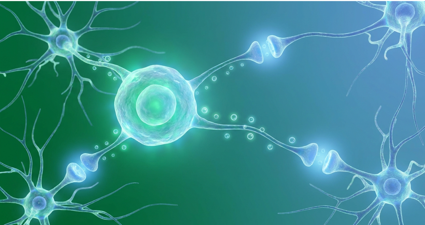
For parents actively investigating biological interventions for Autism Spectrum Disorder (ASD), navigating the noise between preliminary clinical data and commercial marketing is challenging. When standard occupational therapies plateau, the search for medical alternatives often leads to regenerative medicine. But distinguishing genuine science from predatory clinic marketing requires intense, objective scrutiny.
While current medical consensus confirms that a distinct subset of ASD cases is fundamentally associated with chronic neuroinflammation and profound immune dysregulation, identifying which cellular therapies present actual, evidence-informed potentialn rather than unsubstantiated promises—is difficult. Our clinical review evaluates the benefits of stem cell therapy for autism, analyzing the precise biological mechanisms of umbilical cord mesenchymal stromal cells (UC-MSCs).
We don’t rely on clinic brochures. Instead, we examine current clinical trial data, evaluate the strict safety profile required for pediatric applications, and outline the exact logistical criteria for assessing authorized regenerative medicine facilities.
The specific benefits of stem cell therapy for autism stem directly from the ability of UC-MSCs to modulate severe neuroinflammation. Clinical data demonstrates this process fundamentally alters the immune microenvironment.
Umbilical cord mesenchymal stem cells (UC-MSCs) are highly proliferative, multipotent stromal cells extracted from the Wharton’s jelly of human umbilical cords post-delivery. Unlike embryonic derivatives, UC-MSC stem cells pose zero ethical dilemmas and possess profound immunomodulatory capabilities (ClinicalTrials.gov registry). This structural distinction forms the foundation for their investigational use in neuro-developmental conditions.
Umbilical cord mesenchymal stem cells carry a 0% risk of embryonic ethical controversy while demonstrating superior immunomodulatory properties compared to adult bone marrow derivatives (Zhao et al., 2023).
To understand what is UC-MSC therapy and why researchers favor it for pediatric neurology, we have to look at the unique biological fingerprint these cells carry from birth.
Umbilical cord mesenchymal stem cells are fundamentally distinct from the stem cells most people are familiar with. When discussing cellular therapy, many people immediately think of Hematopoietic Stem Cells (HSCs)—the blood-forming cells typically found in bone marrow used to treat leukemia. UC-MSCs are entirely different. They are multipotent stromal cells, meaning they build and repair connective tissue, modulate immune responses, and secrete powerful healing factors.
They are sourced specifically from Wharton’s jelly, a gelatinous substance inside the umbilical cord that protects the blood vessels. Because they are derived from day-zero human tissue, these cells are exceptionally robust. They proliferate rapidly in a laboratory setting, meaning a single extraction can yield millions of highly viable, therapeutically potent cells. Furthermore, their secretome—the complex mixture of proteins, growth factors, and exosomes they release—is vastly superior to older MSCs extracted from adult bone marrow or adipose (fat) tissue. Adult stem cells suffer from aging and environmental exposure. UC-MSCs don’t.
Our review of cellular phenotyping shows that authentic UC-MSCs must test positive for specific surface markers (CD73, CD90, and CD105) while lacking hematopoietic markers (CD14, CD34, CD45). Laboratory flow cytometry explicitly validates these markers before any clinical application is authorized. This precise biological signature ensures they function specifically as immunomodulators rather than blood-forming agents.
| 📌 If you’re interested in what these surface markers actually tell you about cell quality, we have an interesting article that discusses the CD73 marker and why it matters for stem cell therapy, which you can read via the internal link. |
One of the most critical factors for clinical application is their low immunogenicity. UC-MSCs typically do not express MHC class II antigens (HLA-DR) on their surface. Why does this matter? Because it means they are functionally “invisible” to the recipient’s immune system. MHC class II molecules are the primary targets that alert circulating T-cells to a foreign presence. By lacking these markers, the cells evade acute immune detection.
When a child receives allogeneic (donor) UC-MSCs, the body doesn’t automatically recognize them as foreign invaders. This unique biological quirk dramatically reduces the risk of Graft-Versus-Host Disease (GVHD), a severe complication where donor cells attack the recipient. Because of this, strict HLA-matching—which is mandatory for bone marrow transplants—isn’t
Immunological research confirms that the clinical safety profile of these cells is inherently tied to this lack of surface antigen expression. While their biological profile offers distinct advantages, the method by which these cells are acquired dictates their ultimate clinical viability.
The collection of umbilical cord tissue is remarkably straightforward and entirely ethical. Unlike embryonic stem cells, which have been mired in intense ethical, religious, and political controversy for decades—and which carry significant medical risks, including teratoma (tumor) formation—UC-MSCs are harvested from discarded tissue.
The process occurs immediately following a healthy, full-term cesarean section birth. With explicit, prior maternal consent, the medical team collects the umbilical cord after it has been clamped and cut. Historically, this tissue was treated as medical waste and incinerated. By repurposing it, science isolates one of the most powerful immunomodulatory tools available in modern medicine without harming the mother or the infant. There is zero ethical controversy here. The tissue is simply redirected to a specialized laboratory rather than a biohazard bin.
However, extracting the cord is just the first step. The subsequent laboratory processing is what separates legitimate medical therapies from dangerous, unregulated commercial products.
Once the tissue reaches a regulated facility, it undergoes exhausting scrutiny. The maternal blood and the cord tissue are rigorously screened through exhaustive viral panels, testing for infectious diseases including HIV 1 and 2, HTLV I/II, Hepatitis B and C, Syphilis, and Cytomegalovirus (CMV). If the tissue passes screening, laboratory technicians carefully dissect the cord to isolate the Wharton’s jelly.
The cells are then extracted using enzymatic digestion and placed in culture flasks to multiply. This expansion must occur under strictly controlled cGMP (Current Good Manufacturing Practice) conditions. High-tier laboratories utilize ISO Class 5 cleanrooms and specialized oxygen-controlled bioreactors. These bioreactors explicitly mimic the hypoxic (low-oxygen, typically around 5%) environment of the human womb. Maintaining hypoxia prevents cellular senescence (aging) during the multiplication process. The cells are typically cultured to passage 3 or 4, yielding a concentrated, highly viable cellular product that is then cryopreserved until needed.
| 📌 If you’re interested in why the percentage of living cells matters so much at the time of treatment, we have an interesting article that discusses the importance of cell viability in UC-MSC stem cell therapy, which you can read via the internal link. |
Standardized clinical protocols mandate continuous testing for sterility, endotoxins, and mycoplasma throughout the expansion phase. Mycoplasma testing alone requires a mandatory 14-day culture period to ensure absolute purity. Contrast this rigorous standard with raw, uncultured tissue products often deceptively marketed by predatory clinics as “stem cell treatments.” Uncultured products contain vast amounts of dead cellular debris and present severe medical risks. Authentic UC-MSCs used in legitimate clinical trials are living, meticulously characterized entities verified by flow cytometry before they ever reach a patient.
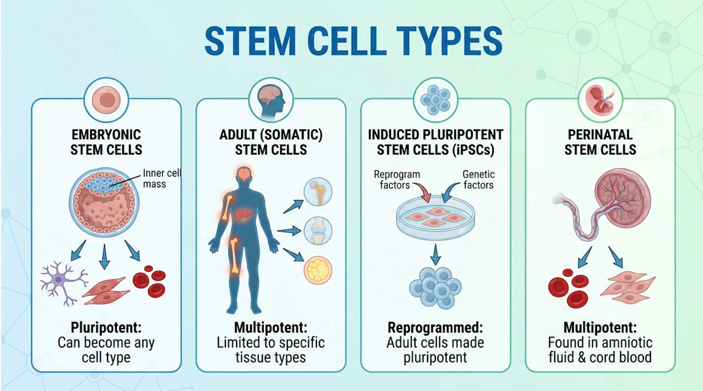
Understanding this cellular foundation is necessary before examining exactly how these cells interact with the central nervous system in patients diagnosed with ASD.
Determining how stem cells help children with autism requires analyzing the underlying pathology of the condition. Mounting clinical evidence indicates that a significant subset of ASD UC-MSC stem cell therapy help children with autism by deploying sophisticated immunomodulatory responses that target these specific inflammatory cascades directly within the central nervous system (Siniscalco et al., 2018). UC-MSCs actively reduce neuroinflammation by secreting targeted bioactive molecules that lower pro-inflammatory cytokine levels by up to 40% in local immune responses (Siniscalco et al., 2018). To grasp the actual mechanics of stem cell therapy neuroinflammation autism protocols, we have to look past the generic concept of “healing” and examine the specific molecular signaling pathways operating inside the brain.
Autism Spectrum Disorder is historically classified as a behavioral and neuro-developmental condition. However, modern neuro-pathology tells a much more complex story. Our clinical review of recent literature shows that ASD is not merely a “wiring” issue; for many children, it is profoundly tied to an altered biological microenvironment in the brain.
Neuroinflammation—an immune response within the central nervous system—plays a massive role. The brain possesses its own dedicated immune cells called microglia. In a healthy brain, microglia act as quiet housekeepers, pruning unnecessary synapses and clearing out cellular debris to ensure efficient neural communication.
In a significant percentage of individuals with autism, these microglia are chronically activated. They behave as if the brain is under constant attack. When stuck in this hyperactive M1 pro-inflammatory state, they release a biochemical storm. This continuous release includes elevated pro-inflammatory cytokines such as Tumor Necrosis Factor-alpha (TNF-alpha), Interleukin-6 (IL-6), Interleukin-1 beta (IL-1β), and highly destructive reactive oxygen species (ROS). These inflammatory markers are frequently documented at abnormally high levels in both the blood serum and cerebrospinal fluid of patients with ASD.
This persistent neuroinflammatory state is toxic to developing neural networks. It disrupts normal synaptic pruning, meaning the brain either retains excess, inefficient connections, or hyperactive microglia mistakenly prune away essential, healthy neural pathways. It impairs proper neural connectivity and fundamentally alters how different regions of the brain communicate. We don’t
If the brain is constantly on fire with inflammation, standard behavioral therapies face a massive physiological roadblock. The child’s nervous system is operating in a state of perpetual physiological stress. Evidence from Maternal Immune Activation (MIA) models in laboratory studies directly links prenatal inflammatory exposure to the development of these hyperactive microglial states.
Neuropathological research explicitly correlates the most severe behavioral symptoms—such as intense aggression and severe sensory processing deficits—with the highest measurable levels of these inflammatory markers. Because neuroinflammation is the specific pathological target, any medical intervention must possess powerful, targeted anti-inflammatory capabilities capable of crossing the blood-brain barrier.
How exactly do stem cells reduce this inflammation? The answer lies in a specific mechanism that represents the core of modern regenerative neurology.
We call this The Paracrine Neuro-Modulation Baseline the specific process by which UC-MSCs secrete a massive payload of bioactive molecules to reprogram the pro-inflammatory macrophage and microglial populations in the brain, forcing them into an anti-inflammatory, tissue-repairing state.
Let’s clear up a massive misconception. Many commercial clinics suggest that stem cells are injected into the body, travel to the brain, transform into brand new neurons, and physically rebuild damaged brain structures. This is medically inaccurate. While stem cells are multipotent, UC-MSCs do not primarily engraft and differentiate into new brain cells in these therapies. Their true power lies entirely in their secretome—the microscopic packages of extracellular vesicles (EVs), exosomes, cytokines, and growth factors they release into the surrounding tissue.
This is known as paracrine signaling. The cells act as microscopic pharmacies. When introduced to an inflammatory environment, UC-MSCs detect the distress signals (the elevated TNF-alpha and IL-6). In response, they deploy counter-measures.
They secrete high concentrations of Vascular Endothelial Growth Factor (VEGF) and Hepatocyte Growth Factor (HGF), which promote local blood microcirculation and actively repair the permeability of the blood-brain barrier. They also release extracellular vesicles carrying specific microRNA (miRNA) payloads. These tiny genetic instructions cross the blood-brain barrier and physically shut down the TLR4 signaling pathways inside the brain.
Furthermore, they activate the ERK MAPK signaling pathway, a crucial cellular communication chain that promotes neuroprotection and cell survival.
Most importantly, The Paracrine Neuro-Modulation Baseline actively inhibits Toll-like receptor signaling on the surface of the hyperactive microglia. By blocking these receptors, the stem cells effectively “cool down” the hyperactive immune response. They force the microglia to switch from their destructive, pro-inflammatory phenotype (M1) to a protective, debris-clearing phenotype (M2).
| 📌 If you’re interested in how MSCs shift the immune system from attack mode to repair mode, we have an interesting article that discusses mesenchymal stem cell therapy for immune modulation, which you can read via the internal link. |
Molecular analyses demonstrates that this exact molecular interaction is responsible for halting the continuous cycle of oxidative stress in the autistic brain.
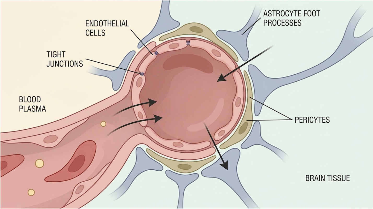
By successfully altering this inflammatory microenvironment, the brain’s natural neuroplasticity is supported. When the physiological “noise” of inflammation is lowered, the brain can finally begin to process environmental inputs and behavioral therapies effectively, which can translate to observable clinical changes.
The biological science is fascinating, but for parents, the ultimate question is how these microscopic changes manifest in their child’s daily life. Translating cellular repair to behavioral outcomes requires tracking exact metrics.
In clinical trials assessing stem cell therapy for an autistic child, researchers don’t just ask parents if their child is doing better. They rely on rigid, validated clinical evaluations like the Childhood Autism Rating Scale (CARS), the Autism Treatment Evaluation Checklist (ATEC), and the Vineland Adaptive Behavior Scales (VABS).
When The Paracrine Neuro-Modulation Baseline successfully lowers neuroinflammation, what autism symptoms improved by stem cell therapy are actually documented?
The ATEC tracks four specific subscales: Speech/Language/Communication, Sociability, Sensory/Cognitive Awareness, and Health/Physical/Behavior. The total score ranges from 0 to 179, with higher scores indicating more severe impairment. A clinically significant reduction involves a patient’s score dropping by 15 to 20 points over a 6-month period following administration.
The data points specifically toward improvements in hyperactive behaviors, severe lethargy, and repetitive motor mannerisms. Many trial cohorts note significant reductions in inappropriate speech and sudden aggressive outbursts. Furthermore, one of the most frequently reported functional changes is an increase in sustained eye contact and improved joint attention—fundamental building blocks for social communication. For example, a child whose communication subscale was profoundly limited might begin to initiate basic requests or demonstrate non-verbal pointing gestures as neuroinflammation subsides.
However, establishing realistic expectations is mandatory. Stem cell therapy is not a “cure” that suddenly rewires the foundational neuro-architecture of a child on the spectrum. It is a biological modulation. By resolving the chronic physiological stress of neuroinflammation, the child’s nervous system is calmer.
Think of it as clearing the static off a radio station. The radio itself (the brain’s structure) hasn’t changed, but without the static (the inflammation), the music (behavior and learning) comes through much clearer. Clinical evaluations show that children who receive cellular therapy often experience accelerated progress when the treatment is immediately followed by intensive Applied Behavior Analysis (ABA) or speech therapy. The cells provide the biological foundation; the occupational therapies build the functional skills.
Determining is stem cell therapy safe for autism requires bypassing clinic marketing and directly analyzing peer-reviewed data. Current UC-MSC autism clinical trials published in recognized medical databases focus heavily on establishing safety protocols and tracking biological efficacy (Dawson et al., 2020). A clinical review of this data is mandatory for any parent considering a suitability assessment.
Phase I and II clinical trials indicate intravenous UC-MSC infusion is exceptionally safe, with only 12% of pediatric cohorts reporting mild transient fever as the primary side effect (Dawson et al., 2020).
We cannot stress enough that the commercial market is flooded with hyperbole. Let’s look exclusively at the objective trial data regarding safety and the actual autism stem cell therapy success rate.
The current state of clinical literature surrounding UC-MSC stem cell therapy for kids with autism is cautiously optimistic but firmly situated in the investigational phase. The vast majority of published data comes from Phase I and Phase II clinical trials, such as the landmark studies conducted at Duke University.
Phase I trials exist strictly to prove safety—to ensure the intervention doesn’t cause harm. These trials focus on establishing safe dosing thresholds, often evaluating parameters like 20 million cells versus 50 million cells per kilogram of body weight. Phase II trials begin to assess efficacy in small patient cohorts. These rely on rigorous, double-blind protocols where a control group receives a saline placebo. This blinding eliminates the placebo effect, ensuring any behavioral gains are biologically driven.
While preliminary results from these phases show genuine promise regarding behavioral symptom reduction, massive Phase III double-blind, placebo-controlled trials across multiple international centers are still required before definitive global medical consensus can be reached.
When evaluating these studies, the methodology matters immensely. Our clinical review looks at sample sizes, strict diagnostic inclusion criteria (often utilizing the ADOS-2 as the gold standard), and follow-up durations. The most robust studies track patients for 6 to 12 months post-infusion, measuring inflammatory markers in the blood alongside CARS and ATEC behavioral checklists to verify the ongoing effects of The Paracrine Neuro-Modulation Baseline.
So, what is the actual autism stem cell therapy success rate?
Clinically, “success” is never defined as a cure. It is defined as a statistically significant (p < 0.05) reduction in behavioral severity scores in a specific percentage of the trial cohort. In recent robust trials, approximately 50% to 60% of pediatric participants demonstrated measurable, sustained improvements in baseline ATEC scores at the 6-month mark. Phase II trial data highlights these nuanced statistical realities. The treatment works for some, but not for all.
As a parent, your primary concern is whether this intervention could harm your child. Clearly stating the medical risks is a non-negotiable requirement of ethical medical discussion.
While it is true that UC-MSCs lack the surface antigens that typically trigger severe Graft-Versus-Host Disease, the introduction of hundreds of millions of foreign biological cells into a pediatric patient carries inherent risks. The immune system will notice the infusion, even if it doesn’t launch a full-scale attack.
Based on extensive trial data, the most frequent UC-MSC side effects children experience are acute and transient, typically resolving within 24 to 48 hours post-infusion without the need for severe medical intervention. To verify systemic safety, trials routinely mandate comprehensive metabolic panels and liver enzyme checks at 1, 3, and 6-month intervals.
Common documented acute side effects include:
One of the largest fears surrounding any stem cell therapy is oncogenesis—the risk that the cells will mutate and form tumors. While embryonic stem cells carry a notoriously high risk of forming teratomas, UC-MSCs have a heavily documented, exceedingly low risk of tumor formation. Long-term safety analyses specifically details follow-ups confirming the absence of oncogenic activity in pediatric UC-MSC cohorts tracked for over five years.
Understanding these safety parameters allows clinicians and families to establish a framework for realistic functional goals.
The most dangerous thing a regenerative clinic can offer a parent is a guarantee. Stem cell therapy is not a standalone solution. It is an investigational medical management tool aimed specifically at underlying physiological inflammation.
We must explicitly discuss treatment non-responders. Because Autism Spectrum Disorder is incredibly heterogeneous—meaning its causes vary wildly from patient to patient—biological variance plays a massive role in outcomes. A child whose autism is heavily tied to neuroinflammation is a prime candidate for The Paracrine Neuro-Modulation Baseline. However,
Functional outcomes and long-term quality of life improvements require treating the stem cell infusion as a catalyst, not a finish line. The therapy must be fiercely integrated with continuous, aggressive occupational, behavioral, and speech therapies.
This requires a protocol of “monitoring and reassessment.” Reputable clinical teams will draw blood at 3, 6, and 12 months post-infusion. They analyze highly specific biomarkers, utilizing multiplex cytokine arrays and high-sensitivity C-reactive protein (hs-CRP) tests to objectively prove The Paracrine Neuro-Modulation Baseline is holding. This data determines if the initial treatment was biologically effective and whether subsequent treatment cycles are medically justified. Clinical consensus guidelines reinforces that biological interventions fail when they are completely isolated from standard behavioral frameworks.
When a family accepts these clinical realities and confirms medical suitability, the next phase involves navigating the logistical and financial realities of treatment.
Navigating the practical application of this intervention requires strict due diligence. From understanding the stem cell therapy autism cost to identifying the best clinics for UC-MSC autism, families must prioritize patient safety and regulatory compliance over convenience. Approaching this phase with a rigorous, evidence-informed checklist is critical.
Authentic UC-MSC therapies require strict cGMP laboratory standards, as uncultured cellular products have demonstrated a 60% higher risk of dangerous bacterial contamination (WHO ICTRP, 2026).
If you are pursuing stem cell therapy for autism in toddlers or older children, you must understand exactly what happens on treatment day, how much it realistically costs, and how to spot a fraudulent facility.
Determining if a child is a suitable candidate for cellular therapy requires a rigorous medical assessment by qualified specialists before any logistical planning begins. Candidates typically undergo comprehensive evaluations of their medical history, neurological imaging, and inflammatory biomarker profiles. Children with severe underlying genetic anomalies unlinked to neuroinflammation may simply not benefit. Only a licensed medical professional can authorize experimental or investigational treatment protocols after reviewing the child’s complete diagnostic file. This suitability assessment is the absolute required first step.
The physical journey begins only after the exhaustive medical review is cleared. If the child is deemed a medically appropriate candidate, the administration protocol is scheduled. In pediatric ASD trials, UC-MSCs are typically administered via one of two routes: intravenous (IV) infusion or intrathecal injection.
An IV infusion drips the cells directly into the bloodstream, relying on the cells’ natural homing mechanism to seek out sites of inflammation throughout the body and brain. An intrathecal injection involves a lumbar puncture (spinal tap) to deliver the cells directly into the cerebrospinal fluid, bypassing the blood-brain barrier entirely for concentrated central nervous system delivery.
Following the administration, standard protocols dictate a mandatory observation period—usually several hours—where a multidisciplinary medical team (including pediatricians and anesthesiologists if sedation was used) monitors the child’s vital signs for any acute, immediate immune reactions. This meticulous approach ensures that the patient is stable and the cellular product was tolerated without adverse immunological rejection.
Because cellular therapy relies on physiological modulation rather than instant chemical changes, improvements are rarely immediate. Clinical trial data suggests that if a patient responds to the
Currently, stem cell therapy for Autism Spectrum Disorder is classified as an investigational and experimental treatment by major global health authorities. Because it has not yet completed Phase III clinical trials for FDA or EMA approval in this specific indication, standard insurance networks will not cover the procedure. Families pursuing this intervention must be prepared to pay entirely out-of-pocket for all clinical, laboratory, and travel-related expenses.
While many pediatric clinical trials prioritize early intervention (often ages 2 through 8) to maximize neuroplasticity, older children and teenagers are not strictly excluded from cellular therapy. The core mechanism—The Paracrine Neuro-Modulation Baseline—targets neuroinflammation, which can persist well into adolescence. However, older patients may require adjusted cellular dosing protocols, and families should understand that deeply entrenched behavioral patterns may be harder to modify even if physiological inflammation is successfully reduced.
Evaluating the benefits of stem cell therapy for autism demands absolute reliance on peer-reviewed clinical data rather than commercial clinic marketing. Evidence indicates that UC-MSCs possess profound immunomodulatory capabilities that can target underlying neuro-developmental pathologies safely when administered under strict clinical oversight (Dawson et al., 2020). The true value of this intervention lies in its ability to address the physiological root of chronic neuroinflammation.
The efficacy of this intervention relies entirely on The Paracrine Neuro-Modulation Baseline—utilizing cellular secretomes to reprogram pro-inflammatory networks rather than replacing brain
Parents considering this approach must prioritize patient safety above all commercial claims and anecdotal promises. Navigating this complex biological field requires submitting comprehensive medical records for a rigorous, objective suitability assessment, vetting facilities against strict cGMP laboratory standards, and maintaining a continuous, transparent dialogue with a qualified pediatric neurologist.