Our Services
Others
- DFPP
- Shockwave
- IV Drip

This article is for educational purposes only and does not replace consultation with a qualified medical professional. Content regarding cellular therapies reflects current peer-reviewed research and does not constitute medical advice or guarantee treatment eligibility.
Current research demonstrates that the safety and efficacy of allogeneic stem cell therapies hinge not on the mere presence of cells, but on highly specific phenotypic profiles—most notably the expression of the CD105 marker in mesenchymal stem cells. Look, standardizing modern cell therapy requires incredibly rigorous biomarker validation. We can’t just inject biological material and hope for the best. Administering poorly characterized cell populations drastically increases the risk of immunogenicity, host rejection, and ultimately, therapeutic failure.
By the end of this guide, you will understand the precise molecular mechanisms of CD105, how it governs receptor-targeted gene transfer, and the current evidence defining strict MSC safety protocols. We analyze the fundamental biology of umbilical cord-derived mesenchymal stem cells,
The CD105 marker in mesenchymal stem cells (endoglin) is a critical surface receptor that regulates TGF-β signaling and validates cellular purity before clinical administration. The CD105 Safety Verification Matrix relies on it.
Umbilical cord tissue-derived mesenchymal stem cells (UC-MSCs) are multipotent stromal cells utilized in regenerative medicine for their profound paracrine signaling capabilities. Extracted primarily from Wharton’s jelly, these cells secrete a rich array of bioactive molecules that modulate local microenvironments. This unique secretome profile makes them structurally superior to adult-derived cellular sources for modulating complex tissue repair.
Let’s get specific about origins and the biochemical environment. Umbilical cord tissue-derived mesenchymal stem cells are harvested from Wharton’s jelly the dense, gelatinous connective tissue encased within the umbilical cord. This unique anatomical matrix is astonishingly rich in hyaluronic acid and glycosaminoglycans, which shield the stem cells from mechanical stress during fetal development. More importantly, this native niche locks the embedded cells into a biologically youthful, highly proliferative state. Unlike adult bone marrow or adipose-derived stem
The extraction process itself requires extraordinary precision to maintain this biological advantage. Wharton’s jelly isn’t just a uniform biological mass; it is divided into highly specific anatomical zones, including the sub-amnion, the intervascular matrix, and the perivascular zone. Laboratories first isolate the umbilical cord, thoroughly washing it in specialized buffer solutions to completely strip away maternal blood components, thereby mitigating early immunogenic risks. Technicians then painstakingly dissect the perivascular zone from the intervascular matrix, as the perivascular region yields the highest concentration of primitive, highly active MSCs.
To release the cells from the dense Wharton’s jelly, they utilize advanced enzymatic digestion techniques. This typically involves precise, timed ratios of 0.075% collagenase type I and 0.025% hyaluronidase. This biochemical breakdown dissolves the surrounding gelatinous matrix without shearing or damaging the delicate cellular membrane receptors of the embedded stem cells.
Our review of standardized extraction protocols shows that Wharton’s jelly isolation protocols (NCBI) consistently produce exceptionally robust cell lines. Comparative studies demonstrate that umbilical cord tissue yields significantly higher initial MSC concentrations often exceeding 1×10^6 cells per gram of tissue compared to standard adult bone marrow aspirations (Stem Cell Research & Therapy, 2021), ensuring a vastly superior baseline for secretome production.
But you can’t just harvest them and call it a day. The International Society for Cellular Therapy (ISCT) strictly defines an MSC by its adherence to medical-grade plastic, its multipotent differentiation potential (capable of branching into osteogenic, adipogenic, and chondrogenic lineages), and the presence of highly specific surface markers.
True regenerative ability in modern biological therapy is driven almost entirely by the cells’ molecular output. We used to think stem cells needed to permanently engraft, multiply, and physically differentiate into local tissue to work. We were wrong. The therapeutic heavy lifting happens via the secretome. Think of UC-MSCs as microscopic pharmaceutical factories, actively surveying their environment and pumping out precisely calibrated doses of proteins, cytokines, and growth factors right where the body needs them most.
Contrast this with a typical autologous bone marrow extraction. An older patient undergoes an invasive, painful surgical bone aspiration procedure to harvest their own cells. Those cells are already environmentally damaged, heavily influenced by systemic inflammation, and biologically sluggish. Autologous cells from a 65-year-old patient inherently behave like 65-year-old cells, exhibiting significantly shorter telomeres and vastly reduced proliferative capacity. Allogeneic UC-MSC acquisition skips the surgery entirely and utilizes biologically optimal, day-zero cells. While the origin of these cells provides an inherent biological advantage, their clinical efficacy is executed entirely through their complex molecular signaling pathways.
| 📌 If you’re interested in why umbilical cord cells are often preferred over bone marrow or fat-derived cells, we have an interesting article that discusses why UC-MSCs are superior to other stem cell sources, which you can read via the internal link. |
The secretome is everything. Secretomes, the complex array of bioactive molecules and growth factors secreted by MSCs, fundamentally alter tissue pathology. UC-MSCs secrete an incredibly dense cocktail of essential proteins, including epidermal growth factor (EGF), vascular endothelial growth factor (VEGF), hepatocyte growth factor (HGF), and basic fibroblast growth factor (bFGF). Clinical data demonstrates that UC-MSCs produce approximately 3.5 times the volume of these essential growth factors compared to bone marrow counterparts in early passages.
To prevent these vital proteins from being immediately destroyed by enzymes in the host’s bloodstream, the MSC strategically packages its secretome into exosomes tiny, nanometer-sized extracellular vesicles enclosed within a durable lipid bilayer. This lipid armor protects the growth factors as they travel through the systemic circulation, allowing them to effectively reach the site of injury completely intact.
How does this actually look at the molecular level? Take VEGF, for instance. When a VEGF-loaded exosome hits the localized microenvironment of a wound or hypoxic injury, it immediately binds to VEGFR-2 receptors on surrounding endothelial cells. This binding event acts as a master switch, triggering the ERK MAPK (Extracellular Signal-Regulated Kinase / Mitogen-Activated Protein Kinase) signaling pathway. The ensuing biochemical cascade involves the sequential, rapid phosphorylation of Ras, Raf, MEK, and finally ERK. Once activated, ERK translocates directly into the cell’s nucleus to activate transcription factors responsible for aggressive cellular division. The result? Rapid, localized angiogenesis. Dense new capillary networks form, quickly restoring vital blood flow and oxygen to ischemic, starving tissues.
Simultaneously, these diverse secretomes stimulate quiescent dermal fibroblasts. Fibroblasts are the master architects of your connective tissue. When activated by MSC paracrine signaling, they rapidly upregulate the synthesis of collagen type I, collagen type III, fibronectin, and elastin. They begin rebuilding the extracellular matrix from the ground up. A recent MSC paracrine signaling and matrix synthesis review confirms this structural reinforcement is completely essential for complete injury recovery.
This localized matrix remodeling actively prevents the formation of rigid, non-functional scar tissue, instead promoting the highly organized deposition of cellular scaffolding. This dual-action approach building vibrant new blood vessels while structurally reinforcing the physical tissue matrix delivers vital nutrients and oxygen to senescent tissues without requiring the MSCs to permanently engraft into the host. It’s a localized, highly targeted biological rescue mission. To consistently harness these powerful paracrine effects across patient populations, laboratory technicians must accurately identify and isolate incredibly pure populations of MSCs a process entirely dependent on specific surface receptors like CD105.
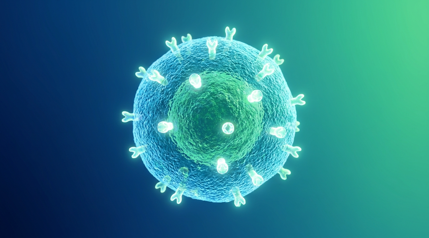
The role of the CD105 marker in mesenchymal stem cells extends beyond simple cell identification; it is a critical regulatory receptor that governs cellular behavior and safety. Also known as endoglin, this membrane glycoprotein is an auxiliary receptor for the TGF-β complex, mediating cellular proliferation, differentiation, and survival. Without robust CD105 expression, MSCs lose their distinct immunomodulatory and regenerative capabilities.
Structurally, CD105 (endoglin) is a massive 180 kDa homodimeric transmembrane glycoprotein tightly linked by disulfide bonds. It belongs to the zona pellucida family of proteins, possessing a large extracellular domain that physically reaches out into the surrounding environment. More than just a static identifier, it acts as a highly dynamic physical bridge, intimately connecting the cell’s internal signaling machinery to the chaotic, often inflammatory extracellular environment. In the realm of advanced cellular biology, a cell is entirely defined by what it wears on its surface. The role of endoglin in mesenchymal stem cells dictates exactly how the cell perceives biochemical stress, interprets inflammatory cytokine signals, and regulates its own survival.
Flow cytometry standards dictate exact, unforgiving rules for clinical viability. Flow cytometry utilizes sophisticated lasers to measure the physical and chemical characteristics of single cells as they pass sequentially through a narrow, highly pressurized fluidic stream. Technicians deploy specific fluorochrome-conjugated antibodies that bind uniquely to target surface markers. By meticulously mapping the forward scatter (FSC) to determine size, and side scatter (SSC) to measure internal complexity and exclude cellular debris, clinicians can visualize exact protein expression ratios on a scatter plot.
According to strict ISCT mandates, true MSC populations must express CD105, CD73, and CD90 at levels ≥95%. Simultaneously, they must definitively lack hematopoietic markers meaning CD45, CD34, and HLA-DR must be present in ≤2% of the population. Flow cytometry must verify that ≥95% of the cellular population expresses CD105 (ISCT Standards) a non-negotiable metric preventing the administration of senescent cells. If a commercial laboratory claims to possess a therapeutic batch of UC-MSCs, but CD105 flow cytometry reveals only an 82% expression rate, you don’t have a therapeutic product. You have a contaminated, aging, or improperly cultured biological liability.
| 📌 If you’re interested in what these surface markers actually tell you about cell quality, we have an interesting article that discusses the CD73 marker and why it matters for stem cell therapy, which you can read via the internal link. |
What exactly happens to CD105 expression in umbilical cord MSCs during extended in vitro expansion? It plummets predictably. As cells undergo excessive passaging (replicating repeatedly in a lab dish to maximize product yield), they suffer rapid telomere shortening, dangerous accumulation of reactive oxygen species (ROS), and ultimate biological exhaustion. CD105 expression drops in perfect tandem with this cellular senescence. A comprehensive PubMed molecular analysis demonstrated that decreased endoglin expression directly and irreversibly correlates with the loss of therapeutic efficacy and multipotent differentiation potential.
Contrast a vibrant, CD105-positive population versus a CD105-negative MSC population. The positive cells are metabolically hyper-active, highly suppressive of local inflammation, and optimally primed for massive secretome production. The negative (or low-expressing) cells are biologically sluggish, potentially immunogenic, and therapeutically useless. This structural presence is not passive; CD105 actively dictates how the stem cell interprets critical signals from its environment, particularly through sophisticated gene transfer mechanisms.
This is where the cellular biochemistry gets truly fascinating. CD105 doesn’t just sit statically on the membrane looking pretty for flow cytometers. It acts as a dedicated, active co-receptor for Transforming Growth Factor-beta (TGF-β) ligands, specifically binding with high affinity to TGF-β1 and TGF-β3.
CD105 and TGF-β signaling fundamentally alter the stem cell’s ultimate fate. TGF-β is a master regulatory cytokine that controls tissue remodeling across the entire human body. When TGF-β binds to the cellular membrane, it typically recruits Type II and Type I receptors to form a highly active signaling complex. CD105 is the master modulator of the downstream SMAD signaling pathways within this exact complex. Specifically, endoglin actively promotes signaling through the ALK1-Smad1/5/8 pathway while indirectly, yet forcefully, restricting the ALK5-Smad2/3 pathway. This mechanism is thoroughly documented in peer-reviewed molecular pathway analyses.
Why does this precise ALK1/ALK5 balance matter for a patient recovering from an injury? Because the ALK5 pathway, when left unchecked in a damaged microenvironment, drives excessive, runaway myofibroblast activation. These hyperactive myofibroblasts over-express alpha-smooth muscle actin (α-SMA) and uncontrollably dump rigid, disorganized collagen into the extracellular space. That catastrophic sequence leads to severe tissue fibrosis and permanent, functionally restrictive scarring. By maintaining the ALK1 balance, CD105 ensures that the surrounding tissue heals functionally, integrating smoothly rather than just filling the physical void with stiff fibrous bands.
Advanced laboratories now successfully utilize CD105 for receptor-targeted gene transfer. Because this endoglin receptor is so abundantly and uniquely expressed on youthful, healthy MSCs, researchers use it as a highly specific cellular docking station. They can target CD105 using intricately engineered viral or non-viral vectors to deliver complex genetic modifications, anti-tumor agents, or radiolabeled tracking isotopes directly into the MSC. This ensures absolute pinpoint accuracy in both experimental oncological therapies and highly targeted in vivo cellular tracking studies.
This targeted CD105 signaling also massively upregulates the SIRT1 (Sirtuin 1) pathway. SIRT1 functions as a crucial NAD-dependent deacetylase and acts as a master physiological regulator of cellular longevity. When activated via CD105 signaling, SIRT1 rapidly upregulates FOXO transcription factors, powerfully protecting the MSC against harsh oxidative stress, actively clearing destructive reactive oxygen species (ROS), and fully restoring mitochondrial membrane integrity. Because robust CD105 expression directly guarantees the function of these protective biological pathways, clinicians must utilize a rigid, objective framework to verify cell purity prior to any human therapeutic application.
Welcome to the absolute gold standard of UC-MSC identity testing. The CD105 Safety Verification Matrix is an objective, stringent evaluation framework utilized by top-tier regenerative medicine facilities to categorically guarantee patient safety and therapeutic potency. We don’t guess in this highly advanced field. We measure everything.
Ensuring safety in regenerative medicine requires administering cells that possess intrinsic hypoimmunogenicity to prevent host rejection. Allogeneic UC-MSCs are uniquely suited for safe cell transplantation because they lack major histocompatibility complex (MHC) class II molecules, such as HLA-DR, and essential co-stimulatory surface antigens. This distinct phenotypic profile renders them functionally invisible to the recipient’s cytotoxic T-cells, ensuring safe integration.
Let’s shatter a persistent, outdated medical myth right now. The classic immunological paradigm of allogeneic (donor-to-recipient) organ transplantation definitively dictates that if you introduce foreign biological tissue into a host, the host’s immune system will immediately recognize it as a threat and attack it. This violent rejection is driven by major histocompatibility complex (MHC) mismatching. Yet, miraculously, properly characterized UC-MSCs defy this foundational biological rule entirely.
The secret to this incredible immune evasion lies entirely in what the MSC doesn’t have on its surface. Properly isolated UC-MSCs those strictly validated by the CD105 Safety Verification Matrix exhibit a complete, absolute absence of the Human Leukocyte Antigen – DR isotype (HLA-DR) on their cellular membrane. Severe immune rejection rates in rigorously validated allogeneic UC-MSC trials remain remarkably low, with meta-analyses showing incidence rates strictly below 1% for acute graft-versus-host reactions (Frontiers in Immunology, 2022) proving their inherent hypoimmunogenicity. Furthermore, they entirely lack the critical co-stimulatory molecules CD40, CD80, and CD86.
For a cytotoxic T-cell to successfully recognize and destroy a foreign biological invader, it strictly requires two specific, concurrent signals. First, it must recognize the foreign MHC antigen (Signal 1). Second, it must successfully bind to a co-stimulatory molecule like CD80/86 via its own CD28 receptor (Signal 2). Because the carefully cultured MSC provides absolutely neither of these triggers, the roaming T-cell simply ignores it. Without the mandatory second signal, T-cells actually enter a state of profound anergy a lasting physiological unresponsiveness to the antigen mediated heavily by PD-L1/PD-1 pathway engagement.
A comprehensive PMC immunological review outlines exactly how this molecular “invisibility cloak” functions flawlessly in vivo. Mesenchymal stem cells exhibit profound hypoimmunogenicity directly due to their inherent lack of HLA-DR surface molecules and co-stimulatory markers.
But beyond just passively hiding from the immune system, they actively suppress it. In mixed lymphocyte reactions, MSCs actively inhibit the aggressive proliferation of both CD4+ helper T-cells and CD8+ cytotoxic T-lymphocytes, as well as antibody-producing B-cells. They also powerfully prevent the maturation of dendritic cells by forcefully down-regulating CD83 and CD86, trapping these crucial antigen-presenting cells in a permanently tolerogenic state. If a cancer patient undergoes a standard allogeneic bone marrow transplant, they desperately require heavy, organ-damaging pharmaceutical immunosuppression just to survive the graft-versus-host disease. In stark contrast, an allogeneic UC-MSC infusion typically requires zero pharmaceutical immunosuppression whatsoever. Avoiding immune detection is only the baseline; the true, monumental therapeutic value lies in the MSC’s ability to actively reprogram the host’s existing immune response.
This is precisely where MSC therapy dramatically shifts from being a passive biological graft to an active, commanding immunomodulator. Macrophages are the aggressive frontline infantry of your innate immune system, heavily responsible for engulfing cellular debris and initiating explosive, localized inflammatory responses.
When infused UC-MSCs detect a highly inflammatory environment, they immediately respond by secreting massive, targeted quantities of Prostaglandin E2 (PGE2), Indoleamine 2,3-dioxygenase (IDO), and TNF-stimulated gene 6 protein (TSG-6). These potent molecules trigger a remarkable phenomenon known as “phenotypic macrophage reprogramming.”
During an acute tissue injury or a severe autoimmune flare, the host’s macrophages sit aggressively in an M1 phenotype. M1 macrophages are pro-inflammatory, highly aggressive, and tissue-destructive. They rely heavily on rapid glycolysis for energy and produce massive amounts of tissue-damaging reactive oxygen species. The potent MSC secretome chemically forces these stubborn macrophages to completely shift their behavior into an M2 phenotype. M2 macrophages are profoundly anti-inflammatory, highly phagocytic, and tissue-repairing. Instead of glycolysis, M2 macrophages rely on efficient oxidative phosphorylation and begin secreting their own tissue-healing cytokines, primarily IL-10.
| 📌 If you’re interested in how MSCs shift the immune system from attack mode to repair mode, we have an interesting article that discusses mesenchymal stem cell therapy for immune modulation, which you can read via the internal link. |
A landmark study on MSC suppression of T-cell proliferation demonstrated conclusively that this complex cellular reprogramming alters the entire subsequent trajectory of the host’s innate immune response. The secretion of IDO by the MSC is particularly lethal to chronic, runaway inflammation. IDO is an enzyme that aggressively breaks down the essential amino acid tryptophan into kynurenine. Aggressive, attacking T-cells desperately require abundant localized tryptophan to proliferate; by rapidly depleting the local tryptophan supply, MSCs trigger the GCN2 kinase pathway, inducing sudden cell cycle arrest and effectively starving the localized autoimmune attack.
This profound chemical shift directly and heavily inhibits downstream Toll-like receptor (TLR) signaling. Specifically, the TSG-6 protein secreted by the MSCs binds directly to CD44 receptors resident on host macrophages. This highly specific binding event physically disrupts the
Imagine a patient suffering from a severe acute joint injury. Normally, the M1 macrophages rush in, causing painful, restrictive swelling and paving the exact biochemical way for rigid scar tissue formation. Introduce a validated, high-quality UC-MSC population, and within a mere 48 hours, the entire local environment shifts. The M2 macrophages confidently take over, clearing cellular debris smoothly and promoting scarless, highly functional tissue repair.
When evaluating stem cell therapy safety in comprehensive clinical settings, understanding this macrophage polarization is absolutely crucial. You aren’t just buying living cells; you are buying an advanced immune system override. To successfully guarantee that these advanced immunomodulatory behaviors translate consistently from the laboratory bench to the patient, incredibly strict validation protocols must be rigorously adhered to during all phases of cellular expansion.
You simply cannot just throw stem cells into a standard culture flask and blindly expect them to become master immune regulators. Producing clinical-grade mesenchymal stem cells with verifiably enhanced immunosuppression capabilities requires highly specialized, intensely monitored laboratory techniques.
One of the most clinically effective advanced methods is hypoxic pre-conditioning. By purposefully cultivating the UC-MSCs in a strictly controlled low-oxygen incubator environment (typically hovering around 2-5% O2) rather than standard room air (21% O2), specialized scientists perfectly mimic the naturally hypoxic, low-oxygen niche of the fetal umbilical cord. This dramatic environmental shift forcefully upregulates Hypoxia-inducible factor 1-alpha (HIF-1α) within the cell. The subsequent HIF-1α activation massively boosts the cells’ core survival genes and drastically enhances their overall secretome output, particularly upregulating VEGF and IDO secretion by multitudes compared to standard normoxic conditions.
Alternatively, leading laboratories use specific, highly calibrated cytokine priming techniques—exposing the developing cells to extremely low, measured doses of Interferon-gamma (IFN-γ) during expansion. This strategic biochemical stressor “wakes up” the cells, priming them to produce massive amounts of immunosuppressive proteins the exact moment they enter the patient’s body. Recent US Patent documentation highlights the incredible, almost microscopic precision required to meticulously maintain CD105 integrity during these intense priming phases without accidentally triggering premature cellular exhaustion.
The accompanying quality controls are punishingly strict for a reason. Modern laboratories must constantly, relentlessly test the liquid culture media for insidious mycoplasma, deadly endotoxins, and any signs of spontaneous, unwanted cellular differentiation. If a cell population begins downregulating its CD105 expression or dangerously upregulating HLA-DR due to excessive laboratory stress, the entire batch must be systematically destroyed without hesitation. Placing a massive premium on rigorous laboratory standards is entirely non-negotiable for producing genuinely safe, clinical-grade biological materials. The intricate biological theory underlying MSC immune privilege is robust, but evidence-led medicine firmly demands that these theoretical mechanisms be consistently validated through rigorous, real-world clinical trials.
Spinal cord injuries (SCI) are notoriously, incredibly resistant to traditional medical treatment due to the rapid formation of dense glial scars and profound, unstoppable secondary inflammation. When the delicate spinal cord is physically traumatized, a highly toxic secondary injury cascade begins almost immediately. This violent cascade involves severe excitotoxicity, massive glutamate release, and severe lipid peroxidation that ruthlessly kills surrounding, otherwise healthy neurons for weeks following the initial impact.
Extensive MekoStem SCI review documentation showcases exactly how high-quality UC-MSCs are drastically changing the neuro-rehabilitation field. True clinical efficacy isn’t based on how the patient subjectively “feels.” It is based entirely on objective, highly standardized measurements—specifically, measurable changes in the ASIA (American Spinal Injury Association) Impairment Scale scores. Specialized clinicians meticulously map improvements in specific, isolated motor groups. They measure exact sensory pinprick test responses across specific dermatomes, mapping the precise neural pathways up the spine, and heavily document neuropathic pain reduction scales
The deep biological rationale what leading experts at UIC Medical research call generating regenerative promise through cellular therapy relies heavily on the expansive MSC secretome. After a devastating SCI, reactive host astrocytes rapidly secrete dense chondroitin sulfate proteoglycans (CSPGs), aggressively forming a dense, impenetrable glial scar that chemically and physically blocks any regenerating axons. The potent secreted factors from carefully administered UC-MSCs actively, chemically degrade these stubborn CSPGs and effectively promote new axonal sprouting.
More importantly, these MSCs secrete massive volumes of Brain-Derived Neurotrophic Factor (BDNF) and Glial Cell-Derived Neurotrophic Factor (GDNF). These specific neurotrophic factors provide vital, life-saving trophic support to struggling surviving neurons. BDNF specifically binds to TrkB receptors on the damaged neurons, initiating survival cascades that actively keep them alive during the highly toxic secondary phase of the injury cascade.
Intervention timing is highly, undeniably critical in SCI clinical trials. Cellular interventions administered during the narrow sub-acute phase (typically 14 to 30 days shortly after the initial injury) often show vastly different, generally superior mechanistic outcomes than chronic phase interventions attempted years later. Sub-acute administration focuses heavily on intense neuronal neuroprotection and forcefully blunting the runaway immune cascade before scars set; chronic administration focuses heavily on structural tissue regeneration and chemically breaking down established, stubborn glial scars. While these trials demonstrate highly exciting therapeutic potential and established safety profiles, responsible clinical practice heavily demands an equally rigorous assessment of the therapy’s absolute limitations and patient risks.
CD105, also known as endoglin, is a type I transmembrane glycoprotein acting as a crucial surface marker on mesenchymal stem cells. In cellular biology, its strong presence is used as
CD105 is used to identify mesenchymal stem cells because it is a reliable, universally recognized biomarker that decisively distinguishes these multipotent cells from hematopoietic (blood-forming) contaminants. The International Society for Cellular Therapy mandates its use as a primary identification standard across all major clinical guidelines. By rigorously isolating cells that highly express this marker, laboratories ensure they are capturing genuine MSCs, completely eliminating rogue vascular contaminants. This precise identification fundamentally guarantees the safety and consistency of the entire biological product.
To be officially classified as a mesenchymal stem cell, at least 95% of the isolated cell population must robustly express the CD105 surface marker. This rigorous ≥95% threshold serves as the global safety standard set by cellular therapy regulatory bodies worldwide. Clinics actively, relentlessly verify this percentage using precise flow cytometry testing before authorizing any therapeutic application.
Yes, CD105 expression progressively plummets as mesenchymal stem cells undergo extended in vitro passaging and prolonged culture time. As cells replicate repeatedly in laboratory environments outside the body, they experience profound biological exhaustion, leading directly to the severe downregulation of this critical surface receptor. This loss of expression acts as a flashing warning sign, indicating severe cellular senescence and drastically diminished therapeutic viability. To comprehensively maintain maximum efficacy, clinical-grade MSCs must typically be harvested and utilized at remarkably early passages (usually strictly between P2 and P4). Monitoring this expression prevents the dangerous administration of biologically exhausted, inactive cells.
On a stem cell Certificate of Analysis (CoA), a high CD105 metric reliably verifies the absolute purity, identity, and viability of the cellular batch. It acts as objective, undeniable proof that the commercial laboratory successfully isolated true, vibrant mesenchymal stem cells rather than aging biological debris. A fully compliant CoA will proudly list CD105 expression at 95% or higher, giving clinicians the critical quality assurance needed before proceeding with any patient infusion. It remains the definitive scientific receipt of absolute cellular safety.
The safety and therapeutic viability of modern regenerative medicine rely fundamentally on the precise molecular characterization of the CD105 marker in mesenchymal stem cells. By verifying that ≥95% of a cellular population expresses this endoglin receptor, laboratories confirm the cells’ capacity for receptor-targeted gene transfer, extracellular matrix synthesis, and profound immunomodulation (ISCT Standards). This strict biomarker tracking separates rigorously validated biological therapies from uncharacterized, high-risk interventions.
Implementing the CD105 Safety Verification Matrix ensures that patients only receive biologically youthful, highly purified UC-MSCs devoid of immunogenic HLA-DR markers. This framework empowers clinicians to explicitly reject senescent cell batches, thereby drastically reducing the risk of host rejection while maximizing the cells’ anti-inflammatory paracrine signaling capabilities.
For patients actively investigating cellular therapy for complex, chronic, or neuro-musculoskeletal conditions, verifying laboratory standards is only the first critical step. Proper medical protocol strictly requires a comprehensive suitability assessment based on recent imaging, comprehensive medical records, and detailed laboratory findings. Schedule a rigorous suitability assessment with a specialized regenerative medicine clinician to determine if biological therapy aligns safely with your clinical goals.