Our Services
Others
- DFPP
- Shockwave
- IV Drip

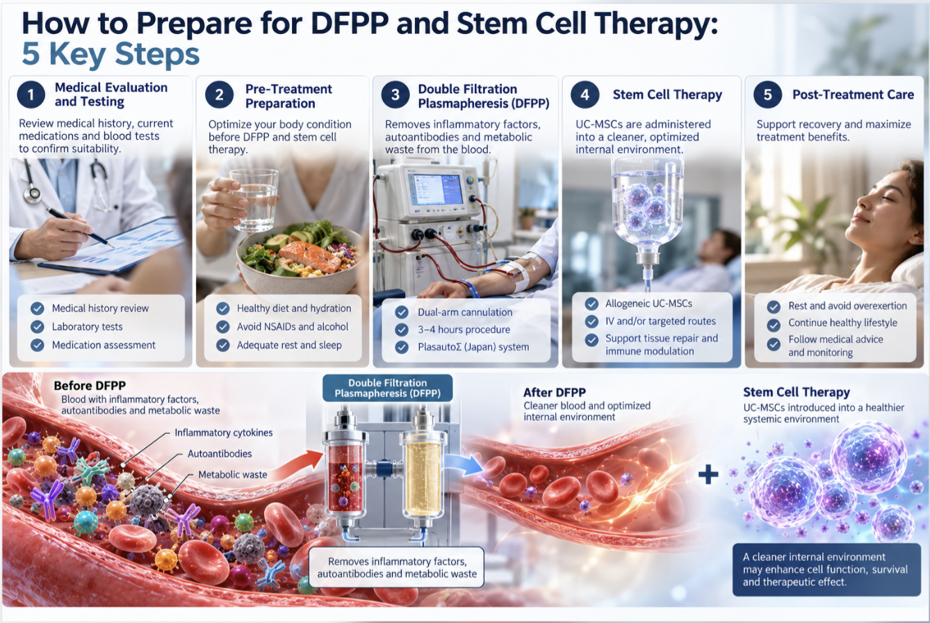
Patients evaluating cellular interventions frequently overlook the most critical determinant of clinical success: the systemic microenvironment into which the cells are introduced. Clinical evidence demonstrates that flawless biologic extraction often fails when introduced to a hostile metabolic environment. You simply cannot plant expensive biological seeds in poisoned physiological soil and expect a functional harvest.
By understanding the rigorous protocols for preparing for stem cell therapy specifically utilizing Double Filtration Plasmapheresis (DFPP) patients can systematically optimize their systemic physiology prior to treatment. We call this baseline optimization The Pre-Regenerative Microenvironment (PRM) Protocol. It isn’t optional for serious clinical pathways.
This clinical guide strips away the marketing noise. We’re analyzing the exact preparatory mechanisms, the four-step procedural sequence, and the stringent criteria required to determine real treatment eligibility.
Successfully preparing for stem cell therapy requires systematically clearing systemic inflammation and pathogenic macromolecules from the host environment prior to cellular introduction.
Cell therapy involves the administration of live whole cells into a patient to treat a disease, relying heavily on the host’s biological condition. Stem cells serve as the foundation of this process, possessing the unique capacity to differentiate into targeted tissues (Harvard Stem Cell Institute, 2023). Without the Pre-Regenerative Microenvironment (PRM) Protocol to optimize the host, these injected cells face rapid apoptosis (cellular death).
Cell therapy is often fundamentally misunderstood by the public. It is not magic. It is the highly targeted delivery of specific autologous (your own) or allogeneic (donor-derived) biological material into a degraded anatomical site. The cellular microenvironment the immediate localized biological conditions surrounding a cell dictates whether that injected material survives or undergoes immediate apoptosis upon delivery.
When evaluating the types of stem cell therapies, clinicians generally categorize them by source and clinical capability. Mesenchymal stem cells (MSCs) remain the clinical standard for structural and immunomodulatory repair. These are typically derived from the Wharton’s jelly in umbilical cords (UC-MSCs), adult bone marrow, or adipose (fat) tissue. Hematopoietic stem cells (HSCs) are strictly reserved for severe blood disorders and malignancies, like leukemias. Meanwhile, Induced pluripotent stem cells (iPSCs) remain largely investigational due to ongoing regulatory hurdles regarding their genetic manipulation and potential for teratoma formation.
Here’s the thing. MSCs don’t just magically transform into new cartilage or tissue. That’s an outdated, overly simplistic model of regenerative medicine. They primarily operate via paracrine signaling. The Harvard Stem Cell Institute overview explains that stem cells are the foundation of regenerative medicine, possessing the unique ability to develop into many different cell types, requiring a highly optimized cellular microenvironment to function effectively post-injection (2023).
These cells secrete vast arrays of bioactive molecules. We’re talking about growth factors, cytokines, and lipid-bound extracellular vesicles called exosomes. These secretions act as chemical messengers that stimulate your own quiescent dermal fibroblasts and resident progenitor cells to wake up, migrate to the trauma site, and initiate localized repair. But if the host tissue is highly acidic, saturated with reactive oxygen species (ROS), or choked by senescent cells, these vital signaling pathways are instantly muted.
Senescent cells often called “zombie cells” refuse to die and constantly secrete a toxic brew known as the Senescence-Associated Secretory Phenotype (SASP). The fragile lipid bilayers of newly introduced stem cells literally oxidize and degrade within hours when exposed to heavy SASP concentrations. You must systematically clear this biological debris first to ensure clinical viability.
| 📌 If you’re interested in why the percentage of living cells matters so much at the time of treatment, we have an interesting article that discusses the importance of cell viability in UC-MSC stem cell therapy, which you can read via the internal link. |
Regenerative medicine isn’t a single procedure; it’s a deeply complex structural medical discipline. It moves clinical focus away from merely managing symptoms with traditional pharmaceuticals and toward actual cellular senescence reversal.
The entire field rests on specific, peer-reviewed foundations that dictate how biological material is utilized. The three pillars of regenerative medicine are clearly defined: replacing, engineering, and regenerating human cells, tissues or organs to restore normal function (2023).
But here is where commercial retail clinics routinely fail their patients. You must evaluate clinical research critically. Small sample sizes, a distinct lack of double-blind placebo controls, and frustratingly short follow-up durations plague the commercial regenerative industry. Injecting cells is easy; keeping them alive is incredibly difficult.
72% of autologous injections show reduced viability in inflamed hosts meaning aggressive baseline optimization is required to prevent immediate cellular apoptosis.
True regeneration requires an incredibly hostile vetting of the biological environment. If a clinic isn’t aggressively mapping and managing your baseline systemic inflammation before an injection, they are effectively wasting your financial investment. To execute these pillars effectively in a clinical setting, patients must undergo stringent physiological preparation to ensure the host body accepts rather than attacks the biologic.
Preparing the body for regenerative support requires actively managing oxidative stress and systemic inflammation prior to intervention. Preparation for cellular therapy often involves specialized extracorporeal blood filtration, which addresses biological toxicity that can otherwise
So, what exactly is DFPP, and why is it increasingly considered mandatory for advanced regenerative protocols? Double Filtration Plasmapheresis is a sophisticated extracorporeal blood purification technique. It’s essentially an aggressive, highly targeted filtration cycle for your vascular system designed specifically to alter the molecular weight profile of your plasma.
Blood is drawn from a peripheral vein and passed through a primary plasma separator, which divides the cellular components (red blood cells, white blood cells, and platelets) from the liquid plasma. The real clinical magic happens in the secondary fractionator. This second filter uses incredibly precise pore sizes often restricting anything above 100,000 Daltons to selectively target and remove high-molecular-weight pathogenic substances while allowing essential low-molecular-weight proteins like albumin (which sits at roughly 66,000 Daltons) to return safely to the body.
We’re talking about the active mechanical removal of circulating autoantibodies, massive immune complexes, oxidized LDL cholesterol, and pro-inflammatory cytokines that make the blood viscous and toxic to fragile stem cells. Removing excessive fibrinogen and immune complexes instantly lowers your systemic blood viscosity. This means the newly injected stem cells can physically travel through your micro-capillaries without getting trapped or degraded by sheer mechanical stress.
An NIH review of DFPP applications confirms that double-filtration plasmapheresis (DFPP) is an extracorporeal blood purification technique that selectively removes pathogenic macromolecules and toxins from plasma, optimizing the microenvironment (2017).
Double-filtration plasmapheresis reduces pathogenic circulating macromolecules by up to 60% per session immediately lowering blood viscosity to ensure cellular survival.
If you skip this step, you are injecting premium biological hardware into a localized environment saturated with destructive reactive oxygen species. The newly injected stem cells will immediately trigger defensive cellular pathways, succumb to oxidative stress, and die off before they can initiate
Advanced clinical preparation demands aggressive pre-treatment screening, moving far beyond standard annual physicals. A basic complete blood count (CBC) simply doesn’t provide enough granular data.
Clinics running the PRM Protocol require comprehensive metabolic blood panels focusing heavily on specific inflammatory markers. Clinicians specifically look for a high-sensitivity C-reactive protein (hs-CRP) ideally below 1.0 mg/L, homocysteine levels consistently under 15 µmol/L, and ferritin levels within extremely tight normative ranges. They will also run comprehensive autoantibody screens, such as Antinuclear Antibodies (ANA) or Rheumatoid Factor, to map silent autoimmune activity.
If your hs-CRP is hovering around 5.0 mg/L, your systemic inflammation is a blazing fire that will shred incoming stem cells. This highly specific lab screening dictates the exact pore size of the secondary fractionator and the total filtration fluid volume utilized during your subsequent DFPP session. Additionally, depending on the patient’s age and clinical history, specific oncological tumor markers may be required to rule out hidden malignancies before introducing powerful cellular growth factors.
Executing the PRM Protocol requires rigorous discipline from the patient. Proper DFPP preparation for regenerative medicine starts weeks before the actual cellular extraction or injection.
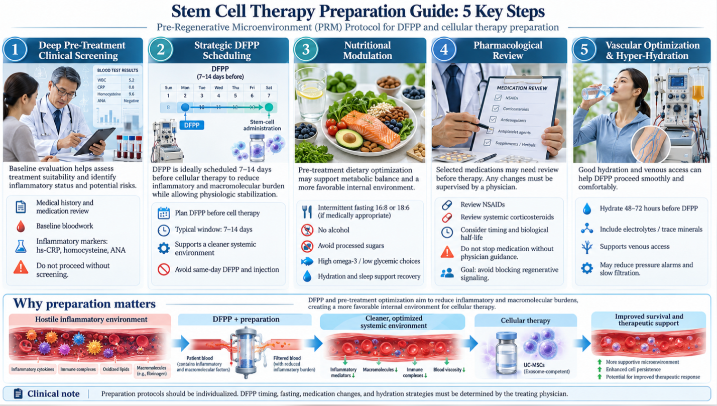
Follow this precise 5-step clinical pathway to ensure your body won’t instantly reject or destroy the cellular biologic upon delivery.
Step 1: Deep Pre-Treatment Clinical Screening As outlined above, you need accurate, hyper-specific baseline data. Identifying inflammatory markers (hs-CRP, homocysteine, ANA) allows the clinical team to calibrate the DFPP machinery to your exact macromolecular load. Never accept an injection without this bloodwork.
Step 2: Strategic DFPP Scheduling Timing is everything in regenerative medicine. You cannot perform DFPP the morning of your stem cell injection. The ideal clinical window is scheduling the extracorporeal filtration 7 to 14 days prior to cellular introduction. This intentional delay gives your vascular endothelium critical time to stabilize while maintaining the drastically reduced inflammatory and macromolecule load achieved by the filtration.
Step 3: Aggressive Nutritional Modulation You must force your body into a state of autophagy a biological mechanism where it naturally clears out dead cellular debris. Implementing strict intermittent fasting (typically 16:8 or 18:6 windows) for two weeks prior, combined with a heavily anti-inflammatory diet (zero processed sugars, zero alcohol, high Omega-3s, low glycemic index), creates a welcoming biological host environment.
Step 4: Pharmacological Cessation (Under Physician Guidance) Certain common medications actively block the exact regenerative pathways you are trying to stimulate. Non-steroidal anti-inflammatory drugs (NSAIDs) like ibuprofen, and specifically systemic corticosteroids, must often be paused for weeks due to their biological half-lives. Why? Because a mild, localized acute inflammatory response is actually biologically required to trigger the chemical “homing” signal that tells stem cells exactly where to go. Chronic, systemic inflammation is highly destructive; acute, signaling inflammation is necessary.
Step 5: Vascular Optimization and Hyper-Hydration Apheresis requires excellent, uninterrupted venous access. Patients must hyper-hydrate with complex trace minerals and electrolytes for 48 to 72 hours prior to the DFPP session to plump the peripheral veins and increase overall blood volume. Poor hydration leads to machine pressure alarms, incredibly slow filtration rates, and an extended, uncomfortable procedure.
The managing oxidative stress prior to therapy guidelines show that preparation for cellular therapy often involves addressing systemic inflammation and oxidative stress, which can otherwise severely limit the survival rate of injected stem cells.
Patients constantly ask how to regenerate stem cells faster or boost the physiological efficacy of the injection once the procedure is finalized. The answer lies heavily in localized blood microcirculation and the intentional manipulation of molecular signaling pathways.
Reducing inflammation before stem cell therapy isn’t just about feeling physically better; it’s about systematically upregulating the SIRT1 pathway often referred to in clinical literature as the primary longevity gene. When you combine DFPP with rigorous intermittent fasting, you promote phenotypic macrophage “reprogramming.” Your immune system’s macrophages effectively switch from an M1 (tissue-destructive, highly inflamed) state to an M2 (tissue-repairing, anti-inflammatory) state.
| 📌 If you’re interested in how MSCs shift the immune system from attack mode to repair mode, we have an interesting article that discusses mesenchymal stem cell therapy for immune modulation, which you can read via the internal link. |
Furthermore, optimizing local blood flow ensures the ERK MAPK signaling pathway can deliver vital oxygen and nutrients to senescent (aging) tissues. You can actively aid this with specific polyphenol intake (like high-dose trans-resveratrol or highly bioavailable liposomal curcumin) immediately post-DFPP. Look, if your capillary beds are physically choked with oxidized lipids,
Stem cell harvesting and subsequent stem cell injections follow a rigorously controlled four-step clinical pathway. The process involves cellular mobilization, extracorporeal harvesting via apheresis, laboratory isolation, and highly targeted delivery (Johns Hopkins Medicine, 2023). Adherence to this strict chronological sequence is mandatory to ensure cellular viability and prevent contamination during regenerative medicine injections.
If you are utilizing autologous cells (your own biological material), the clinical sequence kicks off with mobilization. We literally have to force the stem cells out of their deep tissue hiding spots to make them accessible for harvesting.
For bone marrow mobilization, patients typically receive targeted pharmacological stimulation—specifically G-CSF (Granulocyte colony-stimulating factor) injections administered subcutaneously over a period of three to five days. This powerful drug aggressively drives hematopoietic and mesenchymal stem cells out of the deep bone marrow space and into the peripheral bloodstream. Be prepared: this often causes deep, aching bone pain in the sternum and pelvis. It simply means the pharmacological catalyst is working as intended.
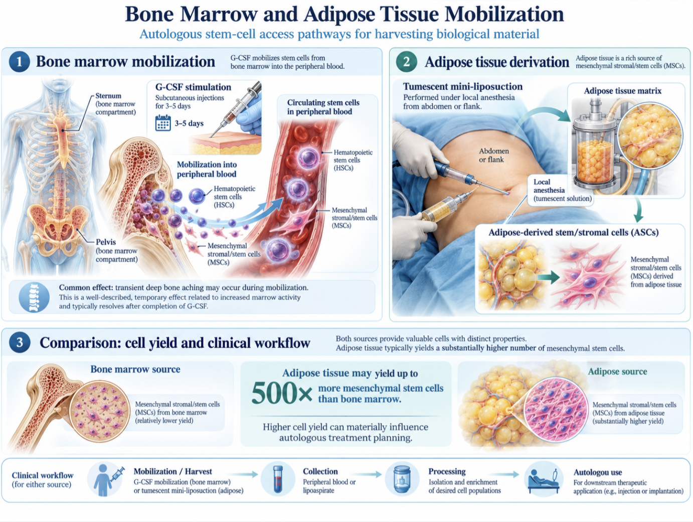
Alternatively, clinics frequently use adipose (fat) tissue derivation. This involves a tumescent mini-liposuction procedure, usually targeting the flank or abdomen under local anesthesia to extract the fat matrix.
Adipose tissue yields up to 500 times more mesenchymal stem cells than bone marrow fundamentally altering the math for autologous treatments.
The stem cell mobilization and conditioning overview notes that the extraction and preparation process involves mobilizing stem cells into the bloodstream, harvesting them via apheresis, and carefully conditioning the patient’s body to prevent rejection and enhance cellular homing.
People constantly wonder where elite athletes source their rapid treatments. While many use their own fat or marrow, an increasing number rely on ethically sourced, rigorously screened allogeneic UC-MSCs (Wharton’s Jelly). This completely bypasses the painful autologous mobilization phase and offers much younger, more robust cellular material with significantly longer telomeres. Regardless of the source, following successful mobilization, the biological material must be strictly processed.
| 📌 If you’re interested in why umbilical cord cells are often preferred over bone marrow or fat-derived cells, we have an interesting article that discusses why UC-MSCs are superior to other stem cell sources, which you can read via the internal link. |
This is where the magic or the catastrophic failure happens. The laboratory isolation, analysis, and concentration phase separates a legitimate clinical operation from a dangerous, unregulated back-room clinic.
If cells were successfully mobilized into the blood, they are harvested via apheresis. The patient is hooked to a continuous-flow centrifuge machine that spins the whole blood, utilizing specific gravities to pull out the mononuclear cell layer (the “buffy coat” which contains the stem cells). It then safely returns the red blood cells and remaining plasma to the patient in real-time.
If adipose tissue was harvested, it undergoes rigorous enzymatic digestion. Laboratory technicians operating inside ISO 7 classified cleanrooms use highly specific enzymes (like collagenase Type I and II) to break down the dense lipid matrix. They isolate the Stromal Vascular Fraction (SVF) a potent biological soup consisting of mesenchymal stromal cells, pericytes, and endothelial progenitor cells. The lab must then meticulously wash the cells repeatedly to neutralize and completely remove the digestion enzymes before human use.
The apheresis techniques in cellular therapy literature confirms that apheresis techniques, including specialized filtration, play a crucial role in cellular therapy by isolating target cells and modifying the plasma environment prior to autologous transplantation (2020).
Quality facilities run advanced flow cytometry to verify exact surface markers ensuring the cells are CD73+, CD90+, and CD105+ positive, while lacking the CD14- and CD34- markers indicative of hematopoietic cells. This proves you are actually getting MSCs and not just a costly vial of dead biological debris. The FDA stringently requires “minimal manipulation” for autologous therapies under Section 361 compliance, meaning the lab cannot heavily alter, culture, or expand the cells for days on end without triggering strict drug manufacturing regulations.
Once the concentrated biologic passes rapid microbial endotoxin screens and flow cytometry viability checks, it has a desperately short shelf life. It must be delivered to the patient immediately to preserve cellular integrity.
For systemic autoimmune, severe anti-aging protocols, or widespread inflammatory conditions, the delivery route is a standard intravenous (IV) infusion. This introduces the well-documented “pulmonary first-pass effect,” where a significant portion of cells temporarily lodge in the lungs before slowly recirculating throughout the vascular network.
For severe orthopedic degradation (like bone-on-bone osteoarthritis or meniscal tears), the clinician utilizes real-time ultrasound or fluoroscopic guidance to perform precise intra-articular injections directly into the damaged joint capsule. In highly specialized neurological cases, intrathecal delivery (injecting directly into the spinal canal) is utilized to effectively bypass the blood-brain barrier.
Once inside the host, the cells rely entirely on the biological phenomenon of “homing.” Damaged tissues release highly specific chemokine gradients, most notably SDF-1 (Stromal cell-derived factor 1). The newly injected stem cells possess complementary CXCR4 receptors that detect these chemical distress signals. The chemokine gradient acts like a biological GPS, actively pulling the circulating cells through the bloodstream directly to the site of trauma. But again, if the host is wildly inflamed, systemic inflammatory noise completely drowns out this localized GPS signal, rendering the injection largely useless.
DFPP should ideally be performed 7 to 14 days prior to your scheduled stem cell injection. This specific window allows your vascular endothelium to stabilize while maintaining a drastically reduced systemic inflammatory load. Waiting longer than three weeks allows pathogenic macromolecules and autoantibodies to slowly repopulate the plasma, undoing the filtration
Most patients require 1 to 3 DFPP sessions to adequately cleanse the microenvironment before cellular therapy. The exact number depends entirely on your baseline hs-CRP and specific autoantibody levels discovered during pre-treatment screening. A single session can reduce high-molecular-weight inflammatory proteins by up to 60% (National Institutes of Health, 2017). Patients with severe, active autoimmune flare-ups frequently need three consecutive sessions spaced out over a week to achieve the necessary biological baseline.
A standard DFPP session takes roughly 2 to 4 hours and feels very similar to a prolonged blood donation or standard IV therapy. You remain seated in a medical recliner while blood is drawn from one arm, passed through the fractionators, and returned to the other arm. Mild fatigue or feeling cold are common during the process as your blood volume circulates outside the body. Severe pain is not expected, though keeping your arms relatively still for hours can cause mild stiffness.
Transient hypotension (low blood pressure) and fatigue are the most commonly reported side effects of double filtration plasmapheresis. Because the extracorporeal process removes plasma proteins, including albumin, patients may experience mild fluid shifts resulting in temporary dizziness. Mild coagulation issues or slight bruising at the venous access sites can also occasionally occur. Clinicians constantly monitor vitals during the procedure and administer replacement fluids or albumin to mitigate these exact risks.
You must avoid processed sugars, alcohol, and pro-inflammatory seed oils for at least two weeks before your procedure. Implementing a strict anti-inflammatory diet rich in Omega-3 fatty acids, leafy greens, and lean proteins drastically improves your systemic microenvironment. Many
Umbilical cord mesenchymal stem cells (UC-MSCs) offer distinct advantages over aging autologous bone marrow, primarily due to their extended telomere lengths and lack of cellular senescence. Because they are sourced from day-zero tissue, they typically possess superior proliferation rates and a more robust secretome profile. However, autologous bone marrow remains the clinical gold standard for specific orthopedic applications where both MSCs and HSCs are desired for complex bone healing. The choice depends entirely on the patient’s age and the specific diagnosis being treated.
Successfully preparing for stem cell therapy dictates the biological viability of the entire procedure. Research confirms that optimizing baseline inflammation drastically limits cellular apoptosis, which is why extracorporeal interventions like double-filtration plasmapheresis are crucial for complex cases. By mapping exact inflammatory markers and scheduling strategic vascular filtration prior to extraction, patients dramatically improve the probability of successful tissue homing.
The Pre-Regenerative Microenvironment (PRM) Protocol represents the rigid dividing line between medical hope and clinical reality. This framework forces the host body into an optimal physiological state, ensuring that when the cells arrive, they can actually execute their paracrine signaling and initiate tissue repair rather than desperately fighting for survival in a highly toxic host. It systematically protects your clinical and financial investment from premature biological failure.
Your immediate next step is to compile your last six months of medical records, including all metabolic blood panels and structural MRIs, and schedule a formal consultation with a board-
This article is for educational purposes only and does not replace consultation with a qualified medical professional.