

Important factors for bone health are mechanical loading from habitual movement, walking, and exercise. In the absence of this physical stimulation, bones undergo loss of density and structural strength over time and risk fragility fractures. Now, researchers from the University of Hong Kong have identified a molecular mechanism that may explain how skeletal tissue senses movement and exerts its bone-protective effects. They also established Piezo1, a mechanosensitive protein of bone marrow mesenchymal stem cells as an essential “exercise sensor” in guiding these cells towards forming bone and not fat. The findings, which were published in Signal Transduction and Targeted Therapy, could guide the development of therapies that replicate some molecular effects of exercise for people who cannot remain active.
- Why Age-Related Bone Loss Becomes a Major Clinical Problem
The Global burden of Osteoporosis and age-related bone loss remains to be a major health challenge, especially in the elderly population. Fragility fractures (i.e. fractures due to low trauma) are common, debilitating and expensive in terms of health sector costs as they cause pain leading to loss of mobility and independence. According to World Health Organization data cited in the HKU announcement, about one in three women and one in five men older than 50 will suffer an osteoporotic fracture. It’s a particular burden among aging populations like that of Hong Kong, where osteoporosis is common in elderly women and men, the same release says.
With age, bone density decreases and porosity increases. Mesenchymal stem cells can differentiate into bone-forming or fat in the marrow. As we age, this equilibrium can become increasingly unbalanced, resulting in a higher ratio of these stem cells dividing into adipocytes compared with osteoprogenitors. And as marrow fat increases, the internal bone environment becomes less supportive of skeletal strength, and the cycle of progressive weakening can reach a point where reversal by current treatment alone may become impossible.
- Piezo1 as a Biological Sensor of Physical Activity
The Hong Kong researchers concluded that Piezo1 is a molecular switch that responds to mechanical cues associated with physical movement. In experimental models, activity-induced Piezo1 activation limited marrow fat accumulation and enhanced new bone formation. In Piezo1’s absence, a different pattern emerged: marrow stem cells were nudged toward fat formation, and bone loss increased. This positions Piezo1 as a critical mechanistic bridge between physical activity and the preservation of bone.
This finding is significant because it elucidates, on a cellular level, why exercise protects bone. Rather than seeing exercise as just a sort of lifestyle prescription, the study suggests that bones react via a specific — definable — signaling cascade. That raises the prospect that researchers could someday devise treatments that trigger similar pathways even in bedridden patients, or those who are frail or constrained by chronic disease. The future use remains investigational, but the mechanistic information is important.
- Stem Cell Fate, Bone Formation, and Marrow Fat
At the heart of the study is how marrow stem cells determine whether to become bone-supporting cells or fat-storing ones. The HKU team found that Piezo1 promotes healthier lineage commitment through mechanical stimulation, favouring bone-related differentiation. In the absence of that signal, marrow stem cells were more likely to embark on an adipogenic pathway, which leads to weaker skeletal tissue as time passes.
The loss of Piezo1, they found, was also associated with an increase in the release of inflammatory mediators such as Ccl2 and lipocalin-2, both of which further tipped the marrow environment toward fat accumulation and away from bone formation. In their work in the lab, prevention of these signals reverted towards a more favorable skeletal environment. This indicates that Piezo1 is not working in isolation, but rather as a component of a more extensive network linking mechanical loading, inflammation, stem-cell fate and the integrity of bone.
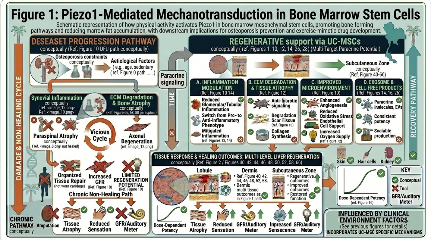
Schematic representation of how physical activity activates Piezo1 in bone marrow mesenchymal stem cells, promoting bone-forming pathways and reducing marrow fat accumulation, with downstream implications for osteoporosis prevention and exercise-mimetic drug development.
- Translational Significance for Osteoporosis Care
Modern osteoporosis treatment still relies heavily on exercise, fall prevention, nutrition and medications to reduce resorption or strengthen bone. But many older adults, bedridden patients and people with chronic illness cannot move sufficiently to derive the full bone benefits of movement. Which is why the notion of an “exercise-mimetic” therapy is attracting interest: It could mimic some part of the bone-preserving biology of exercise at the molecular level.
The researchers themselves characterize this work as a preliminary step toward identifying targets for intervention, not something that’s imminently ready to be used in the clinic. That is to say, the detection does not imply that such a pill that would substitute physical activity for bone health already exists. What it does mean is that the biological pathway has become clearer, which clarity may help lead to the development of drugs targeted at fragile-bone populations down the road.
- Concluding Perspective
It was the most salient mechanobiological finding as to how exercise protects the skeleton. By identifying Piezo1 as the vital molecular sensor that connects movement to bone-preserving stem-cell behavior, the HKU study is paving a new path for osteoporosis research going forward. The long-term goal is to develop therapies that enable bones to retain some of activity’s benefits, even when patients are unable to move with sufficient intensity on their own. For now, the finding can be considered a scientific and translational advance but not yet a therapy. Still, it offers an interesting template for future drugs to fight bone loss.