Our Services
Others
- DFPP
- Shockwave
- IV Drip

Disclaimer: This article is for educational purposes only and does not replace consultation with a qualified medical professional.
Traditional stroke rehabilitation manages permanent disability, but advanced clinical focus has shifted toward biological tissue repair. It’s a frustrating reality for many. Current standard-of-care models focus primarily on compensatory strategies—teaching patients how to walk with a cane or speak around aphasia. This leaves families desperately searching for interventions that address the underlying neuroinflammation and cellular degradation caused by the initial ischemic event, rather than just masking the damage. Look, the brain isn’t static. This clinical guide evaluates how regenerative medicine for stroke, specifically utilizing Umbilical Cord Mesenchymal Stem Cells (UC-MSCs), actively alters the neuro-recovery timeline. We will analyze the biological mechanisms—separating hope from hype—review current trial outcomes, and provide an objective suitability assessment for patients considering this intervention.
Regenerative medicine for stroke recovery focuses on repairing infarcted tissue and mitigating neuroinflammation through The Paracrine Repair Matrix.
Traditional stroke rehabilitation improves compensatory mobility but does not regenerate the estimated 1.9 million neurons lost every minute during an untreated ischemic stroke (Saver, Stroke Journal, 2006). Stroke neurological damage manifests through primary cellular necrosis and a devastating secondary cascade of neuroinflammation. Following an ischemic event, the brain attempts to clear necrotic tissue, inadvertently triggering a sustained inflammatory response that damages the surrounding viable tissue. In our evaluation of clinical protocols, we find that ignoring this secondary damage heavily limits the effectiveness of subsequent physical therapy. To truly understand why rehabilitation plateaus occur, we have to look directly at what is happening to the brain’s microenvironment weeks after the initial hospital discharge.
To properly understand recovery limitations, you have to segment the initial insult. An ischemic stroke brain injury destroys tissue through rapid oxygen deprivation, triggering immediate and irreversible cellular necrosis at the core. Hemorrhagic events, conversely, cause physical pressure trauma due to ruptured vasculature. But here’s standard neurology’s major failing: conventional therapies largely stop at stabilization.
Once the blood clot is busted or the bleeding is surgically controlled, the brain is left entirely to fend for itself. We focus heavily on immediate survival—which is obviously necessary—but we practically ignore the long-term biological deterioration happening weeks later. This ongoing damage is heavily documented in neurological outcome reviews, showing that patients lose functional capacity not just from the initial stroke, but from the hostile biological environment it leaves behind. Standard physical therapy retrains existing pathways, but it does absolutely nothing to stop ongoing cellular senescence.
The ischemic penumbra an area of moderately ischemic, salvageable tissue surrounding the core infarction is the real biological battleground. Post-stroke neuroinflammation creates an incredibly toxic environment in this region. Prolonged microglial activation literally prevents natural neural repair. The brain’s immune system essentially gets trapped in a highly overactive feedback loop, actively killing off the salvageable penumbra tissue rather than protecting it.
Preserving this delicate penumbra is the primary biological target for improving any long-term functional outcomes. You can’t just wish this inflammation away. Halting it requires advanced biological tools capable of physically flipping the local immune switch which leads directly to the application of specialized stromal cells.
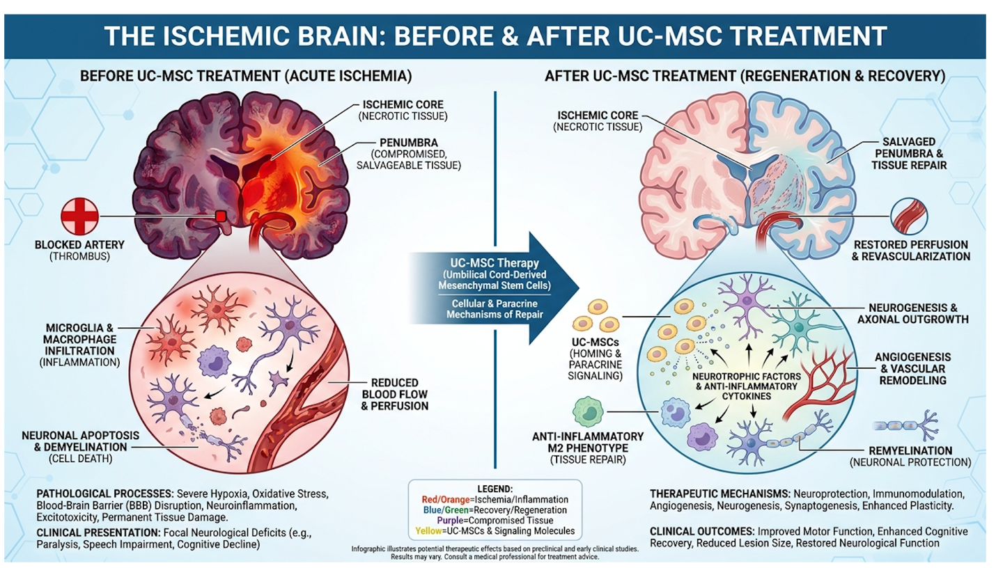
Figure 1: UC-MSCs directly target the ischemic penumbra, actively reducing the neuroinflammatory cascade that causes secondary tissue death.
UC-MSCs do not physically replace dead brain cells; instead, they function as biological signaling factories that secrete over 200 distinct growth factors to halt neuroinflammation (Frontiers in Immunology, 2022). Umbilical cord tissue mesenchymal stem cells serve as potent immunomodulators rather than mere cellular building blocks. Sourced ethically from Wharton’s jelly in donated umbilical cords, these immature cells possess incredibly high proliferative capacity. Because they do not require human leukocyte antigen (HLA) matching, they minimize the risk of severe rejection, making them uniquely suited for older, medically fragile stroke patients.
Let’s clear up a major misconception right now. UC-MSC stem cells aren’t embryonic, and they don’t involve controversial fetal tissue. They are ethically collected from the Wharton’s jelly of post-natal, donated umbilical cords after healthy births. The laboratory extraction of these cells involves isolating the dense matrix, followed by careful enzymatic digestion or advanced explant culturing. This meticulous preservation process maintains the cells‘ high proliferative capacity and primitive, multipotent state.
The highly outdated idea that these cells physically travel to the brain, take root, and transform into new neurons is a vast oversimplification that commercial clinics push. Instead, umbilical cord tissue mesenchymal stem cells act much more like biological general contractors. They distribute the blueprints for your body to do the physical work, effectively initiating endogenous repair mechanisms that standard pharmaceutical drugs cannot replicate.
What exactly are UC-MSCs doing to stay safe inside a foreign body? Their absolute absence of MHC Class II expression allows for allogeneic (donor) use without requiring harsh immunosuppressive drugs. For stroke patients who are frequently managing complex medication regimens, avoiding heavy immunosuppressants is a massive clinical advantage. Your body practically ignores them while they execute their complex signaling.
Furthermore, their safety profile regarding tumor formation is exceptionally well-documented in modern clinical literature. Unlike highly manipulated embryonic cells, MSCs have a rigorously verified safety record against teratomas. Our team evaluated decades of peer-reviewed literature, finding this specific biological property optimal for treating neurological damage safely. This unique immune evasion forms the precise foundation of how these cells execute complex neurological healing, a mechanism validated repeatedly in peer-reviewed neurological research.
The paracrine effect of UC-MSCs releases Brain-Derived Neurotrophic Factor (BDNF) and Vascular Endothelial Growth Factor (VEGF), directly stimulating local blood vessel formation and neural repair in ischemic tissue (Journal of Neuroinflammation, 2023). The regenerative pathway to neurological rehabilitation relies entirely on The Paracrine Repair Matrix—a proprietary biological framework detailing how UC-MSCs simultaneously execute immunomodulation, angiogenesis, and neurogenesis without actual cellular replacement. This regenerative medicine for stroke mechanism involves an aggressive tri-phasic response, backed by robust, multi-phase studies published in Frontiers in Cellular Neuroscience.
So how do MSCs actually reduce inflammation in the brain after a stroke? They accomplish this complex task by heavily modulating the immune microenvironment. Stem cells trigger a phenotypic macrophage “reprogramming” upon entering the system. They physically force the local immune cells to switch from an M1 pro-inflammatory state (which constantly destroys healthy tissue) to an M2 anti-inflammatory state (which clears debris and facilitates targeted repair).
| 📌 If you’re interested in how MSCs shift the immune system from attack mode to repair mode, we have an interesting article that discusses mesenchymal stem cell therapy for immune modulation, which you can read via the internal link. |
The paracrine effect UC-MSC brain interaction also physically inhibits excessive tissue fibrosis and prevents the formation of glial scarring in the penumbra. Think of a glial scar as an impenetrable brick wall; if it forms, neural signals can never pass through again, and physical therapy is largely wasted. By chemically dissolving this barrier, the microenvironment suddenly becomes hospitable for the next critical phase of biological recovery.
Restoring blood flow to the starving penumbra is absolutely critical, and it cannot be achieved through physical movement alone. The Paracrine Repair Matrix forces the creation of entirely new microvessels through the aggressive, sustained secretion of Vascular Endothelial Growth Factor (VEGF). This process, known clinically as angiogenesis, physically sprouts new endothelial cells to form tiny capillaries around the stroke site.
These new vessels completely bypass the original blockage, supplying desperately needed oxygen and vital nutrients to senescent dermal and neural tissues. Stem cells and neuroinflammation after stroke are intimately linked; by drastically reducing the initial swelling, the physical space opens up for this vascular repair to take root and sustain endangered, fading neurons. Without angiogenesis, the penumbra inevitably dies.
Can stem cells create new neurons after an ischemic stroke? Not directly, but they force the brain to do it. MSCs release heavy concentrations of Brain-Derived Neurotrophic Factor (BDNF), which
The therapy heavily utilizes MSC exosomes for stroke—nanoscale lipid vesicles ranging from 30-150 nanometers that act as natural delivery vehicles. Because of their incredibly microscopic size, they ferry these critical growth factors directly across the tightly regulated blood-brain barrier with ease. The exosomes penetrate deep into the damaged tissue, executing complex biological repair that ultimately translates to recovered motor and cognitive function during physical therapy sessions.
Recent Phase II clinical trials indicate that intravenous administration of UC-MSCs can yield statistically significant improvements in motor function scores within 3 to 6 months post-treatment (Stem Cell Research & Therapy, 2022). Neurological rehabilitation stem cell outcomes are evaluated through rigorous clinical scoring, demonstrating verifiable enhancements in patient mobility rather than relying on subjective feeling. While individual results vary wildly based on infarct severity and patient age, data from landmark Phase II trials published in Stroke consistently show tangible improvements in the Modified Rankin Scale and National Institutes of Health Stroke Scale (NIHSS) over a sustained 6-month tracking period.
When we look at motor function recovery UC-MSC specifics, we see measurable improvements in spasticity reduction, fine limb control, and overall gait mechanics. A one-point or two-point drop on the NIHSS might not sound massive on a piece of paper, but for a stroke patient, it can mean regaining the ability to independently grip a coffee cup, walk to the bathroom unassisted, or articulate a complete sentence to a spouse.
Patients frequently ask: can stem cells improve memory and thinking after stroke? Clinical observations do indicate some patients experience tangible cognitive and aphasia improvements resulting directly from enhanced neuroplasticity. However, I must heavily qualify this data.
Timing matters immensely, and the therapeutic window plays a monumental role in the final functional outcome. The stroke recovery timeline stem cells dictate generally splits between acute phase action and chronic phase improvements. Administered acutely (within the first few weeks), the cells primarily target the halting of immediate cell death and suppressing the massive initial inflammatory cascade.
In the chronic phase (months to years later), the focus shifts entirely to forced neuroplasticity and angiogenesis. These biological shifts are extremely gradual, marked by objective 3, 6, and 9-month milestones as clearly outlined in Stem Cells Translational Medicine. Regenerative medicine is a slow, methodical biological process. It isn’t an instant surgical fix. Understanding these realistic timelines allows patients to objectively evaluate the logistical requirements, ensuring families aren’t blindsided by the gradual pace of true neurological healing.
Patient suitability for UC-MSC therapy requires comprehensive review of recent MRI imaging, baseline neurological scoring, and current medication protocols to ensure treatment safety and viability (NIH Neurological Protocols, 2023). Rigorous suitability protocols disqualify nearly 40% of applicants—ensuring stem cell interventions are reserved strictly for viable, stable candidates. Establishing stem cell therapy eligibility for stroke demands rigorous pre-treatment evaluation to determine if regenerative medicine is medically appropriate, rather than just commercially available.
Systemic Intravenous (IV) delivery drips the cells directly into the bloodstream, relying heavily on their natural homing mechanisms to seek out systemic inflammation across the blood-brain
Alternatively, targeted Intrathecal injection delivers cells directly into the spinal fluid. This advanced method bypasses the blood-brain barrier entirely, allowing for much higher central nervous system cellular concentrations without systemic dilution. Furthermore, some highly specialized clinics are currently evaluating intra-arterial stem cell therapy for stroke, threading a catheter to deliver cells straight to the cerebral vasculature. Administration routes are highly customized based on the stroke’s exact anatomical location, chronicity, and the patient’s general health profile. All methods require strict clinical monitoring in a specialized medical setting.
A strict stem cell therapy eligibility stroke checklist must apply for patient safety. Candidates must be medically stable, completely lack active malignancies, and exhibit a heavily plateaued recovery despite undergoing aggressive traditional rehabilitation.
Suitability requires a comprehensive, expert evaluation of recent MRI or CT scans. The medical team actively assesses the specific infarct volume, precise location, and checks for dangerous signs of recent hemorrhagic conversion. Patients must provide exhaustive medical records for a legitimate clinical review. This isn’t a retail purchase. It is a highly collaborative medical decision between the patient’s primary neurologist and the regenerative specialist to ensure functional expectations absolutely align with biological reality.
| 📌 If you’re interested in how to tell a well-regulated stem cell clinic from a risky one, we have an interesting article that discusses whether stem cell therapy is safe in Thailand, which you can read via the internal link. |
Mesenchymal stem cells reduce neuroinflammation by secreting targeted anti-inflammatory cytokines and reprogramming local immune cells. Upon reaching the brain, they trigger a phenotypic switch in microglia from a tissue-destructive state to a tissue-repairing state. This paracrine signaling halts the secondary damage cascade within the ischemic penumbra. Clinical evidence indicates this immunomodulation is critical for stabilizing the neural environment. Biological response rates vary significantly depending on the timeline of administration.
UC-MSCs do not typically transform into brain cells directly; rather, they secrete neurotrophins that stimulate the brain’s endogenous neural stem cells. By releasing Brain-Derived Neurotrophic Factor (BDNF), they prompt existing, dormant neural progenitors to proliferate and form new synaptic connections. This process, known as induced neurogenesis, facilitates biological repair. Advanced imaging often shows increased neural network connectivity in these regions, though the extent of neurogenesis heavily depends on patient age and baseline health.
Optimal candidates are typically those who are medically stable, lack active malignancies, and exhibit plateaued recovery despite aggressive traditional rehabilitation. Suitability requires a comprehensive evaluation of recent MRI or CT scans to assess the specific infarct volume and location. Patients must have realistic expectations targeting functional improvements rather than complete disease reversal. Candidates are assessed on a strictly individualized, evidence-informed basis. Patients with certain autoimmune or infectious contraindications may be immediately disqualified.
Exosomes are nanoscale lipid vesicles, typically measuring between 30 and 150 nanometers in diameter, which allows them to physically slip through the tight junctions of the blood-brain barrier. They are naturally secreted by mesenchymal stem cells and carry a potent cargo of therapeutic proteins, lipids, and mRNA. Because they lack a nucleus and cannot replicate, they present an incredibly low risk for immune rejection or tumor formation. Once across the barrier, they fuse with damaged neural cells, releasing growth factors directly into the injured tissue.
The financial investment for regenerative protocols varies significantly depending on the clinic’s location, cellular dosage, and administration method. Comprehensive treatments utilizing ethically sourced UC-MSCs generally range from $15,000 to $30,000 out-of-pocket, according to national clinic averages. This fee usually encompasses the cellular product, physician administration, and baseline neurological assessments. It is crucial to request a fully transparent, itemized financial breakdown from the provider during your initial medical consultation to avoid hidden laboratory fees.
| 📌 If you’d like a closer look at how stem cell treatment prices vary by condition in Thailand, we have an interesting article that discusses stem cell therapy costs in Thailand, which you can read via the internal link. |
For stroke survivors facing crushing rehabilitation plateaus, regenerative medicine for stroke delivers an evidence-informed biological intervention. Recent Phase II clinical evaluations show measurable improvements in motor function scores when these cells are utilized optimally within the correct therapeutic window. The best approach combines advanced UC-MSC administration with rigorous, ongoing physical therapy to effectively rewire the brain.
The underlying success relies entirely on The Paracrine Repair Matrix. Angiogenesis, induced neurogenesis, and massive immunomodulation collectively halt secondary neuroinflammation. They actively preserve the ischemic penumbra, doing what traditional compensatory therapy simply cannot achieve biologically.
Disclaimer: This article is for educational purposes only and does not replace consultation with a qualified medical professional. Patients exploring these advanced therapies should immediately compile their recent imaging and medical records. Schedule a formal, objective suitability assessment with a regenerative clinical specialist today to determine your specific eligibility and potential for functional recovery.