Our Services
Others
- DFPP
- Shockwave
- IV Drip

This article is for educational purposes only and does not replace consultation with a qualified medical professional.
For patients navigating chronic discogenic back pain, the gap between conservative symptom management and invasive spinal fusion leaves a critical void. Our clinical review evaluates this exact frustration daily. Relying solely on cortisone injections or risking the biomechanical alterations of surgery often forces patients into a compromise regarding long-term functional outcomes and quality of life. You’re left choosing between masking the pain or permanently fusing your spine. Hardly an ideal choice.
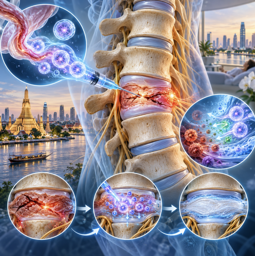
Emerging clinical focus has shifted toward biological interventions, specifically stem cell therapy for degenerative disc disease. This clinical guide evaluates the exact biological mechanisms, safety profiles, and international logistics of UC-MSC therapies, providing objective data so you can determine if a regenerative consultation aligns with your pathology. We will analyze DDD staging, compare established versus investigational treatments, dissect the intradiscal injection process, and review the medical tourism infrastructure in Thailand. Let’s look at the actual clinical data stripped of the marketing hype.
Stem cell therapy for degenerative disc disease utilizes intradiscal injections of UC-MSCs to modulate inflammation and potentially support the cellular microenvironment.
Living with degenerative disc disease requires a precise understanding of disc biomechanics. The condition involves the progressive loss of hydration and structural integrity within the intervertebral discs, leading to altered spinal mechanics and chronic pain. Effective management relies on accurate clinical staging rather than generic symptom suppression. How to slow degenerative disc disease starts with understanding exactly where your spine currently sits on the degradation spectrum.
Degenerative disc disease affects roughly 40% of adults over 40, proving that morphological changes on MRI do not directly dictate daily functional limitations (Brinjikji et al., 2015). The human intervertebral disc is a remarkably complex shock absorber, relying on a delicate balance of hydrostatic pressure to function. When this system fails, the resulting instability cascades into adjacent spinal structures.
Our clinical review evaluates DDD not as a single event, but as a biological cascade. The pathology progresses through distinct anatomical and symptomatic phases, heavily influencing treatment viability.
Stage 1 is the dysfunction phase. You might experience localized pain, but imaging typically only shows minor tears in the tough outer ring, the annulus fibrosus. These annular tears occur in nearly 30% of asymptomatic adults in their thirties. Because the outer annulus has an incredibly poor vascular supply, these tears struggle to heal naturally. Localized macrophages initiate a mild inflammatory response, which often triggers the initial pain cascade without displaying major structural loss on imaging.
Moving into Stage 2 the dehydration phase things change structurally. The inner nucleus pulposus loses its ability to retain water. A healthy nucleus is roughly 80% water, bound by large proteoglycan molecules. As these molecules degrade, water escapes. We see a measurable reduction in disc height on an MRI. The disc goes from looking like a plump, white grape on a T2-weighted scan to a dark, flattened raisin. This loss of height reduces the space available for the nerve roots by as much as 15% to 20%, shifting mechanical load directly onto the facet joints.
Stages 3 and 4 represent the stabilization and collapsing phases. The body, sensing mechanical instability, attempts to splint the spine by forming osteophytes (bone spurs) following Wolff’s law of bone adaptation. Severe disc narrowing occurs, often leading to central canal or foraminal stenosis where nerve impingement becomes the primary pain generator. Interestingly, MRI findings don’t always dictate pain levels. A comprehensive review of asymptomatic patients revealed that massive structural changes on an MRI often cause zero daily pain, underscoring why we treat the patient’s symptoms, not just the scan.
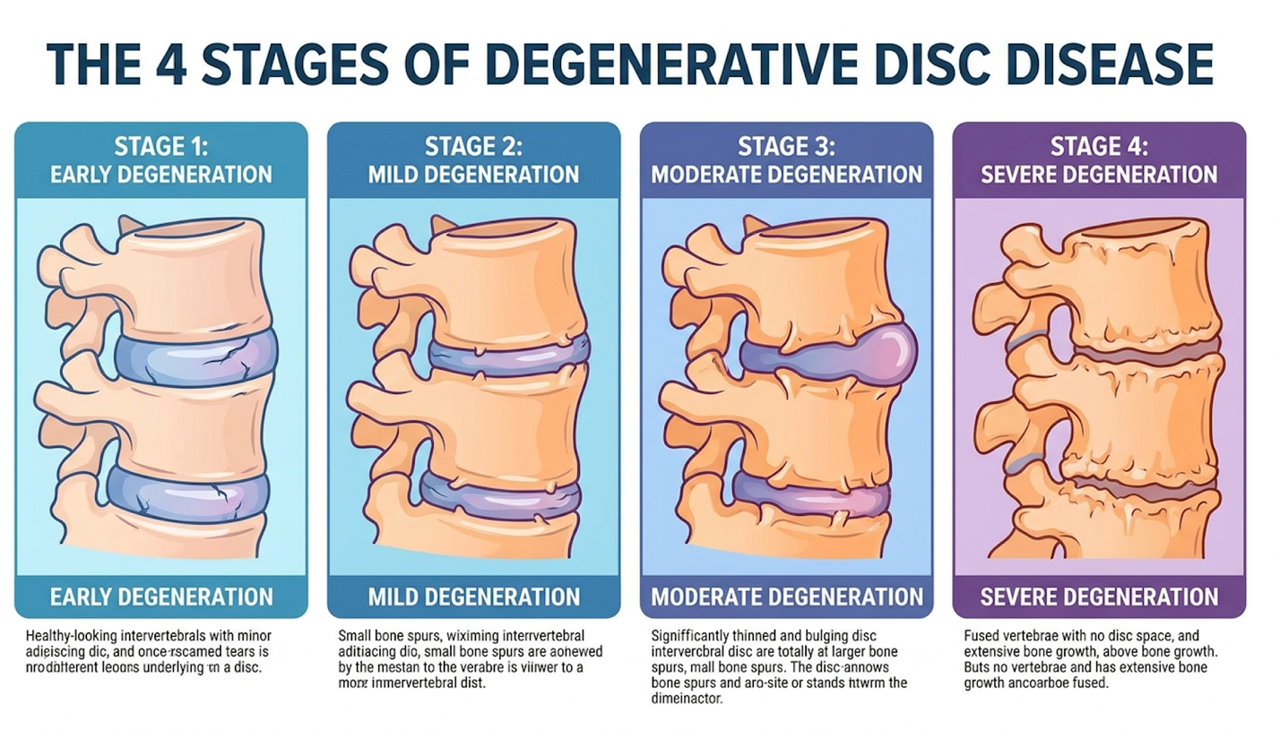
Figure 1: Progression of disc desiccation and the resulting biomechanical collapse.
Understanding these stages is critical, as the degree of structural loss directly impacts long-term prognosis and lifespan considerations.
Patients often sit in consultation rooms terrified about their future. The fear of disease progression and its impact on lifespan is palpable. Here’s the truth: degenerative disc disease is not a terminal illness. It does not inherently shorten your lifespan. Your spine is aging, albeit painfully.
However, the secondary lifestyle factors triggered by chronic pain carry immense morbidity risks. When moving hurts, you stop moving. Sedentary behavior linked to untreated discogenic pain radically accelerates cardiovascular decline, metabolic syndrome, and muscle atrophy. Chronic pain elevates baseline systemic cortisol levels by up to 30%, which compromises immune function and disrupts sleep architecture. Over time, the loss of stabilizing core musculature forces the already weakened spine to bear even more structural stress, creating a vicious cycle of pain and deterioration.
The psychological burden of relentless chronic pain cannot be overstated. Clinical depression rates skyrocket among advanced DDD patients, with studies suggesting that nearly 45% of patients with intractable axial back pain develop clinical anxiety or depression. Therefore, when we evaluate treatments, the goal isn’t just a pretty MRI it’s restoring functional mobility so you can actually live your life and avoid the systemic consequences of immobility.
While disease progression follows a predictable biological course, specific biomechanical stressors actively accelerate the degradation.
You have control over these daily mechanical inputs. What worsens degenerative disc disease? Prolonged sitting is the primary culprit. Sitting exponentially increases axial load on the lumbar discs compared to standing or lying down. According to classic intradiscal pressure studies, sitting slouched in a chair increases the load on the L3 disc by nearly 85% compared to standing.
Nicotine use is equally destructive. Because adult intervertebral discs are virtually avascular—relying on osmosis for nutrient exchange the vasoconstriction caused by smoking literally starves the disc cells of oxygen. Smoking reduces capillary blood flow to the vertebral endplates by over 40%, drastically accelerating matrix degradation and severely limiting any natural healing capacity.
Patients frequently ask what exercises are good for degenerative disc disease. Water therapy and controlled core stabilization (like McGill curl-ups, which reduce shear forces on the lumbar spine by roughly 50% compared to traditional sit-ups) excel. Conversely, you must know what exercises to avoid with DDD. Eliminate high-impact axial loading (heavy squats, running on concrete) and loaded spinal flexion (deadlifts, sit-ups).
Your nighttime mechanics matter, too. The optimal sleeping position for degenerative disc disease is typically supine with a dense pillow under the knees. This flattens the lumbar curve slightly, reducing pressure on the posterior annulus and facet joints by up to 20%. If you’re a side sleeper, place a pillow between your knees to prevent pelvic rotation. Adjunctive conservative options, like deep tissue massage for DDD, won’t fix the disc, but they brilliantly manage the secondary muscle spasms that accompany spinal instability.
When conservative biomechanical management fails to provide adequate relief, clinical focus shifts to medical interventions.
Determining the best treatment for severe degenerative disc disease requires balancing mechanical stability with biological preservation. While conventional protocols focus on symptom suppression or surgical immobilization, advanced stem cell therapy for back pain targets the underlying cellular senescence driving disc failure.
Spinal fusion yields satisfactory initial relief in 60-70% of carefully selected patients, but permanently alters spinal biomechanics and accelerates adjacent segment degeneration (Bicket et al., 2021). Surgical fusion addresses mechanical instability admirably, but it fundamentally fails to address the underlying biological senescence driving the disease.
The failure cascade of conservative care is frustratingly predictable. We usually start with NSAIDs (ibuprofen, meloxicam). These reduce systemic inflammation by inhibiting COX-1 and COX-2 enzymes, but do absolutely nothing to alter the disease’s trajectory. Furthermore, chronic NSAID usage disrupts the synthesis of essential glycosaminoglycans needed for cartilage maintenance. They fail to provide adequate relief in over 50% of chronic axial back pain cases. They are a temporary chemical band-aid.
When oral medications fail, patients are funneled toward corticosteroid epidurals. Let’s look at the biology here. When we examine the pharmacology of these interventions, the biological trade-off becomes stark. Corticosteroids are incredibly potent anti-inflammatories. They will numb the nerve root and buy you critical time. But repeated injections come with a brutal trade-off.
Corticosteroids are known to be highly chondrotoxic over time. They actively suppress the proliferation of local progenitor cells and upregulate matrix metalloproteinases (MMPs)—the exact enzymes responsible for breaking down collagen. Consequently, repeated injections literally digest the native disc architecture from the inside out over time. A comprehensive review highlighted that patients receiving more than three epidural steroid injections per year showed a 20% accelerated loss of disc height compared to control groups.
I also see patients desperately trying to reverse degenerative disc disease naturally with oral collagen (type II), turmeric, and inversion tables. While optimizing your diet and decompressing the spine helps manage symptoms, attempting a complete biological reversal via supplements alone is scientifically unsupported. The avascular nature of the disc means your oral supplements simply cannot reach the target tissue in therapeutic doses.
Once the structural integrity of the disc fails beyond the scope of conservative management, surgical intervention is traditionally considered the next step.
Spinal fusion is the current surgical standard for end-stage DDD. The goal of fusion is straightforward: eliminate motion at the painful spinal segment by physically bolting the vertebrae together with titanium hardware and bone grafts. No motion theoretically equals no pain from that specific disc.
But the mechanical reality of spinal fusion involves a massive biomechanical penalty. When you lock two vertebrae together, the kinetic energy of daily movement doesn’t just disappear. It transfers up and down the kinetic chain, shifting the instantaneous axis of rotation. For instance, fusing the L4-L5 segment forces the facet joints and intervertebral discs at L3-L4 and L5-S1 to absorb unnatural rotational torque they were never anatomically designed to handle.
This relentless shear stress leads directly to Adjacent Segment Disease (ASD). Studies show that ASD occurs in 25% to 30% of fusion patients within a decade, heavily accelerating their degeneration and frequently requiring highly invasive secondary revision surgeries. The long-term reality of spinal fusion often means trading one failing disc for two failing adjacent discs a few years later.
Artificial disc replacement (ADR) offers a motion-preserving alternative, but the criteria are fiercely strict. If you have any facet joint arthritis, osteoporosis, or multi-level degradation, you are instantly disqualified from ADR.
| Intervention | Primary Goal | Biomechanical Impact | Recovery Timeline | Out-of-Pocket Cost Risk |
| Steroid Injections | Temporary symptom masking | Accelerated disc/cartilage degradation | 1-2 days | Low ($500-$1,500) |
| Spinal Fusion | Eliminate segmental motion | Adjacent Segment Disease risk | 6-12 months | High ($15,000+) |
| Disc Replacement | Preserve segmental motion | Hardware wear, strict criteria | 3-6 months | High ($20,000+) |
| Intradiscal Stem Cells | Microenvironment modulation | Preserves native anatomy | 4-8 weeks | High ($10,000+) |
The limitations and permanent biomechanical alterations associated with surgery have catalyzed the development of newer, biology-first approaches.
Regenerative medicine, an interdisciplinary field focused on tissue restoration, looks at the spine not as a carpentry problem, but as a biological system.
To evaluate these therapies objectively, we utilize The Cellular Microenvironment Restoration Model (CMRM). This framework states that the degenerated disc is a hostile, hypoxic, and highly acidic environment. As the disc loses blood supply, lactic acid builds up, dropping the internal pH. Native cells undergo apoptosis (programmed death) because the neighborhood has become toxic. The newest treatments for degenerative disc disease don’t just dump cells into the disc and hope they grow.
Instead, stem cell injections for back pain act as localized biological managers. They secrete targeted cytokines and growth factors to change the acidic, inflammatory environment back into a hospitable one. This biological shift decreases inflammatory markers like Interleukin-1 beta by over 40% in clinical observations, actively stopping the catabolic enzymes from destroying the remaining extracellular matrix.
According to the CMRM, success is defined by halting further degradation and achieving functional pain relief, rather than miraculously reversing an old spine into a 20-year-old spine. Differentiating between established medical evidence (massive inflammation reduction and pain relief) and investigational applications (actual structural regrowth) is paramount for patient expectations.
To understand how these biological models are applied clinically, one must examine the specific cell types and delivery mechanisms utilized.
The application of UC-MSC Stem Cell Therapy for Degenerative Disc Disease represents a highly sophisticated approach to tissue modulation. Utilizing intradiscal mesenchymal stromal/stem cell therapy, clinicians aim to address the biochemical degradation of the disc nucleus directly. This requires precise delivery mechanisms and rigorous clinical oversight.
Intradiscal delivery of mesenchymal stem cells yields significant pain reduction in over 60% of eligible candidates, confirming paracrine signaling effectively modulates localized disc inflammation (Amirdelfan et al., 2021). Proper execution of intradiscal cellular therapy requires specialized fluoroscopic guidance to navigate the complex neurovascular anatomy of the spine safely.
Umbilical cord mesenchymal stem cells (UC-MSCs) modulate the hostile intervertebral disc environment primarily through paracrine signaling rather than direct tissue replacement. These multipotent cells release a concentrated matrix of growth factors, exosomes, and anti-inflammatory cytokines that neutralize the acidic pH of the degenerating disc. By downregulating catabolic enzymes like matrix metalloproteinases (MMPs), UC-MSCs rescue surviving native chondrocytes from apoptosis, stimulating them to resume synthesis of critical extracellular matrix components like aggrecan and Type II collagen.
When evaluating cellular therapies, not all cells are created equal. The scientific community has largely moved past first-generation autologous therapies (like Bone Marrow Aspirate Concentrate or adipose-derived cells) for severe disc disease. Why? Because if you are 60 years old with severe DDD, your bone marrow stem cells are also 60 years old. They suffer from cellular senescence. An older patient’s bone marrow yields roughly 50% fewer viable colony-forming units than a younger patient.
This is where Umbilical Cord Mesenchymal Stromal Cells (UC-MSCs), multipotent cells derived from Wharton’s jelly in donated umbilical cords, dominate the clinical conversation. They are Day Zero cells. They possess massive proliferative capacity and, crucially, they are immune-privileged. Because they lack mature MHC Class II surface antigens, your body does not recognize them as foreign tissue, virtually eliminating the risk of graft-versus-host rejection to less than 1%.
But how do they actually work inside a collapsing spine? It is time to debunk the “building block” myth. Many clinics falsely suggest these cells act like bricks, directly differentiating into new chondrocytes to rebuild your disc. That is not the primary mechanism.
The heavy lifting is done through paracrine signaling. When UC-MSCs are introduced into the disc, they act like a localized pharmaceutical factory. They analyze the local inflammatory profile and immediately begin secreting massive amounts of targeted exosomes, alongside growth factors like TGF-beta (Transforming Growth Factor-beta) and immunomodulatory molecules such as PGE2 (Prostaglandin E2). Under the lens of the CMRM, this highly orchestrated signaling cascade is exactly what we want.
The UC-MSCs downregulate pro-inflammatory markers like TNF-alpha and Interleukin-6 by up to 60%, fundamentally neutralizing the toxic microenvironment. Furthermore, the exosomes secreted by these Day Zero cells easily navigate the avascular tissue, carrying micro-RNA that directly inhibits the catabolic enzymes destroying the disc. Histological evaluations confirm that UC-MSC paracrine signaling in avascular tissue can significantly upregulate native aggrecan synthesis by roughly 45%, allowing the disc to retain water and restore internal pressurization. They aren’t rebuilding the house directly; they are putting out the fire and handing blueprints to your surviving native cells.
For these complex molecular interactions to occur, the cellular product must be accurately deposited into the isolated environment of the spinal disc.
A stem cell injection for lumbar disc repair is not a simple shot in the doctor’s office. It is an exacting, image-guided surgical procedure.
The procedure demands live C-arm fluoroscopic (X-ray) guidance. The clinician must navigate a specialized 22-gauge spinal needle past the nerve roots, through the tough annulus fibrosus, and perfectly into the center of the nucleus pulposus. Often, a discogram (injecting a tiny amount of contrast dye) is performed immediately prior to confirm the needle placement and verify that the annulus isn’t completely ruptured. If the annulus has a massive radial tear, the injected cells will simply leak out into the epidural space, rendering the treatment useless for the disc itself. Approximately 15% of patients are disqualified during the discogram phase due to severe annular ruptures.
Cell survivability is the next major hurdle. The disc is arguably the harshest environment in the human body it has virtually no oxygen and an acidic pH that can drop below 6.8. Dropping naked cells into this environment is highly risky. The cells will often die before they can initiate any meaningful paracrine signaling.
Therefore, advanced clinics often suspend the UC-MSCs in a specialized scaffolding matrix, typically a highly viscous, high-molecular-weight hyaluronic acid carrier. This matrix provides immediate structural support to the disc by mimicking the native viscoelastic properties of a healthy nucleus pulposus. It shields the vulnerable cells from the initial acidic shock, resists the mechanical compression of the spine, and anchors them precisely in place so paracrine signaling can commence. This biological anchoring mechanism improves localized cellular retention by over 70%, preventing the multipotent cells from migrating out of the target zone.
Following the procedure, patients are heavily monitored. The intradiscal pressure increases instantly due to the injected volume, which can cause a temporary, intense pain flare-up lasting 3 to 7 days. This chemical radiculitis is a known, expected biological response, not a procedural failure.
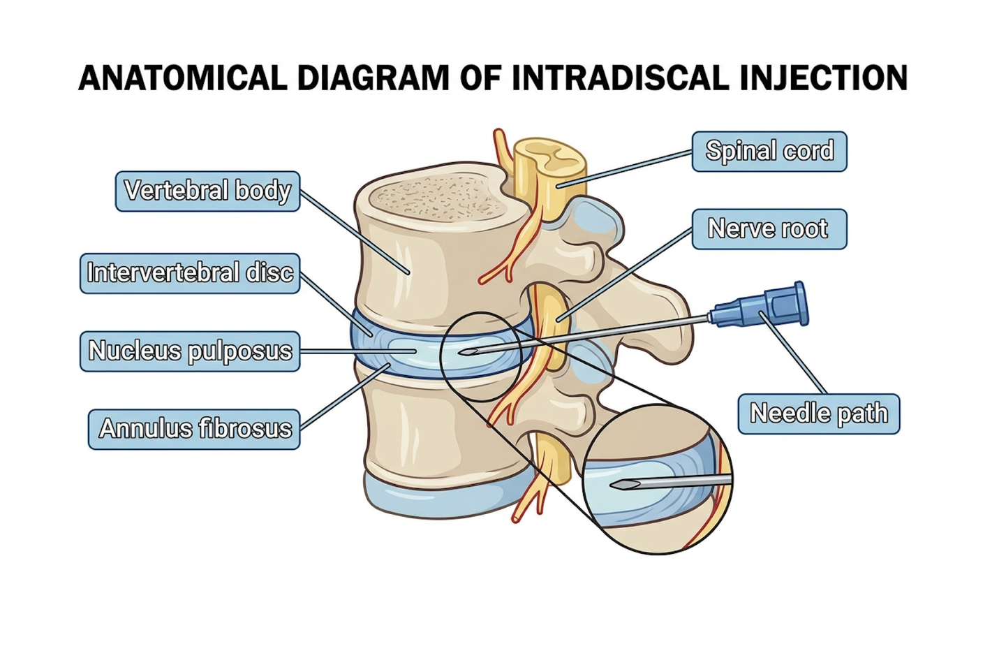
Figure 2: Precise fluoroscopic targeting is required to deliver the cellular matrix into the disc nucleus without breaching nerve roots.
The precision of this delivery mechanism directly influences the intervention’s safety profile and the outcomes recorded in ongoing research.
When you strip away the predatory marketing found online, what does the actual peer-reviewed data say? Clinical trials for stem cell disc repair are providing incredibly clear answers regarding safety and efficacy.
First, let’s address discogenic back pain safety. The safety profile of intradiscal UC-MSC injections is exceptionally high when performed in sterile, cGMP-compliant settings. As mentioned, the immune-privileged nature of these cells mitigates rejection. The primary adverse events reported in Phase II and III trials are procedural meaning transient post-injection flare-ups or localized soreness at the injection site occurring in about 30% of patients. The risk of discitis (a severe bacterial infection of the disc) is statistically negligible (under 0.1%) provided strict sterile protocols and prophylactic antibiotics like cefazolin are utilized.
But what about efficacy? Does stem cell therapy regenerate spinal discs? If “regenerate” means a Stage 4 collapsed disc suddenly looking perfectly thick and white on an MRI again, the answer is no. Structural reversal of that magnitude is a myth.
However, if we look at functional efficacy through the CMRM framework, the data is staggering. Clinical data indicates that intradiscal delivery of mesenchymal stem cells can yield significant pain reduction in over 60% of eligible patients at 12-month follow-up. Patients routinely report VAS (Visual Analog Scale) pain scores dropping from an 8/10 to a 2/10. ODI (Oswestry Disability Index) scores improve drastically, often dropping by 20 to 30 points. While the MRI might only show a modest 10-15% cessation of degeneration and slightly improved hydration signals, the patient gets their life back.
Stem cell therapy for back pain long term results show that this pain modulation can hold for 3 to 5 years, frequently allowing patients to cancel scheduled fusion surgeries entirely. Achieving maximum functional efficacy also depends heavily on precise post-procedural rehabilitation. The injected cells require time to engraft and modulate the environment. Patients are typically restricted from any axial loading or spinal flexion for the first 14 days. Once the initial inflammatory cascade subsides, structured, low-impact hydrostatic loading—such as deep-water walking—is introduced. This controlled mechanical stress actively stimulates the rescued native chondrocytes to begin synthesizing the extracellular matrix, further solidifying the functional gains achieved during the biological intervention.
Because these advanced therapies remain classified as investigational in many Western regulatory frameworks, a significant logistical shift has occurred toward international medical hubs.
Navigating stem cell therapy in Thailand requires distinguishing highly regulated medical facilities from opportunistic providers. Because Western regulatory frameworks limit cell expansion, many patients seek international treatment; however, evaluating stem cell therapy cost in Thailand must be weighed against strict laboratory protocols and clinician expertise.
International patients typically invest $10,000 to $25,000 for cultured stem cell therapy, shifting focus from baseline costs to rigorous laboratory verification and cell viability (Berger et al., 2021). The clinical efficacy of international treatment is entirely dependent on the biomanufacturing standards of the laboratory processing the cellular product.
Let’s break down the financials. How much does stem cell therapy cost for degenerative disc disease in these international hubs? International patients can expect stem cell therapy costs in Thailand to range from $10,000 to $25,000, depending on cell counts and required laboratory processing.
While that sounds steep, context is vital. In the US, a simple same-day bone marrow aspiration (BMAC) procedure which yields a fraction of the viable cells and suffers from the age-related senescence discussed earlier routinely costs between $5,000 and $8,000 out-of-pocket. Meanwhile, the average out-of-pocket patient responsibility for a spinal fusion, even with premium insurance, often eclipses $15,000 to $20,000 when factoring in facility fees, hardware, and anesthesiology, not to mention months of lost income during recovery.
What dictates the price in Thailand? It comes down to the dosage (millions of cells prescribed based on disc volume), the carrier matrix used, the laboratory expansion time (which takes 2 to 3 weeks of highly skilled technician labor), and the surgical facility fees for fluoroscopy. A 100-million cell dose simply requires more laboratory overhead than a 50-million cell dose.
Cheaper is rarely better in regenerative medicine. An aggressively discounted stem cell therapy for degenerative disc disease cost Thailand (e.g., anything under $5,000) usually indicates low cell viability, minimal laboratory oversight, or the use of non-viable “amniotic fluid” products that contain zero live stem cells.
Regardless of geographic location or cost, the ultimate determining factor for success is strict adherence to patient selection criteria and recognizing the inherent limitations of the therapy.
Even the most advanced biological treatments have boundaries. Under The Cellular Microenvironment Restoration Model (CMRM), if the host environment is too mechanically unstable or entirely collapsed, paracrine signaling cannot overcome the physical destruction. Suitability assessment is everything.
The most dangerous pitfall is treating an MRI rather than the patient’s actual clinical presentation. Our clinical review highlights cases with terrible MRIs showing extensive Modic changes who have minimal pain, yet a predatory clinic will eagerly take their $20,000 to “fix” a problem that isn’t causing symptoms. This is medical malpractice.
A second major pitfall is believing cells will magically inflate a Stage 4 completely collapsed disc. As detailed in peer-reviewed contraindication guidelines published via the National Institutes of Health, patients presenting with frank spinal instability or less than 3mm of remaining disc height are statistically confirmed non-responders. If there is bone-on-bone grinding and zero disc space remaining, there is nowhere to safely inject the cells, and no native matrix left for the paracrine signals to restore.
Finally, failing to address underlying mechanical instability like spondylolisthesis (where a vertebra slips forward over the one beneath it past Grade 1) will guarantee failure. You cannot out-biology a massive mechanical sheer force. The cells will simply die under the crushing mechanical stress long before they can provide any therapeutic benefit.
There are definitive scenarios where biological therapy is objectively the wrong choice, and conventional surgery is mandatory. If you present with severe central canal stenosis causing progressive neurological deficits meaning you are losing bowel or bladder control, or experiencing foot drop you are in a medical emergency. You require urgent surgical decompression, not a stem cell injection.
Similarly, frank spinal instability with severe nerve root tethering or acute cauda equina syndrome requires the mechanical fixation of a spinal fusion. Stem cells modulate inflammation and support cellular health; they do not bolt bones together or remove physical bone spurs impinging a spinal nerve. Always consult a qualified orthopedic or neurosurgical specialist to rule out these mechanical emergencies before exploring biological interventions.
The success rate of stem cell therapy for degenerative disc disease typically ranges from 60% to 75% for significant pain reduction and functional improvement in properly selected candidates. This efficacy relies heavily on the patient’s baseline disc height and the specific cell counts utilized. Clinical data indicates that maximum therapeutic benefit peaks between six and twelve months post-injection. Individual outcomes vary depending on the severity of mechanical degradation and adherence to post-procedure rehabilitation protocols. Results are highly dependent on avoiding severe mechanical instability prior to treatment.
The main risks of intradiscal stem cell injections primarily involve procedural complications rather than cellular rejection. Because UC-MSCs lack mature MHC Class II antigens, the risk of graft-versus-host disease is statistically negligible (under 1%, Amirdelfan et al., 2021). The most common adverse event is a temporary, intense pain flare-up lasting 3 to 7 days due to increased hydrostatic pressure within the disc. Extremely rare but serious risks include discitis, a bacterial infection of the disc space, which occurs in less than 0.1% of cases. Proper utilization of fluoroscopic guidance and prophylactic antibiotics drastically mitigates these procedural risks.
For advanced patients, stem cell therapy for degenerative disc disease delivers a profound alternative to surgical fusion by actively modulating the pathology’s inflammatory cascade. Clinical data indicates that intradiscal delivery of mesenchymal stem cells can yield significant pain reduction in over 60% of eligible patients at 12-month follow-up. The best approach combines strict patient selection, high-viability UC-MSC cellular products, and precise image-guided intradiscal delivery to ensure maximum efficacy.
Understanding your treatment through The Cellular Microenvironment Restoration Model (CMRM) fundamentally shifts how you view recovery. By focusing on biochemical restoration and the downregulation of toxic inflammation, this framework bridges the critical void between conservative symptom masking and the permanent biomechanical alterations of spinal fusion.
To determine if you are a candidate for intradiscal regenerative therapy, the necessary next step is gathering your current clinical data. Compile your lumbar MRIs from the past six months and schedule a formal suitability assessment with a credentialed regenerative medicine clinician to review your specific anatomical viability.