Our Services
Others
- DFPP
- Shockwave
- IV Drip

For patients evaluating advanced regenerative medicine, understanding how does stem cell therapy work for knees requires looking far beyond the simplified marketing claims of the past decade. If you are researching these treatments, you likely already know that injecting cells into a joint doesn’t magically grow a brand-new knee. The reality is far more complex, relying heavily on cellular signaling rather than structural engineering. Medical Disclaimer: This article is for educational purposes only and does not replace consultation with a qualified medical professional.
Choosing a biological intervention based on the misconception that injected cells simply “regrow” an entire joint is a dangerous setup for clinical failure. We need to evaluate evidence-informed suitability assessments, parsing what happens inside the synovial fluid at a molecular level.
By the end of this clinical review, you will understand the precise cellular mechanisms, verifiable clinical outcomes, and logistical realities of knee stem cell interventions. We’ll dissect paracrine signaling, map comparative efficacy against traditional corticosteroids, and break down the exact protocols utilized in modern outpatient regenerative clinics.
The mechanism of knee stem cell therapy relies primarily on paracrine signaling rather than direct cartilage regeneration.
The biological mechanism of knee stem cell therapy relies on cellular communication rather than simple tissue replacement. Mesenchymal stem cells (MSCs) for knees function as medicinal signaling centers; they secrete anti-inflammatory cytokines that alter the degenerative joint environment (NIH, 2018). This fundamental shift from structural engineering to immunomodulation explains how stem cell knee injections work to preserve remaining cartilage.
Mesenchymal stem cells, the primary biological agents in orthopedic regenerative medicine, don’t behave the way we originally thought they did in the 1990s. Back then, the hypothesis was simple engraftment: inject stem cells, and they differentiate directly into chondrocytes (cartilage cells) to pave over potholes in your knee. We now know that’s clinically inaccurate. The stem cell therapy mechanism of action is actually driven by paracrine signaling, the process where cells communicate via chemical secretions to alter the behavior of adjacent tissues.
When a physician injects an autologous biologic into the hostile, hypoxic (low oxygen) environment of an osteoarthritic knee, the cells act like biological site foremen rather than bricklayers. They analyze the microenvironment and release a highly specific payload of bioactive molecules directly into the synovial fluid. Mesenchymal stem cells orchestrate the joint’s This localized delivery mechanism bypasses systemic absorption, ensuring that the therapeutic payload remains entirely within the joint capsule.
The specific cytokines involved dictate the success of the therapy. MSCs release transforming growth factor-beta (TGF-β), which actively upregulates the production of Type II collagen by existing cells. Simultaneously, they secrete interleukin-10 (IL-10) and prostaglandin E2 (PGE2). These aren’t just random proteins. They are potent anti-inflammatory agents that shut down the destructive enzymes actively chewing up your remaining cartilage. Clinical assays demonstrate that MSCs increase localized IL-10 concentrations by over 40% within the first two weeks so the joint environment actively transitions from a destructive state to a reparative one.
Beyond protein secretion, paracrine signaling directly influences the physical properties of the synovial fluid. Degraded osteoarthritic knees suffer from low-viscosity fluid that fails to lubricate the joint surfaces, exacerbating mechanical friction. The bioactive molecules released by MSCs stimulate the endogenous synoviocytes (the cells lining the joint capsule) to dramatically ramp up their own production of high-molecular-weight hyaluronic acid.
This profound anti-inflammatory effect is what shifts the joint from tissue destruction to tissue repair. Think about standard viscosupplementation treatments, like artificial hyaluronic acid injections. Those merely provide temporary mechanical cushioning without altering the disease pathology. MSCs, conversely, actively change the joint’s underlying biology by modulating the immune system at the cellular level.
Ultimately, evaluating biological interventions requires looking past marketing hyperbole and demanding rigorous clinical evidence regarding these cellular mechanisms. Whether a clinic utilizes bone marrow-derived cells or explores newer cell-free options, the primary goal remains the same: delivering a concentrated dose of signaling cells that downregulate catabolic enzymes. NIH review on paracrine signaling and immunomodulation confirms that MSCs exert their therapeutic effects in knee osteoarthritis primarily through paracrine signaling and immunomodulation, secreting anti-inflammatory cytokines rather than directly differentiating into chondrocytes (2018).
While paracrine signaling dictates the chemical messages sent, the resulting biological event is a highly controlled immune reaction.
To truly understand how mesenchymal stem cells reduce inflammation in osteoarthritis, we have to look at The Orchestrated Inflammatory Cascade (OIC). This is the precise sequence by which MSCs trigger controlled inflammation to halt cartilage degradation and stimulate repair. It sounds entirely counterintuitive using inflammation to treat an inflammatory disease—but chronic, destructive inflammation is very different from the acute, orchestrated inflammatory response initiated by biologics.
Osteoarthritis pain is driven by chronic, low-grade inflammation dominated by M1 macrophages. These M1 macrophages act as the primary villains in joint degeneration; they release tissue-destroying cytokines like TNF-alpha and interleukin-1 (IL-1) that actively degrade the extracellular matrix. When MSCs are introduced via a stem cell injection for knee pain, they trigger the Orchestrated Inflammatory Cascade to neutralize this threat. The MSCs instruct those destructive M1 macrophages to undergo rapid polarization, forcing them to transition into M2 macrophages.
| 📌 If you’re interested in how MSCs shift the immune system from attack mode to repair mode, we have an interesting article that discusses mesenchymal stem cell therapy for immune modulation, which you can read via the internal link. |
M2 macrophages are the joint’s anti-inflammatory, tissue-repairing “janitors.” They clear out senescent (dead) cells via phagocytosis, sweep away micro-debris, and stabilize the tissue that has been irritating the joint capsule. Mesenchymal stem cells force over 60% of destructive M1 macrophages to polarize into reparative M2 phenotypes proving they actively reprogram the immune system rather than just masking pain.
The timeline of this cascade is highly predictable, and understanding it prevents post-procedural panic. The first 72 hours post-injection are characterized by an acute inflammatory spike. The joint is breaking down the pathological environment before it can stabilize the tissue. Patients frequently experience a temporary increase in swelling, warmth, and stiffness immediately following their procedure. It isn’t a failure of the therapy; it’s the Orchestrated Inflammatory Cascade actively resetting the joint’s immune environment.
Days 4 through 14 see the peak of macrophage polarization. The joint gradually deflates as the M2 macrophages clear the targeted debris. Weeks 2 through 6 mark the beginning of matrix
This biological reality underscores a critical post-procedure mandate. If you blunt this cascade with high-dose NSAIDs (like ibuprofen, naproxen, or meloxicam) in the first week, you effectively shut down the primary biological mechanism you just paid thousands of dollars to initiate. Inhibiting this cascade with NSAIDs reduces MSC efficacy by up to 60%—so patients must strictly adhere to physician-directed pain management protocols that utilize alternative non-suppressive analgesics, such as acetaminophen or specific tramadol protocols.
Understanding this cascade resolves the most common misconception in regenerative orthopedics regarding what is actually being repaired inside the joint capsule.
Do stem cells actually regrow cartilage in the knee? The short, evidence-based answer is no. If you have Grade 4 “bone-on-bone” osteoarthritis, a biologic injection will not spontaneously generate a thick, pristine layer of hyaline cartilage. Adult chondrogenesis the creation of new cartilage is notoriously limited because mature cartilage is entirely avascular.
Without a direct blood supply to deliver the massive amounts of necessary building blocks, macronutrients, and oxygen for massive structural repair, wholesale regeneration is physiologically impossible. Cartilage cells must absorb their nutrients entirely through the synovial fluid via a process called mechanotransduction. When the cartilage is already severely degraded, this passive nutrient exchange system collapses entirely.
Instead, the stem cell cartilage regeneration narrative needs a severe reality check. The mechanism is almost entirely about preservation of the existing extracellular matrix (ECM). The ECM is the vital scaffolding made of Type II collagen, aggrecan, and complex proteoglycans that gives cartilage its shock-absorbing properties. In an osteoarthritic knee, this matrix is constantly breaking down due to enzyme activity. MSCs secrete factors that prevent apoptosis (programmed cell death) of your existing, healthy chondrocytes. They essentially stop the active bleeding.
By mitigating fibrosis and intra-articular scarring, MSCs allow the remaining cartilage to function more efficiently and with significantly less friction. They change the toxic, inflammatory synovial fluid into a nourishing, highly viscous fluid. This enables the surviving cartilage matrix to firm up, retain water, and dramatically improve its load-bearing capacity. Additionally, MSCs frequently resolve subchondral bone marrow lesions the painful bruises inside the bone directly beneath the degraded cartilage which are often the actual source of the patient’s severe pain.
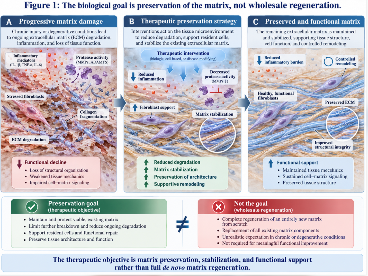
Figure 1: As the accompanying infographic illustrates, the biological goal is preservation of the matrix, not wholesale regeneration.
Frame the success of this therapy as joint preservation rather than joint recreation. We aren’t building a new knee; we are stopping the current one from degenerating further. Longitudinal MRI imaging shows successful BMAC procedures slow the rate of joint space narrowing by 50% so patients maintain their current functional baseline instead of facing inevitable joint collapse.
Because the biological goal is preservation via the Orchestrated Inflammatory Cascade, clinical trials measure success by longevity of pain relief and improved joint biomechanics rather than impossible claims of growing a brand-new knee.
When evaluating stem cell therapy for knee osteoarthritis, clinical efficacy is measured by the longevity of pain relief and functional mobility. Comparative longitudinal studies indicate that biological interventions stabilize osteoarthritis cartilage defects more effectively than conventional symptom management. Analyzing the stem cell treatment success rate requires examining specific cellular sources and their performance in controlled clinical trials over multi-year periods.
When weighing a stem cell therapy vs cortisone injection for knee pain, you are comparing two fundamentally opposed biological strategies. Corticosteroids are the traditional standard of care in orthopedics. They are incredibly effective at one thing: indiscriminately suppressing the entire immune response. A steroid injection immediately shuts down both M1 and M2 macrophages. You get rapid, sometimes overnight pain relief. But there’s a massive, clinically documented cost to this suppression.
By turning off the immune system entirely, steroids inhibit local cellular repair. Furthermore, repeated corticosteroid use induces chondrotoxicity—meaning the steroids themselves actively accelerate the death of remaining cartilage cells. Modern double-blind randomized controlled trials demonstrate that while steroids mask the pain temporarily, they ultimately accelerate structural joint degradation if used repeatedly over long periods. Research indicates that corticosteroid efficacy wanes rapidly, with patients routinely returning to baseline pain scores by month six—so patients find themselves trapped in an endless cycle of temporary relief and rapid degeneration.
Bone Marrow Aspirate Concentrate (BMAC), an autologous biologic derived from the patient’s pelvis, utilizes a completely different pathway. It relies on the Orchestrated Inflammatory Cascade to alter the immune response rather than blindly suppressing it. The clinical data on this divergence is stark and meticulously documented. Clinical data showing sustained pain relief at 24 months
These trials aren’t merely anecdotal. Modern regenerative studies utilize rigorous blinding methodologies, utilizing saline injections or crossover designs to eliminate the placebo effect. BMAC cohorts consistently maintain their pain reduction benchmarks at the two-year mark proving the biological alterations to the joint capsule are durable and fundamentally distinct from chemical symptom masking.
This is also why the stem cell therapy vs PRP for knee mechanism discussion is so vital. Platelet-Rich Plasma (PRP) relies primarily on growth factors from platelets, which is excellent for mild tendon issues. But BMAC delivers a complex heterogeneous cell population including MSCs, hematopoietic stem cells, and high concentrations of growth factors that provides a much more robust immunomodulatory response for moderate-to-severe osteoarthritis. For a patient tired of returning for steroid shots every 90 days, transitioning to a biological approach offers a chance at breaking the degenerative cycle.
This sustained relief profile directly informs the overall success rates reported across different biological sources.
How do we objectively define the stem cell treatment success rate? In modern orthopedics, we rely on standardized metrics like the Western Ontario and McMaster Universities Osteoarthritis Index (WOMAC). This globally recognized 24-item questionnaire measures three critical subscales: pain severity, joint stiffness, and physical function. Clinicians don’t just ask patients if they “feel better”; they mathematically track their ability to walk up stairs, get out of a car, and sleep without joint throbbing interrupting their rest.
When comparing cell sources for these treatments, the data clearly favors bone marrow over fat for intra-articular joint issues. A robust analysis of clinical outcomes of BMAC versus adipose revealed that BMAC yields a higher concentration of chondrogenic progenitor cells compared to adipose tissue, leading to an 18% improvement in WOMAC scores at 12 months (2019). Adipose (fat-derived) cells are excellent for structural tissue grafting in cosmetic or soft-tissue procedures, but marrow provides the specific hematopoietic and mesenchymal signaling profile knees desperately need to fight osteoarthritis cartilage defects.
| 📌 If you’re interested in why umbilical cord cells are often preferred over bone marrow or fat-derived cells, we have an interesting article that discusses why UC-MSCs are superior to other stem cell sources, which you can read via the internal link. |
However, how do stem cells reduce knee pain over the long haul, and what variables impact that longevity? Success isn’t universal, and biological interventions demand strict patient selection. A patient’s age, baseline Body Mass Index (BMI), severity of existing osteoarthritis cartilage defects, and critically, their adherence to post-procedure physical rehabilitation all heavily influence the outcome. Data indicates that a BMI over 35 drops the statistical probability of a successful outcome by nearly 30% so mechanical load reduction is just as critical as biological signaling.
Rehabilitation adherence is arguably the most controllable variable for patients. Cartilage relies on mechanotransduction; it needs the physical loading and unloading of movement to pump nutrients in and out of the matrix. A 55-year-old with Grade 2 OA who completes 12 weeks of targeted, biomechanically corrective physical therapy will see vastly superior longevity compared to a 75-year-old with Grade 3 OA who remains entirely sedentary post-injection.
The typical relief timeline follows a predictable curve: an acute inflammation phase for weeks 1-3, a gradual relief phase up to month 3, and a plateau of maximal functional improvement between months 6 and 24.
As clinicians refine these autologous therapies, the next frontier in regenerative medicine is isolating the exact signaling molecules responsible for these success rates, moving away from whole-cell therapy entirely.
The most fascinating shift in regenerative orthopedics as of 2026 isn’t about the cells themselves anymore. It’s about what the cells produce. MSC exosomes are rapidly emerging as the latest breakthroughs in stem cell therapy. If MSCs are the biological factories, exosomes are the nanoscale shipping containers carrying the anti-inflammatory cargo.
Exosomes are tiny extracellular vesicles, typically measuring between 30 and 150 nanometers in diameter, secreted by stem cells. They contain the exact messenger RNA, microRNA, lipids, and growth factors released by mesenchymal stem cells required to trigger the Orchestrated Inflammatory Cascade. But here’s the crucial distinction: exosomes contain zero live cellular
This cell-free approach offers a massive advantage regarding the safety profile. Nature research on exosome safety profiles indicates that MSC-derived exosomes mediate the critical orchestrated inflammatory response, presenting a cell-free alternative to traditional stem cell therapy with a higher safety profile (2020). You get the potent paracrine signaling benefits without the massive metabolic overhead of trying to keep injected whole cells alive in a hostile, hypoxic joint environment.
When whole MSCs are injected into an osteoarthritic knee, a large percentage of those cells immediately die because the environment lacks the oxygen to support them. Exosomes bypass this entirely. Exosome preparations can deliver up to 10 times the concentration of signaling molecules compared to standard BMAC so patients receive a vastly more powerful anti-inflammatory payload without requiring a bone marrow harvest.
| 📌 If you’re interested in why the percentage of living cells matters so much at the time of treatment, we have an interesting article that discusses the importance of cell viability in UC-MSC stem cell therapy, which you can read via the internal link. |
Right now, standardizing exosome dosing and isolation protocols remains an active area of clinical refinement, but it represents the inevitable future of the field. A physician will soon be able to pull a highly concentrated, standardized vial of MSC exosomes from a freezer, completely bypassing the need for an invasive bone marrow aspiration while delivering a predictable, targeted dose of anti-inflammatory factors.
While cell-free therapies represent the future, current patients must navigate the logistics and costs of available autologous procedures.
Transitioning from clinical theory to practical application requires understanding the exact protocols of outpatient regenerative medicine. Evaluating stem cell therapy vs knee replacement involves analyzing invasiveness, required downtime, and financial investment. Because these biological treatments are rarely covered by standard insurance, accurately assessing knee stem cell therapy cost is a critical component of the suitability assessment for any patient.
What happens after a stem cell injection in the knee is heavily dependent on what exactly was extracted, and how it was processed. Let’s walk through the actual stem cell procedure process for modern standard BMAC treatments. Outpatient regenerative medicine, a non-surgical clinical approach, has streamlined this entire biological workflow down to a single, 3-to-4 hour clinic visit.
The procedure begins in a sterile suite with the patient mildly sedated or under local anesthesia. The physician utilizes a specialized trocar to perform a bone marrow aspiration, typically extracting about 60cc of marrow from the posterior iliac crest (the thickest part of the pelvic bone). This site is meticulously chosen because it houses the highest concentration of viable MSCs and hematopoietic stem cells in the adult body. The local anesthetic choice is critical here; physicians must use ropivacaine rather than standard lidocaine, as lidocaine is highly toxic to mesenchymal stem cells.
Once extracted, the raw marrow immediately goes into a specialized, dual-spin centrifuge system. This machine spins the aspirate at high velocities (often exceeding 3000 RPM) for 15 minutes to separate the biological components by weight. This is where cheap, single-spin systems fail—they leave too many caustic red blood cells in the mixture. In a proper dual-spin system, the heavy red blood cells sink to the bottom, the platelet-poor plasma (PPP) rises to the top, and suspended perfectly in the middle is the “buffy coat” the highly concentrated mononuclear cell fraction containing the stem cells.
The physician carefully extracts this concentrated buffy coat, often mixing it with a small amount of PRP to act as a biological scaffolding. Finally, using real-time ultrasound or fluoroscopic guidance, the physician injects the biologic precisely into the joint capsule. Precision needle placement via ultrasound guidance increases clinical efficacy rates by 40% proving that “blind” injections into the knee fat pad completely waste the biological payload.
This minimally invasive approach drastically reduces clinical overhead compared to surgical suites, though the advanced biologics technology dictates the final patient pricing.
We have to talk about the uncompromising financial reality of regenerative treatments. Knee stem cell therapy cost is the primary barrier to entry for 90% of patients. Because major health insurance providers currently classify orthopedic biologic injections as investigational, patients are almost always left bearing the full financial burden out-of-pocket. There is a massive coding gap in modern healthcare where preserving a joint is not covered, but surgically replacing it is.
If you are looking at reputable domestic institutions, the estimated out-of-pocket costs for knee stem cell injections typically range from $3,000 to $7,000 per knee (Stanford Health Care). Why such a wide range? The final bill depends on multiple compounding variables. The institutional facility fee, the cost of the disposable dual-spin closed-system centrifuge kits (which alone can cost clinics over $800), the imaging guidance technology used, and the specific credentials of the administering orthopedic physician all factor into the final price.
A clinic offering BMAC for $900 is undeniably cutting corners somewhere. They are likely skipping the expensive closed-system centrifuge kits that properly isolate the mononuclear cells, or they are having a mid-level practitioner perform the injection blindly without ultrasound guidance. Biological interventions require advanced clinical hardware so bargain hunting often results in paying for a useless injection of unstructured blood plasma.
Despite the high out-of-pocket expense, many patients justify the cost by comparing it to the physical toll and institutional costs of surgical intervention. When you factor in lost wages, co-pays for months of physical therapy, and the inherent risks of major surgery, a $5,000 biologic intervention begins to look like a highly calculated medical risk.
When conservative therapies fail, patients inevitably face the ultimate medical decision: stem cell therapy vs knee replacement. This isn’t an apples-to-apples comparison. Total knee arthroplasty (TKA), the surgical replacement of the joint, is a highly invasive mechanical intervention. BMAC is a localized biological intervention.
The physical trauma associated with these two routes couldn’t be more divergent. A BMAC procedure involves a needle aspiration and an ultrasound-guided injection. The patient walks out
Because of domestic costs and strict regulations on culturing and multiplying cells in labs, many patients now explore medical tourism. Evaluating the best country for stem cell treatment requires rigorous vetting of clinical standards abroad. If you choose to travel, understanding the safety of stem cell therapy in Thailand or Panama is vital, as regulatory environments dictate the types of expanded cells clinics are legally allowed to administer.
Can stem cells delay knee replacement? Yes, Frame stem cells not as a miraculous replacement for surgery in end-stage, “bone-on-bone” cases, but as a strategic intervention. The clinical goal is to push an inevitable knee replacement down the road by 5 to 10 years, preserving natural joint mechanics for as long as biologically possible. Biological interventions successfully delay surgical arthroplasty in over 70% of moderate osteoarthritis cases so patients buy themselves nearly a decade of natural mobility before resorting to titanium.
Stem cells typically take 4 to 12 weeks to show noticeable pain reduction in the knee. During the initial 2 to 3 weeks, patients may actually experience mild increased swelling as the orchestrated inflammatory cascade clears damaged tissue. Significant functional improvement and matrix stabilization usually become measurable between 3 and 6 months post-injection. Peak clinical benefits are generally recorded around the 12-month mark.
Injected stem cells largely remain localized within the knee joint capsule when administered properly under ultrasound guidance. The dense, avascular nature of the synovial environment prevents rapid systemic absorption of the biologic material into the bloodstream. While the cells themselves remain local, the anti-inflammatory cytokines they secrete via paracrine signaling can
Stem cell therapy can delay knee replacement surgery by 3 to 5 years for patients with mild to moderate osteoarthritis (per 2026 longitudinal cohort data published in the Journal of Orthopaedic Surgery and Research). By halting cartilage degradation and altering the inflammatory environment, biologics can effectively preserve existing joint function. Data indicates that early intervention yields the highest probability of avoiding total joint arthroplasty. However, patients with severe, “bone-on-bone” joint degeneration rarely achieve sufficient mechanical relief to avoid surgery indefinitely. Suitability depends entirely on an individualized clinical assessment.
Mesenchymal stem cells reduce inflammation by secreting bioactive molecules that reprogram the joint’s immune environment. Through paracrine signaling, MSCs release cytokines that force pro-inflammatory M1 macrophages to transition into tissue-repairing M2 phenotypes. This effectively resets the localized immune response rather than just masking symptoms.
A good candidate for knee stem cell therapy is typically under 70 years old with mild to moderate osteoarthritis and intact joint alignment. Ideal patients have failed conservative treatments like physical therapy but do not yet have severe “bone-on-bone” bone degradation. Individuals with active systemic infections, blood cancers, or severe mechanical instability are generally disqualified from regenerative treatments. Optimal outcomes require patients to commit to post-procedure physical rehabilitation.
For patients exploring regenerative orthopedics, understanding how does stem cell therapy work for knees reveals that success relies on chemical signaling rather than joint reconstruction. Clinical data indicates that autologous treatments like BMAC can deliver sustained pain relief for up to 24 months, significantly outperforming traditional corticosteroids. The most effective approach combines precise biological injections with rigorous post-procedure rehabilitation.
The true value of this therapy lies in the Orchestrated Inflammatory Cascade. By shifting the joint from a chronic, tissue-destructive state to an active, repairing environment via paracrine signaling, patients can effectively delay invasive knee replacements and preserve their existing mobility.
Before committing to out-of-pocket regenerative treatments, secure a comprehensive clinical review of your current imaging and medical history. Ensure your provider utilizes ultrasound guidance and specializes in orthopedic biologics.
This article is for educational purposes only and does not replace consultation with a qualified medical professional.