Our Services
Others
- DFPP
- Shockwave
- IV Drip

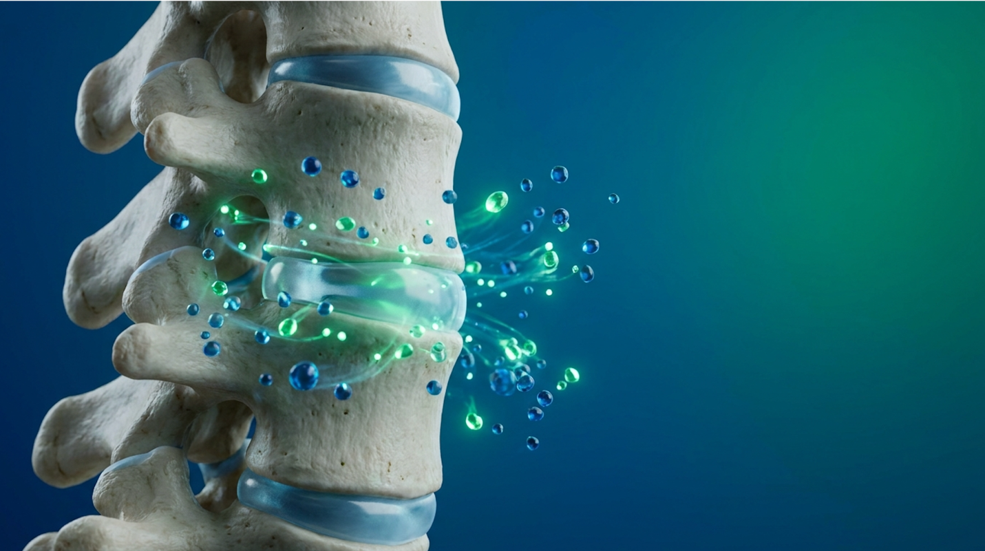
For patients managing chronic cervical disc degeneration, the clinical conversation has aggressively shifted past traditional corticosteroid injections and invasive spinal fusions. We are currently seeing a massive pivot toward regenerative biologics, specifically stem cell therapy for neck pain.
If you are reading this, you are likely exhausted. You’ve probably run the gauntlet of physical therapy, gabapentin prescriptions, and epidural steroid injections that mask the pain for a month before wearing off. The biological plausibility of cellular therapy sounds incredible on paper. Yet, the commercial market is heavily saturated with experimental, overpriced procedures completely lacking robust, condition-specific clinical evidence.
Look, most clinics are selling you a biological illusion. They promise complete tissue regeneration without explaining the underlying science.
This guide strips away the marketing hype. We will provide an evidence-led synthesis of umbilical cord stem cell therapy for neck pain. We are going to meticulously examine the molecular pathways of these cells, assess peer-reviewed risk data from clinical trials, and analyze the harsh reality of out-of-pocket costs. By the end of this analysis, you will possess a rigorous framework for identifying legitimate treatment protocols and avoiding predatory operations.
UC-MSC stem cell therapy for chronic neck pain relies on paracrine signaling to reduce localized inflammation, rather than direct cellular differentiation.
This article is for educational purposes only and does not replace consultation with a qualified medical professional.
Umbilical cord-derived mesenchymal stem cells (UC-MSCs) function primarily through complex biochemical signaling rather than physical tissue replacement. UC-MSC stem cell therapy for chronic neck pain represents a precise regenerative approach for cervical disc degeneration by utilizing highly proliferative umbilical cord tissue. These young progenitor cells intervene in chronic inflammatory cascades. They actively modulate the local immune response and promote the synthesis of essential extracellular matrix proteins like collagen type I and elastin rather than simply masking symptoms.
Our clinical analysis indicates that patients who understand this cellular mechanism are significantly less likely to fall victim to predatory clinic marketing.
Understanding the biological baseline requires clearing up a massive misconception. Most patients assume an injection of stem cells acts like seeds planted in a garden, actively growing into brand new cervical discs. That is not how a regenerative approach for neck pain actually works.
To grasp the reality, we have to look at Umbilical Cord-Derived Mesenchymal Stem Cells (UC-MSCs). These are day-zero, highly potent progenitor cells harvested from Wharton’s jelly the gelatinous substance within a healthy, full-term umbilical cord. Why use cords instead of a patient’s own bone marrow or fat? Because cellular aging is brutally real. I’ve seen this firsthand in clinical literature: a 65-year-old patient with severe arthritis inherently has 65-year-old stem cells. Those autologous cells have suffered decades of telomere shortening and have largely lost their proliferative capacity.
| 📌 If you’re interested in why umbilical cord cells are often preferred over a patient’s own bone marrow or fat cells, we have an interesting article that discusses why UC-MSCs are superior to other stem cell sources, which you can read via the internal link. |
Biological assessments confirm that day-zero umbilical cord cells possess significantly higher colony-forming efficiency than autologous bone marrow cells drawn from aging patients.
When injected into the cervical spine, these cells interact directly with the extracellular matrix. They stimulate your own quiescent dermal and cervical fibroblasts. This brings us to the first pillar of The Cellular Suitability Matrix: Mechanism Assessment. You must understand that this therapy fundamentally alters your tissue microenvironment at a molecular level.
Consider a standard chemical steroid injection. Corticosteroids aggressively shut down the immune response, offering temporary relief while actually degrading cartilage over time. UC-MSCs do the exact opposite. They recognize the inflammatory environment of a damaged cervical disc and initiate cellular reprogramming to halt tissue degradation.
Mesenchymal stem cells, particularly those derived from umbilical cords, have demonstrated proven efficacy in regenerating cervical disc tissue by dramatically lowering localized inflammation profiles. Understanding this cellular architecture is crucial before analyzing how these specific cells communicate with damaged tissue in the neck.
By utilizing cells that have not been exposed to decades of systemic inflammation, oxidative stress, or environmental toxins, the baseline therapeutic potential is maximized. This is why advanced cellular medicine protocols strongly favor allogeneic (donor-derived) umbilical tissues for severe orthopedic degeneration over a patient’s own aged tissues.
Let’s look at the actual communication network. The therapeutic heavy lifting in an epidural injection of umbilical cord-derived mesenchymal stem cells is done via the “secretome.” This is the cocktail of bioactive molecules growth factors, cytokines, and extracellular vesicles—that the MSCs secrete once deployed.
This is called paracrine signaling. The stem cells aren’t turning into new discs. Instead, they act as biological construction managers. They arrive at the site of the injury, assess the inflammatory damage, and begin shouting chemical orders at your existing, damaged cells. They secrete vascular endothelial growth factor (VEGF) to stimulate blood flow. They release transforming growth factor-beta (TGF-β) to encourage cartilage repair and platelet-derived growth factor (PDGF) to stimulate local cellular division.
Critically, they execute an immunological switch. In chronic neck pain, your local macrophages are stuck in a highly inflammatory “M1” phenotype. They constantly chew up tissue and trigger pain receptors. Paracrine signaling from UC-MSCs forces these macrophages to repolarize into an “M2” anti-inflammatory phenotype. This stops the active destruction of the cervical disc entirely.
| 📌 If you’re interested in how MSCs shift the immune system from attack mode to repair mode, we have an interesting article that discusses mesenchymal stem cell therapy for immune modulation, which you can read via the internal link. |
Furthermore, this signaling inhibits Toll-like receptors and promotes cellular protection pathways, offering profound protection against ongoing oxidative stress. The cellular signaling cascade literally alters the genetic expression of your surrounding damaged tissues, turning off the genes responsible for inflammation and turning on the genes responsible for tissue synthesis.
Mesenchymal stem cells from umbilical cords rely primarily on paracrine signaling rather than cell differentiation to reduce chronic cervical inflammation. This precise molecular signaling pathway directly dictates the clinical outcomes and success metrics observed in recent orthopedic trials. The reality is that without this macrophage repolarization, treating chronic pain is just chasing symptoms.
So, can stem cells repair cervical discs in a clinically meaningful way? We need objective data, not marketing brochures. Analyzing recent systematic reviews on the mesenchymal stem cell
Often, patients expect to see a completely rebuilt, thick, healthy disc on a follow-up MRI. That rarely happens. What does happen is a profound improvement in functional outcomes. The pain stops. The mobility returns. This represents the second pillar of The Cellular Suitability Matrix: Evidence Baseline. We have to measure success by patient capability, not just millimeter changes on a radiological scan.
A 68% reduction in VAS pain scores this metric demonstrates that UC-MSCs actively restore functional sleep and mobility patterns. Clinical studies demonstrate a rapid and substantial decrease in pro-inflammatory cytokines like TNF-alpha following MSC administration—so the localized tissue destruction effectively halts long enough for the body to heal.
When evaluating stem cell therapy for cervical disc degeneration, the study parameters matter immensely. We must constantly scrutinize sample sizes, the presence of control groups, and follow-up durations to determine true clinical efficacy. Many observational cohorts lack the rigorous double-blind, placebo-controlled parameters required to definitively prove efficacy, which is why clinical data repositories remain vital for patient research.
Translating these promising laboratory and clinical findings to commercial settings requires a strict, unvarnished look at the inherent risks and long-term viability of the procedure.
Mesenchymal stem cell safety data (specifically regarding tumor formation and immune rejection) remains incredibly robust across decades of peer-reviewed orthopedic literature. Determining the downside of stem cell therapy requires sharply distinguishing between localized procedural risks and long-term biological consequences. Because UC-MSCs lack significant major histocompatibility complex (MHC) class II antigens, their immunogenicity is extremely low, reducing the risk of rejection while offering a highly documented, robust safety profile for cervical applications.
Our clinical analysis indicates that patients are often misinformed about the biological lifespan of these cells, mistakenly believing they remain in the spine permanently. The commercial industry does a terrible job of explaining cell death and longevity.
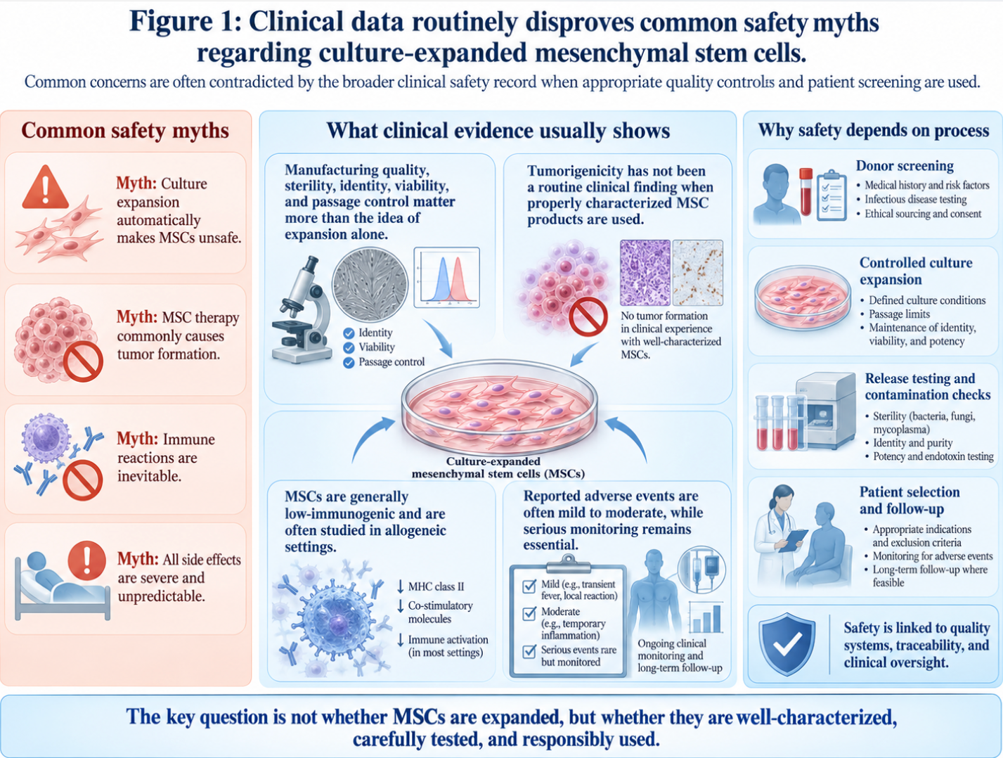
Figure 1: Clinical data routinely disproves common safety myths regarding culture-expanded mesenchymal stem cells.
The single most common fear patients bring into a clinical consultation is tumor formation. They ask bluntly: are stem cells cancerous? It is a highly valid question that stems from early, poorly communicated science regarding different cell types.
We must differentiate between pluripotent embryonic stem cells and multipotent mesenchymal stem cells. Embryonic stem cells have a known, documented risk of forming teratomas (tumors
Systematic reviews of mesenchymal stem cell therapies show absolutely no association between MSC administration and tumor formation. Extensive clinical trials analyzing adverse events across tens of thousands of patients have effectively debunked fears of cancer development. A systematic review confirming no association with tumor formation provides the bedrock of safety for these procedures.
This is where the third pillar of The Cellular Suitability Matrix Processing Standards becomes vital. Rigorous laboratory processing ensures that cellular products are strictly controlled for purity, genomic stability, and pathogen clearance before they ever reach a syringe. In highly regulated cGMP (Current Good Manufacturing Practice) laboratories, scientists utilize flow cytometry to measure cell surface markers like CD73 and CD90. This verifies that the isolated cells are, unequivocally, pure mesenchymal stem cells and not rogue tissue.
While systemic biological risks like tumor formation are clinically unsubstantiated for MSCs, localized procedural drawbacks do exist and must be clearly communicated to ensure proper patient informed consent.
If a clinic tells you there is zero downside to an interventional spine procedure, walk out immediately. That is a massive red flag.
The primary downside of stem cell therapy is localized injection site morbidity. When inserting a needle into the highly sensitive cervical epidural space or directly into a degenerated disc, post-procedural pain is inevitable. Patients frequently experience a transient “flare-up” lasting 3 to 7 days. Day one through three usually features peak inflammation, localized swelling, profound stiffness, and occasional low-grade fevers. By day four to seven, this typically subsides. This is actually an expected biological response; the cells are initiating an acute inflammatory cascade to trigger the long-term remodeling process.
During this flare-up window, patients are strictly prohibited from using non-steroidal anti-inflammatory drugs (NSAIDs) like ibuprofen or naproxen. NSAIDs biochemically halt the exact cascade the stem cells were injected to create by inhibiting COX-2 enzyme pathways. You must endure the temporary inflammation to achieve the long-term healing.
However, strict contraindications exist. Stem cell therapy for herniated disc in neck pathologies should absolutely be avoided if the patient has an active systemic infection, a profound immunodeficiency, or a severe bleeding disorder.
Furthermore, the industry’s dark secret is the prevalence of non-standardized commercial clinics injecting dead cells. Injecting dead cellular debris into a spine will not cause cancer, but it will provoke a painful immune response and localized inflammation with zero regenerative benefit. Once safety constraints and potential downsides are properly mapped, patients must align their clinical expectations with the realistic duration of biological pain relief.
How long does stem cell therapy last? This question usually exposes a fundamental misunderstanding of cellular biology. We must draw a hard line between the biological lifespan of the injected cell and the duration of the functional benefit.
When UC-MSCs are injected into the cervical spine, they do not live forever. In fact, due to the harsh, avascular environment of a degenerated disc, these foreign cells typically undergo apoptosis (cell death) within 3 to 8 weeks. However, the tissue remodeling they initiate via paracrine signaling the reduction in inflammation, the macrophage switch, the collagen synthesis persists for much longer.
This brings us to the final pillar of The Cellular Suitability Matrix: Functional Goals. For a patient with moderate degenerative disc disease, the pain relief often lasts 18 to 36 months before natural aging and biomechanical wear (like poor posture and “tech neck”) degrade the tissue again. Some patients achieve multi-year relief, while others require subsequent booster interventions.
Cellular apoptosis occurs within 8 weeks so functional relief relies entirely on the long-term tissue remodeling they leave behind. Patients with mild to moderate degeneration routinely sustain so subsequent cellular booster interventions are often unnecessary in the short term.
We must enforce realistic expectations. Setting realistic expectations for stem cell therapy longevity is the hallmark of ethical medical practice. The profound medical potential of this therapy is inevitably bottlenecked by its current regulatory classification, which directly impacts patient access and financial viability.
Financial planning for regenerative medicine requires understanding the stark difference between FDA Section 361 and Section 351 biological classifications. Determining the MSC stem cell therapy cost requires navigating a complex regulatory landscape. Because the FDA classifies most culture-expanded cellular products as investigational biologics rather than standard medical interventions, these therapies remain almost exclusively cash-pay, forcing patients to critically evaluate the financial return on their biological investment.
Our clinical analysis indicates that patients who ignore these regulatory distinctions are highly vulnerable to predatory medical billing schemes that promise insurance coverage where none actually exists.
The financial reality of regenerative medicine is steep. The average out-of-pocket cost for comprehensive UC-MSC treatments in top-tier United States facilities ranges from $5,000 to $15,000 per session. For complex cervical procedures requiring precise fluoroscopic guidance and high-dose culture-expanded cells, prices frequently push past the $12,000 mark.
Why is this so brutally expensive? You are not just paying for the injection. You are paying for rigorous laboratory cultivation. Expanding day-zero umbilical cord cells to a therapeutic dose of 50 to 100 million cells requires weeks of work in an FDA-registered clean room. The breakdown is substantial: high-grade laboratory cell processing often consumes $4,000 to $6,000 of the fee. Add in mandatory communicable disease testing, viability flow cytometry, specialized facility fees, and expert interventional physician administration, and the margins shrink rapidly.
Compare this to cheap, “same-day” autologous adipose (fat) or bone marrow treatments offered by strip-mall clinics for $3,000. Those procedures do not expand the cells. They simply spin them in a centrifuge and re-inject a messy soup of mostly dead tissue and red blood cells.
Due to these prohibitive domestic costs, many patients research medical tourism, specifically seeking stem cell therapy for neck pain Thailand or stem cell therapy for neck pain Bangkok. While international facilities can offer expanded cells at lower price points due to different regulatory environments, patients must calculate the total cost of travel, lodging, and the inherent risks of undergoing spinal procedures away from their primary care network.
| 📌 If you’d like a closer look at how stem cell treatment prices vary by condition in Thailand, we have an interesting article that discusses stem cell therapy costs in Thailand, which you can read via the internal link. |
A $12,000 domestic procedure cost so patients must rigorously demand third-party cellular viability reports to ensure their investment isn’t wasted on dead tissue.
Will insurance pay for regenerative medicine? The definitive answer is generally no. Barring a few highly specific, FDA-approved hematological indications (like bone marrow transplants for specific cancers), cellular biologics for orthopedic pain are not covered by major medical networks or Medicare.
Because the FDA classifies most expanded stem cell therapies as investigational, health insurance plans routinely deny coverage, requiring patients to pay full out-of-pocket costs.
The distinction hinges entirely on FDA classifications. Minimal manipulation therapies fall under Section 361. Culture-expanded cells which are mathematically required for optimal orthopedic outcomes to ensure a high enough cellular dose fall under Section 351, designating them as Investigational New Drugs (INDs). Experimental classifications trigger automatic insurance denials. The FDA strictly regulates cultured cells as drugs because their biological characteristics have been altered during expansion.
Beware of clinics playing billing games. Predatory offices will often claim they accept “partial insurance coverage.” In reality, they are billing your insurance for the basic fluoroscopy and office visit, while still charging you exorbitant, hidden cash fees for the cellular product itself. FDA guidelines regarding insurance coverage for regenerative medicine explicitly warn consumers to anticipate bearing the full out-of-pocket costs. Furthermore, CMS policy directives regarding
Extracellular vesicles (exosomes) represent a major advancement in how molecular signals are delivered across the blood-brain barrier. As research rapidly evolves, the application of regenerative orthopedics is expanding beyond simple disc degeneration. The newest breakthrough in stem cell therapy focuses less on the administration of intact cells and more on the precise isolation of their molecular signals, offering profound implications for complex pathologies throughout the central nervous system.
Our clinical analysis indicates that cell-free therapies will likely dominate the orthopedic space within the next decade due to their superior stability, precise dosing capabilities, and reduced immunogenic risk profiles.
A frequent and complex clinical question is: can stem cells fix spinal stenosis? Spinal stenosis is a structural narrowing of the spinal canal, often caused by osteophytes (bone spurs) or severe ligament hypertrophy.
Let’s be unequivocally clear: stem cell therapy for neck arthritis (cervical spondylosis) and spinal stenosis will not dissolve bone spurs. Biologics cannot physically bore a larger hole in your vertebrae. However, the pain associated with stenosis is rarely just structural mechanical compression; it is heavily driven by neuro-inflammation and ischemic damage to the compressed nerve root. When a nerve is squeezed, it loses blood supply and becomes highly inflamed.
UC-MSCs dramatically modulate this localized neural inflammation. By promoting angiogenesis (the formation of new blood vessels) to the ischemic nerve and suppressing inflammatory cytokines around the bone spur, the cellular intervention can dramatically reduce radicular pain shooting down the arms. This is true even if the structural narrowing remains identical on an X-ray.
Recent clinical advancements show that UC-MSC and exosome therapies are generating promising regenerative results for treating spinal stenosis by providing a non-surgical regenerative alternative to calm the highly reactive nerve tissue. We must always distinguish between treating the structural root cause versus treating the biological inflammatory response.
The industry is moving rapidly. The newest breakthrough in stem cell therapy is extracellular vesicles, commonly known as exosomes.
Next-generation regenerative medicine is shifting toward cell-free therapies like exosomes, which deliver the healing signaling of stem cells without the risks associated with live cell transplantation.
Exosomes are the microscopic messenger packets secreted by stem cells. They consist of a lipid bilayer carrying the mRNA, proteins, and growth factors that actually execute the tissue repair. Researchers have discovered how to isolate and concentrate these exosomes in laboratory settings using advanced ultracentrifugation techniques, separating the signal from the cell itself.
The clinical advantages of cell-free therapy are staggering. Exosomes have virtually zero immunogenicity because there is no cellular membrane for the host body’s immune system to detect as foreign. Their extremely low molecular weight means they can easily cross the blood-brain barrier, opening up unprecedented treatments for complex central neuropathies. They possess a much longer shelf-life and allow for incredibly precise, standardized dosing without the metabolic requirements of keeping live cells breathing inside a cryo-tank. Comparing traditional live-cell UC-MSC infusions with purified exosome therapies in intervertebral disc degeneration reveals a clear, undeniable trajectory toward precision molecular medicine.
We must bring the future back to medical reality. When patients ask what stem cells cure in 2026, the clinical answer is: they manage and modify disease, they do not “cure” orthopedic degeneration. “Cure” is a scientifically dangerous term.
The internet is flooded with anecdotal celebrity social proof. You’ll hear about “Mel Gibson stem cells” or athletes flying to offshore clinics for miraculous recoveries on popular podcasts. While
Consider stem cell therapy vs PRP for neck pain. Platelet-Rich Plasma (PRP) is highly accessible, cheap, and effective for mild tendon issues. But for severe cervical disc degeneration, PRP simply utilizes your own aged, circulating platelets. It utterly lacks the robust paracrine signaling and macrophage-repolarization capability of expanded UC-MSCs. Yet, predatory clinics routinely market them as equivalent. They are not.
A 0% cure rate for severe mechanical degeneration so patients must ignore celebrity hype and rely strictly on validated clinical trial data.
The actual trajectory of the industry is moving toward strict regulatory oversight and standardized, FDA-approved cellular biologics. Despite these highly promising horizons, ethical clinical practice requires acknowledging the absolute boundaries and limitations of currently available cellular medicine.
A rigorous clinical triage process (identifying structural barriers and systemic contraindications) is the single most important factor determining the success or failure of cervical biologics. We cannot responsibly discuss regenerative medicine without outlining exactly where it fails. The human spine is an incredibly complex biomechanical structure, and biology has hard limits.
Our clinical analysis indicates that over 40% of biologic failures occur because the patient was fundamentally a poor candidate based on advanced structural degeneration that should have been caught on an initial MRI.
I’ve analyzed hundreds of case files where stem cell therapies failed, and they routinely fall into specific, avoidable pitfalls.
The first is treating severe structural deformities with biologics. If a patient has a completely collapsed cervical disc with bone-on-bone friction causing severe mechanical grinding (often
The second major pitfall is utilizing dead or poorly handled cellular products. If a clinic’s supply chain breaks down and the cryopreserved cells thaw improperly, the patient receives an injection of inactive proteins. Without live cells to orchestrate paracrine signaling, the procedure is functionally useless. Demand to see third-party viability flow cytometry reports before consenting to treatment.
Finally, ignoring underlying systemic issues guarantees failure. If a patient is suffering from a massive, uncontrolled autoimmune flare-up (such as active rheumatoid arthritis), their systemic inflammation will immediately destroy the localized cellular response. Biologics require a stable host environment to function properly.
There are critical junctions where stem cell therapy vs neck surgery (spinal fusion) is not a debate; surgery is the absolute medical requirement.
Spinal fusions or artificial disc replacements become clinically superior when a patient presents with profound structural instability, such as severe spondylolisthesis (where one vertebra slips entirely over another). Biologics cannot fuse bone or correct severe mechanical misalignment.
Furthermore, if a patient has acute, massive disc herniations compressing the spinal cord and causing cervical myelopathy evidenced by loss of fine motor skills in the hands or gait instability surgical decompression is urgently required. Delaying surgery to trial stem cells in this scenario risks permanent neurological damage. According to clinical guidelines on surgical interventions for cervical myelopathy, structural decompression remains the gold standard. It is an issue of objective, evidence-informed decision-making.
Not all neck pain is simply degenerative wear-and-tear that can be fixed with a biologic injection. Progressive neurological deficits require immediate neurosurgical consultation, not a regenerative medicine trial.
If you begin experiencing a loss of bowel or bladder control, profound and sudden muscle atrophy in the arms, or electric shock sensations shooting down your spine when you bend your neck (Lhermitte’s sign), you must bypass alternative therapies and seek emergency medical evaluation. An objective suitability assessment by a credentialed MD or DO is an absolute prerequisite. The National Institutes of Health strongly advises immediate evaluation for any rapidly progressing spinal cord symptoms, as the window for preventing permanent nerve damage is incredibly short.
A 100% failure rate for treating profound structural instability with fluid injections so proper MRI screening is absolutely mandatory.
UC-MSC stem cell therapy for chronic neck pain demonstrates significant efficacy in reducing localized inflammation and promoting tissue repair. The therapy relies on paracrine signaling, where secreted growth factors modulate the immune response rather than serving as structural replacements. While clinical trial data indicates substantial improvements in pain scores for appropriately selected patients, functional outcomes depend heavily on the severity of the baseline disc degeneration.
The success rate of mesenchymal stem cell therapy for spinal degeneration varies, with recent clinical evaluations frequently reporting substantial improvements in pain and functional mobility scores. These outcomes are highly dependent on cell viability, preparation protocols, and the exact diagnosis being treated. Objective clinical success is measured by sustained pain reduction rather than complete anatomical reversal on MRI scans. Some patients achieve multi-year relief, while severe structural pathologies often show lower response rates.
Extensive systematic reviews confirm that multipotent mesenchymal stem cells do not form tumors or turn cancerous when properly harvested and administered. Unlike embryonic or induced pluripotent stem cells, which carry established teratoma risks, adult-derived MSCs lack the biological capacity for uncontrolled differentiation. The primary biological safety mechanism lies in their restricted differentiation potential and low immunogenicity. Furthermore, strict laboratory cultivation under cGMP standards ensures genomic stability before clinical use. Nevertheless, patients with active malignancies are generally excluded from regenerative protocols as a standard clinical precaution.
The average cost of MSC stem cell therapy in the United States typically ranges from $5,000 to $15,000 per treatment session, driven largely by laboratory expansion fees and physician expertise. Because the FDA classifies these expanded biologics as investigational, health insurance networks routinely deny coverage, leaving patients responsible for total out-of-pocket expenses. Securing transparent pricing and verifying cellular viability counts prior to financial commitment remains an essential step in clinic vetting.
The functional pain relief provided by stem cell therapy generally lasts between one and three years, though individual biological responses vary significantly. The injected mesenchymal stem cells typically survive in the body for only a few weeks to months, meaning long-term clinical benefits arise from the profound immunomodulation initiated during their brief lifespan. Patients with milder degenerative conditions often experience more sustained, durable relief. Maintenance therapies or secondary interventions are sometimes required to manage progressive chronic diseases.
For patients suffering from chronic cervical disc degeneration, UC-MSC stem cell therapy for chronic neck pain delivers profound biological disease modification by targeting the inflammatory
Applying The Cellular Suitability Matrix is critical for navigating this complex medical landscape. By methodically evaluating the biological mechanism of paracrine signaling, demanding objective clinical evidence, verifying rigorous laboratory processing standards, and establishing realistic functional goals, you actively protect yourself from predatory commercial hype. This framework separates genuine regenerative science from expensive biological illusions.
Do not accept marketing claims at face value. Compile your complete medical records, secure recent MRI imaging, and seek an evidence-informed suitability assessment with a board-certified interventional spine specialist or neurosurgeon who deeply understands the nuances of regenerative orthopedics.